LICHEN - INTRODUCTION, COMPOSITION, MORPHOLOGICAL TYPES, THALLUS STRUCTURE, REPRODUCTION
A. INTRODUCTION
Lichens
are dorsiventrally thallophytic plants which are formed due to close
association of an alga and a fungus. The fungal component of lichen is called mycobiont and the algal component is
called photobiont. The two organisms live together in intimate
connection forming a compound thallus called consortium.
In
a symbiotic association between the two partners, the fungus lives
saprophytically on algal cells and obtains food from them. On the contrary alga
gets benefited by fungi in providing water and nutrients. Fungus also protects
algal cells from high light intensity.
B. COMPOSITION
OF LICHEN
Lichen
consists of two main components, photobiont
and mycobiont.
Photobiont:- The algal component of
lichen is called photobiont. The
photobiont is mainly Cyanophyceae and rarely Chlorophyceae. Altogether 37
genera have been identified as lichen photobiont (Hawksworth and Hill, 1984).
These algae belong to blue-green (Cyanophyta) and green (Chlorophyta) algae.
Lichen forming blue-green algae are Nostoc,
Anabaena, Gloeocapsa, Stigonema, Chroococcus, Scytonema, etc. Common green
algae are Chlorella, Cephaleuros,
Trentepohlia, Trebouxia, etc.
Mycobiont:- The fungal component of
lichen is called mycobiont. The
mycobiont mainly belong to Basidiomycotina and rarely Deuteromycotina. Most of
the Ascomycetous lichens belong to Discomycetes or Pyrenomycetes. Dictyonema, Multiclavula, Omphalina,
etc., are some of the species of lichen forming genera of Basidiomycotina.
There are 55 species of lichen forming Deuteromycotina of which Blarneya hibernica is the most common.
C. MORPHOLOGICAL
TYPES OF LICHEN
Lichens
exhibit several morphological types. Hawksworth and Hill (1984), describe the
following types of lichens –
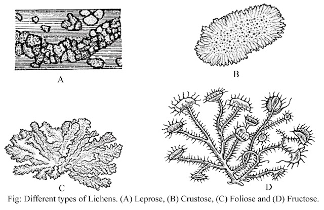
1. Leprose lichen:- This type is the
simplest, here the thallus organization is very simple. The fungal hyphae
doesnot envelop the algal cells throughout. This type of lichen grows
superficially over the substratum and provides a powdery appearance and
therefore is called leprose. Example – Leparia
incana.
2. Crustose lichen:- In this type, the lichen thallus
is very closely adhered to the substratum and provides a crust-like appearance
on rocks, soil, tree barks, etc. The algal cells are covered by a distinct
layer of fungal cells. It is very difficult to separate the thallus from the
substratum.
Some
variations of crustose lichens are as follows –
(a)
Placodioid – Here the entire surface
of the thallus is radially striate and contains raised marginal tissues.
Example – Lecanora, Caloplaca, etc.
(b) Squamulose
– Here the outer surface contains overlapping scale-like squamules. Example – Psora.
3. Foliose lichen:- In this type, the
thallus is flat, dorsiventral, leaf-like, well branched with irregular and
lobed margin. The thallus is attached to the substratum with the help of
rhizoid-like structure called rhizines.
Externally they look like that of crinkled and twisted leaves. Example – Parmelia, Physcia, Peltigera, Collema,
etc.
4. Fruticose lichen:- In this type, the
lichen thallus is vary in shape. They are well branched, erect or pedalous
bushy which provide shrubby structure i.e., twig-like appearance. Example – Usnea, Cladonia, Letharia, etc.
5. Filamentous lichen:- In this type,
the algal component has the main role in the formation of the structure of the
lichen thallus. The algal partner is well developed and filamentous which is
covered by a few fungal hyphae only. The algal partner is dominant and as such
named filamentous by Hawksworth and Hill (1984). Example – Ephebe, Racodium, Coenogonium, etc.
D. STRUCTURE OF LICHEN THALLUS
On
the basis of internal structure of thallus, the lichens are divided into two
groups namely homoiomerous and heteromerous lichens.
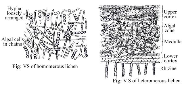
1. Structure of homoiomerous lichen thallus:-
The lichen thallus with the algal component scattered uniformly between the fungal
hyphae throughout is called homioisomerous. In this type, the lichen thallus
shows a simple structure with little differentiation. It consists of loosely
interwoven mass of fungal hyphae with algal cells equally distributed
throughout. Example – Collema, Leptogium,
etc.
2. Structure of heteromerous lichen
thallus:- Most
of the lichen belong to this category. They exhibit considerable
differentiation and layered structure. A VS through a heteromerous lichen
thallus shows the following structures –
(a)
Upper cortex:- It forms the upper surface which is generally thick and
protective. The fungal hyphae in this region grow more or less vertically and
are compactly interwoven to produce pseudoparenchymatous layer. The fungal hyphae
are compactly arranged so that there is no intercellular space between the
hyphae.
(b)
Algal zone:- It is the blue green or green zone which lies immediately
beneath the upper cortex. It consists of a network of loosely interwoven fungal
hyphae with the algal cells of green alga, intermixed with the fungal hyphae.
The algal region is the photosynthetic region of the lichen thallus.
(c)
Medula:- It contains the central core of the thallus. It is less compact
and consists of loosely interwoven hyphae with large spaces between them in
certain regions. The fungal hyphae in this region are scattered and usually
have thick walls. They run in all directions. The central hyphae of the
medullary region usually run longitudinally.
(d) Lower cortex:- It forms the
lower surface of the thallus and is composed of densely compacted hyphae. They
may run perpendicular to the surface of the thallus or parallel to it. Bundles
of hyphae (rhizinae) often arise from the surface of the, lower cortex and
penetrate the substratum to function as anchoring organs. In some lichen
species the lower cortex is absent.
E. REPRODUCTION
The
common methods of reproduction in lichen are by vegetative, asexual and sexual methods –
1. Vegetative reproduction:- Lichen
thallus reproduce vegetatively by various methods. These are –
(a)
Fragmentation:- It consists in the breaking up of the thallus into segments
which are distributed to start new growth. Fragmentation is brought about by ageing and accidental. In ageing the older cells in the basal part of the
thallus die and separate the branches or lobes, each of which grows into new
thallus. Sometimes larger portion of the thallus breaks accidentally from the
parent thallus. Each of these broken parts develops into new individual.
(b) Soredia:- These are small, rounded granules or bud-lie
outgrowths which develop in the form of a grayish-white or grayish-green powder
in extensive patches usually over the upper surface or edges of the thalli of
many species of lichen. Each soridium contains one or few algal cells closely
surrounded by a little weft of fungal hyphae produced by branching of the hypha
from the algal region.
(c)
Isidia:- These are small conical warts developed on the thallus of many
lichens. Isidia are usually constricted at the base and thus can easily be
broken off. Under the favourable conditions, each isidium grows into a new
lichen thallus.
Lichen thallus also reproduces
vegetatively by phyllidia, blastidia,
schizidia, hormocysts, goniocysts, etc.
2. Asexual reproduction:- Lichen
thallus reproduce asexually by various methods. These are –
(a)
Conidia:- In many lichens, conidia of different shape and size develop in
multihypal structure known as conidiomata.
The conidiomata are embedded in a flask-shaped structure, pycnidia. Each pycnidium appears to the exterior by means of ostiole. Conidiophores are developed
from the inner lining of the wall of pycnidium. Conidia are produced
terminally, laterally, or intercalary on conidiophores. Each conidium is
colourlss and may be cylindrical or sickle-shaped or filiform in structure.
Each conidium germinates by producing a germ hypha.
(b)
Oidia:- These are spores developed by breaking up of small fragments of
lichen thallus. The oidium germinates to produce lichen thallus.
3. Sexual reproduction:- Sexual
reproduction takes place by the production of specialized male and female
reproductive structures. Male reproductive structure is called spermogonium and the female reproductive
structure is called carpogonium.
(a)
Spermogonia:- In certain species of lichens pycnidia-like structures are
reported to function as spermogonia. Each spermogonium is a flask-shaped
receptacle immersed in a small elevation on the upper surface of the thallus.
It opens by a small pore, an ostiole,
at the surface. The cavity of the spermogonium is filled with the sterile and
fertile hyphae. The fertile hyphae produce minute rounded cells at their tips.
These are the male cells and are called spermatia.
They are non motile and are produced in large numbers. The spermatia are set
free in a slimy mass which oozes out through ostiole.
(b)
Carpogonia:- The carpogonium is a cellular filament. It consists of two
portions – the lower coiled portion and the upper straight portion. The coiled
portion constitutes the ascogonium.
It is multicellular and the cells are uninucleate. In certain species they are
multinucleate. The ascogonium lies deep in the medullary region of the thallus.
The carpogonia either develop from the hyphae or from the medullary region or
from the hyphae deep in the algal layer of the lichen thallus. The straight
upper portion of the carpogonium is called the trichogyne. It is also multicellular. The cells of trichogyne are
elongated and septate. The terminal portion of the trichogyne ends in a long
cell which projects beyond the surface of the thallus.
(c)
Fertilization:- At the time of fertilization, several spermatia are lodge
to the sticky tip of the trichogyne. A few empty spermatia are found at the tip
of the trichogyne - this seems that
their protoplast have migrated into the trichogyne. But the actual migration of
the male nuclei down the trichogyne has not been seen.
(d)
Formation of ascocarp and ascospores:- After fertilization several
ascogenous hypha develop from the basal portion of the ascogonium. Asci are
produced at the ends of these freely branched ascogenous hyphae. Two nuclei in
a young ascus fuse to form a diploid nucleus. This diploid nucleus divides and
redivides to form eight haploid nuclei. The asci are uninucleate or binucleate.
These asci bearing structures are ascocarps.
Normally eight ascospores are formed
in each ascus. They vary in colour, shape, size and structure. Ascospores are
unicellular, transversely septate or both transversely and longitudinally
septate.
On liberation each ascospore
germinates by producing hyphal branch. This hypal branch comes in contact with
suitable alga, ultimately lichen thallus is formed after the combined growth of
the fungus and the alga.
Prema Iswary,
Assistant Professor,
Department of Botany.
************


Thank you for the information
ReplyDelete