Clinical Pathology Case Study: Cryptococcus in a dog
Background information
| Age: | 1 year |
| Sex: | Female spayed |
| Species: | Canine |
| Breed: | Whippet |
| History: | Not known |
Clinical signs
- The image is taken from a mass on the right lateral thorax of a young dog.
- No additional history or information about this mass is available.
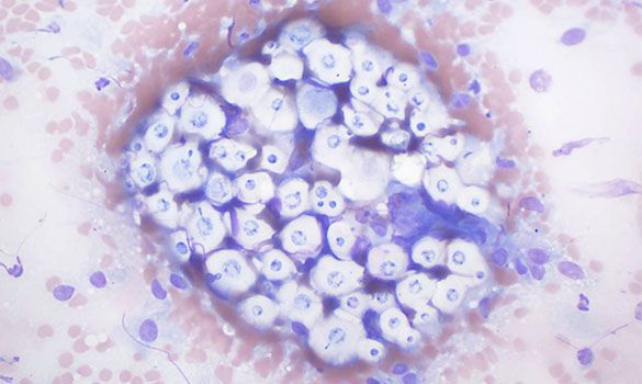
Cytologic description and interpretation
The slides were of relatively low cellularity with small amounts of blood. Nucleated cells consisted of macrophages and neutrophils with fewer small lymphocytes. There were low to moderate numbers of round yeasts admixed among the inflammatory cells. These had a thick clear capsule surrounding a round purple-coloured structure with a distinct cell wall. Narrow-based budding was observed in several organisms.
Interpretation: Findings were consistent with Cryptococcosis.
Discussion
Cryptococcus neoformans is found in the guano of many bird species as well as in decaying plant matter. Cryptococcus gattii, of emerging concern in the last decade in British Columbia, can be found in freshwater, seawater, tree bark, and in the air. Cutaneous infection with Crypcococcus spp. occurs via direct inoculation with fungal spores from the environment.
The distinctive capsule of the yeast is considered protective, helping to prevent destruction by the host immune system. Despite this, cytologic preparations from cutaneous and visceral lesions are often accompanied by a mixed inflammatory infiltrate.
This particular dog had a positive serum antigen titer of 1:2048, supporting systemic exposure to the yeasts. The lump was excised and systemic antifungal therapy was instituted. No further follow-up information was available at time of publication.
About the author
Carrie Flint, DVM, DACVP
Dr. Flint received her DVM in 2006 from Tufts University and completed a residency in clinical pathology at Michigan State University in 2009. She is board-certified in clinical pathology by the American College of Veterinary Pathologists. Dr. Flint joined the IDEXX lab in Delta, British Columbia, in 2009. Her passion for veterinary diagnostics began while working in a laboratory prior to vet school.
Outside of work, she enjoys trivia, baking, and travel, and spending time with her beloved chocolate lab, Lily.
Should you have any questions about this case or wish to discuss the diagnosis in greater detail, please do not hesitate to contact the author.
View clinical pathology case studies
NEW: Dermatophytosis in a dog, by Caroline Piché, DMV, IPSAV, MSc, Diplomate ACVP
Anaplasmosis in a dog, by Julie Webb, DVM, DACVP
Blastomycosis in a dog, by Julie Webb, DVM, DACVP
Cryptococcus in a dog, by Carrie Flint, DVM, DACVP
Cutaneous mycobacteriosis (Leprosy) in a cat, by Natalie Kowalewich, DVM, DACVP
Demodex in a dog, by Brittney Fierro, DVM, DACVP
Feline infectious peritonitis in a cat, by Heidi Peta DVM, MVSc, DACVP
Mammary fibroepithelial hyperplasia in a young cat, by Emmeline Tan, DVM, DVSc, DACVP
Microvascular dysplasia in a dog, by Cathy Monteith DVM, MVSc, DACVP
Severe hyperglobulinemia in a dog, by Sébastien Overvelde, DVM, MSc, DACVP
References
- Cowell RL, Arndt TP. Cowell and Tyler’s Diagnostic Cytology and Hematology of the Dog and Cat, 4th ed. St. Louis: Elsevier Mosby; c2014. Chapter 3, Selected Infectious Diseases; p. 54-55.
- Kidd SE et al. Cryptococcus gattii Dispersal Mechanisms, British Columbia, Canada. Emerg Infect Dis. 2007; 13(1):51-57.
- MacDougall L, et al. Spread of Cryptococcus gattii in British Columbia, Canada, and Detection in the Pacific Northwest, USA. Emerg Infect Dis. 2007; 13(1): 42-50.
- Malik R et al. Infectious Diseases of the Dog and Cat, 3rd ed. St. Louis: Saunders Elsevier, c2006. Chapter 61, Cryptococcosis; p. 584-598.
All case studies were prompted by real submissions to IDEXX Canada pathologists at one of our reference laboratories.
To protect the confidentiality of our customer and clients, the background information in each case has been slightly modified.