Approach Considerations
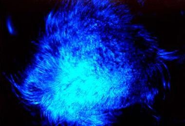
Base the diagnosis on mycologic examination via direct microscopy and culture. Sporadically, the diagnosis can be confirmed by histologic evidence of fungal invasion of the hair. Wood lamp examination may demonstrate a dull green fluorescence. See the image below.
Perform routine direct microscopic examination in 10-20% potassium hydroxide (KOH) solution, with or without dimethyl sulfoxide. This solution helps visualize fungal elements. With favus, hair must be examined immediately after adding KOH solution to observe the bubbling of KOH through the air spaces between hyphae elements. Examination of scutulum fragments shows segmental filaments and spores.
The causative organism is identified on culture, which usually is performed on Sabouraud agar with the addition of cycloheximide and chloramphenicol. These 2 substances inhibit growth of bacteria and other fungi; thus, pure dermatophyte colonies are obtained. In patients who are chronically affected, positive culture results may be difficult to obtain. See the images below.
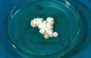 Culture of Trichophyton schoenleinii on Sabouraud agar. Courtesy of Anna Pawlowicz, PhD, and Professor Barbara Raszeja-Kotelba, MD, Dermatology, University School of Poznan, Poland.
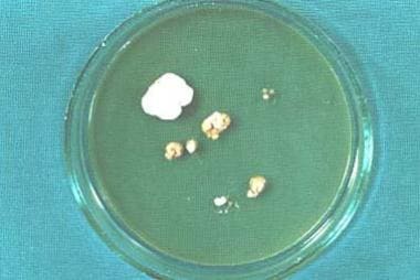 Culture of Trichophyton schoenleinii on Sabouraud agar. Note pleomorphism of the culture. Courtesy of Anna Pawlowicz, PhD, and Professor Barbara Raszeja-Kotelba, MD, Dermatology, University School of Poznan, Poland.
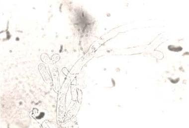 Microculture of Trichophyton schoenleinii shows dichotomic branching and terminal swelling. Light-field microscopy, original magnification X 1000. Courtesy of Anna Pawlowicz, PhD, and Professor Barbara Raszeja-Kotelba, MD, Dermatology, University School of Poznan, Poland.
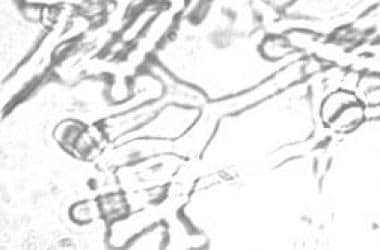 Microculture of Trichophyton schoenleinii shows characteristic dichotomic branching. Light-field microscopy, original magnification X 1000. Courtesy of Anna Pawlowicz, PhD, and Professor Barbara Raszeja-Kotelba, MD, Dermatology, University School of Poznan, Poland.
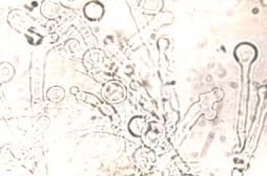 Microculture of Trichophyton schoenleinii shows numerous terminal chlamydospores. Light-field microscopy, original magnification X 1200. Courtesy of Anna Pawlowicz, PhD, and Professor Barbara Raszeja-Kotelba, MD, Dermatology, University School of Poznan, Poland.
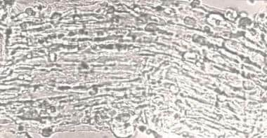 Infected hair filled with hyphae shows bubbles of gas and gas tunnels (light field microscopy, original magnification X 2300).
Grocott staining may detect fungal elements in the crust and hair bulbs, with T schoenleinii diagnosed by polymerase chain reaction–based DNA sequencing using the formalin-fixed, paraffin-embedded tissue samples. [26]
Histologic Findings
Hematoxylin and eosin staining may visualize fungal elements insufficiently; therefore, periodic acid-Schiff staining is recommended. Biopsy sections reveal mycelium in the scutulum and horny layer of the epidermis, which is atrophic. Scutulum borders show well-preserved hyphae; however, the center of the lesion contains degenerating mycelium and granular debris. A moderately severe inflammatory infiltrate is present in the dermis.
-
Tinea favosa of the scalp shows erythematous lesions with pityroid scaling. Some hairs are short and brittle.
-
Favus of the scalp shows extensive lesions with scarring alopecia.
-
Typical fluorescence under Wood lamp examination.
-
Favus, wax montage. Courtesy of Professor Dr Feliks Wasik, Dermatology, Medical University of Wroclaw, Poland.
-
Black man, aged 45 years, with favuslike yellow crusting of scalp. Potassium hydroxide and fungal culture were negative.
-
Culture of Trichophyton schoenleinii on Sabouraud agar. Courtesy of Anna Pawlowicz, PhD, and Professor Barbara Raszeja-Kotelba, MD, Dermatology, University School of Poznan, Poland.
-
Culture of Trichophyton schoenleinii on Sabouraud agar. Note pleomorphism of the culture. Courtesy of Anna Pawlowicz, PhD, and Professor Barbara Raszeja-Kotelba, MD, Dermatology, University School of Poznan, Poland.
-
Microculture of Trichophyton schoenleinii shows dichotomic branching and terminal swelling. Light-field microscopy, original magnification X 1000. Courtesy of Anna Pawlowicz, PhD, and Professor Barbara Raszeja-Kotelba, MD, Dermatology, University School of Poznan, Poland.
-
Microculture of Trichophyton schoenleinii shows characteristic dichotomic branching. Light-field microscopy, original magnification X 1000. Courtesy of Anna Pawlowicz, PhD, and Professor Barbara Raszeja-Kotelba, MD, Dermatology, University School of Poznan, Poland.
-
Microculture of Trichophyton schoenleinii shows numerous terminal chlamydospores. Light-field microscopy, original magnification X 1200. Courtesy of Anna Pawlowicz, PhD, and Professor Barbara Raszeja-Kotelba, MD, Dermatology, University School of Poznan, Poland.
-
Infected hair filled with hyphae shows bubbles of gas and gas tunnels (light field microscopy, original magnification X 2300).


