Presentation
History and Physical Examination
In the standing position, the medial malleolus is unduly prominent, and the heel and hindfoot are angled laterally, relative to the calf (see the image below). A common finding is subfibular tenderness due to impingement. There may be concomitant hindfoot deformity, more commonly planovalgus than cavovarus.
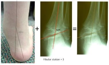 One needs to differentiate between ankle valgus (shown here) and hindfoot valgus. It is imperative to obtain a standing anteroposterior radiograph of the ankle when evaluating foot problems.
Proximally, there may be concomitant genu valgum with a corresponding increase in the intermalleolar distance (see the image below). When the etiology is neuromuscular, the patient may have muscle weakness, imbalance, or contractures.
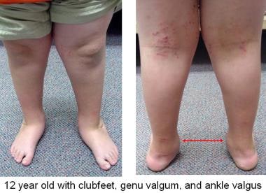 Patients may have valgus at more than just the hindfoot and ankle. This boy with congenital clubfeet has genu valgum compounding his gait problems.
Media Gallery
-
Normal ankle alignment. The lateral distal tibial angle (LDTA) is 87º, and the fibular physis is at or distal to the level of the plafond, which is horizontal and, thus, perpendicular to gravity.
-
Malhotra classified progressive ankle valgus, which is directly proportional to the degree of fibular physis elevation (stage 0 = normal). The described triad of fibular physis elevation, wedging of the lateral tibial epiphysis, and ankle tilt may be accompanied by horizontal expansion of the fibular epiphysis (impingement), medial clear space widening, and avulsion injuries of the tip of the medial malleolus.
-
Lateral impingement may be due to ankle valgus, hindfoot valgus, or both. This is an extreme example.
-
One needs to differentiate between ankle valgus (shown here) and hindfoot valgus. It is imperative to obtain a standing anteroposterior radiograph of the ankle when evaluating foot problems.
-
Patients may have valgus at more than just the hindfoot and ankle. This boy with congenital clubfeet has genu valgum compounding his gait problems.
-
Transmalleolar screws, though easy to insert, may be difficult to remove. Shown here are two complications: screw breakage and intra-articular migration of the screw head, reflecting the drawbacks of imposing a rigid restraint on a dynamic and growing physis.
-
This patient (see also image below) failed to return for follow-up for 24 months following medial malleolar epiphysiodeses. There is obvious iatrogenic varus with tenting of the physes and risk of premature closure.
-
These screws were removed (with difficulty) on an urgent basis (see also image above).
-
This patient had asynchronous medial malleolar epiphysiodeses. The screw on the left could not be retrieved. His opening wedge osteotomy to correct iatrogenic varus collapsed into a nonunion, necessitating salvage with a Taylor spatial frame. This unfortunate sequence would not have happened with an eight-Plate.
-
A drawback of the intraphyseal fulcrum is the rigid constraint of the physis. Correction is relatively slow and inefficient when compared to the flexible, extraphyseal eight-Plate.
-
The nonlocking eight-Plate is placed superficial to the intact periosteum. As lateral growth occurs, the screws diverge, permitting safe and gradual correction of the valgus deformity. The ground reaction force moves medially, toward the center of the ankle. The distal tibial physis can expand and grow laterally; the articular cartilage is spared from harmful shear forces.
-
The fibula may not respond in a synchronous manner. However, as lateral impingement is alleviated, symptoms abate and there are no functional consequences. In children, it is not necessary to lengthen the fibula or fuse it to the distal tibia.
-
Through a 2.5-cm incision, one can place a Keith needle into the distal tibial physis, preserving the periosteum. Center the eight-Plate on the physis, and secure it with the 4.5-mm cannulated screws (either 16 or 24 mm). Place the epiphyseal screw first, with care to avoid the ankle joint or physis.
-
Fluoroscopic sequence showing the steps. The 24-mm screws are preferable if there is enough space to insert them.
-
Sick physes are not a contraindication to medial malleolar epiphysiodesis, even with screws. Note the remodeling of the distal tibial epiphysis as the ground reaction force is restored to neutral and the plafond rendered horizontal.
-
Ankle valgus is relatively common in children with previously operated clubfeet. While these feet may be presumed to be overcorrected, ankle films may reveal ankle valgus and lateral impingement. If the feet are flexible, it may be preferable to deliberately overcorrect into 5º of ankle varus before removing the plates. Continue to observe the child annually until maturity, and repeat as needed.
-
Presenting with an anterolateral bow and initially intact fibula, this child went on to a fibular fracture/pseudarthrosis and ankle valgus by age 3 years. Note the medial widening. It is not necessary to fix, bone-graft, or lengthen the fibula, nor is it helpful to create a tibia-fibular synostosis.
-
The medial malleolar screw was placed at age 4 years, and over the ensuing 2 years, the valgus corrected into slight varus. This procedure was repeated at age 7 years and again at 9 years, employing the eight-Plate.
-
This 12-year-old boy with hemiplegia underwent a rotational supramalleolar osteotomy. Despite the fibula being left intact, he drifted into valgus over the ensuing year.
-
This 10-year-old boy demonstrates the stigmata of hereditary multiple exostoses, with concurrent knee and ankle valgus. These deformities were managed by eight-Plates applied to the distal medial femora and distal medial tibiae. The deformities corrected over the ensuing year, and the plates were then removed.
-
This 9-year-old patient with spina bifida had progressive and symptomatic ankle valgus. One year following eight-Plate insertion, it is evident that the 16-mm screws are losing their grip. The goal was to achieve slight varus overcorrection.
-
In this patient (see also image above),the plates were moved distally and resecured with 24-mm screws.
of
22


