Laboratory Studies
Cell culture
Definitive diagnosis of sporotrichosis at any site requires the isolation of S schenckii or the other species in a specimen culture from a normally sterile body site.
The organism can be recovered with fungal culture from sputum, pus, subcutaneous tissue biopsy, synovial fluid, synovial biopsy, bone drainage or biopsy, and cerebrospinal fluid (CSF).
The concentration of organisms in synovial fluid and, particularly, CSF is often low. Therefore, repeated large-volume cultures may be necessary for diagnosis of sporotrichosis.
Occasionally, S schenckii (cigar-shaped yeast) can be visualized in biopsied tissue specimens that are stained with periodic acid-Schiff, Gomori methenamine-silver, or immunohistochemical stains.
Granulomatous inflammation is common; this is occasionally accompanied by the presence of an asteroid body, but this picture is not specifically diagnostic for sporotrichosis.
Serological techniques
Antibody measurement techniques are available. [30]
Such tests demonstrate significant interlaboratory variability in sensitivity and specificity; therefore, they should rarely serve as the sole basis for diagnosis of sporotrichosis.
They can be useful to raise diagnostic suspicion and to inspire more aggressive attempts to acquire appropriate specimens for culture.
The ratio of CSF to serum antibody titer may suggest the presence of sporotrichotic meningitis (CSF antibody titer higher than serum antibody titer). [31]
Polymerase chain reaction (PCR)–based techniques for diagnosis of sporotrichosis have been described but are not available for routine use.
Imaging Studies
Radiography: Conventional radiographic imaging of the chest (in patients with suspected pulmonary sporotrichosis) and other involved areas (in patients with suspected osteoarticular sporotrichosis) is warranted but does not enable etiologic diagnosis. Chest CT scanning is supportive but not specifically diagnostic of sporotrichosis.
Procedures
Arthrocentesis: Patients with subacute or chronic arthritis should undergo diagnostic arthrocentesis. Sporotrichotic arthritis causes the general findings of an inflammatory arthritis (leukocytosis), with no crystals or growth on routine bacterial cultures.
Synovial tissue biopsy: The diagnostic yield of synovial tissue biopsy for histology and culture is better than that of synovial fluid culture alone in patients with suspected sporotrichotic arthritis.
Bone biopsy: Bone biopsy for histopathology and culture is useful and may be necessary for diagnosis of sporotrichal osteomyelitis.
Full-thickness skin biopsy: Culture of exuded pus from cutaneous lesions can be diagnostic; however, full-thickness skin biopsy for histology and culture may improve diagnostic yield.
Bronchoscopy with bronchoalveolar lavage for culture and transbronchial biopsy for histopathology may be required to establish the diagnosis of pulmonary sporotrichosis.
Histologic Findings
Sporotrichosis is characterized histopathologically by granulomatous inflammation with occasional asteroid bodies. The yeast form of the organism can be demonstrated, with considerable difficulty, in biopsy samples. See the image below.
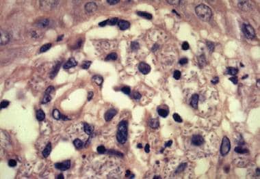 Cigar-shaped yeast of Sporothrix schenckii in tissue macrophages in a biopsy specimen.
-
This photo depicts cutaneous disseminated sporotrichosis in a patient with AIDS before and after therapy. Courtesy of Leonard N. Slater, MD.
-
Photomicrograph that shows the conidiophores and conidia of the fungus Sporothrix schenckii. Courtesy of CDC Public Health Image Library.
-
Lymphocutaneous sporotrichosis.
-
Lymphocutaneous sporotrichosis.
-
Lymphocutaneous sporotrichosis.
-
Cigar-shaped yeast of Sporothrix schenckii in tissue macrophages in a biopsy specimen.
-
Ulcerated lesion in the cheek of a child. Note the satellite lesions. Courtesy of Todd Mollet, MD, University of Texas Southwestern Medical Center.


