Report on the Radiolaria/Plates1

From Wikisource
Jump to navigation Jump to search
1584836Report on the Radiolaria — Plates 1-10Ernst Haeckel

PLATE 1.

Legion SPUMELLARIA.

Order COLLOIDEA.

Family Thalassicollida.


PLATE 1.
Thalassicollida.
Diam. Page.
Fig. 1. Actissa princeps, n. sp., × 300 13
The entire living Spumellarium. c, The spherical central capsule containing finely granulated protoplasm, which is radially striated in the cortical zone; v, spherical vacuoles enclosed by the protoplasm; n, the spherical nucleus in the centre; l, the concentric nucleolus; f, the radial pseudopodia which pierce the calymma or the (yellowish) jelly-envelope of the central capsule and arise from the granular sarcomatrix.
Fig. 1a. Half of the central capsule of another specimen, in which the original central nucleus is cleft into numerous small nuclei, × 400
Fig. 1b. Half of the central capsule of another specimen, filled up by flagellate spores, × 400
Fig. 1c. Eight isolated flagellate spores, × 800
Fig. 2. Thalassolampe maxima, n. sp., × 8 17
The entire living Spumellarium. c, The big spherical central capsule; a, the large alveoles filling the central capsule and surrounding a central nucleus; f, the pseudopodia piercing the extracapsular calymma.
Fig. 2a. The nucleus alone, with numerous nucleoli, × 30
Fig. 3. Thalassopila cladococcus, n. sp., × 20 17
c, The big central capsule; a, numerous large alveoles contained in the central capsule; k, oil globules, many of which are placed in the radially striped cortical zone; the nucleus placed centrally, is covered with numerous radial apophyses or cæcal sacs. f, The radially striped calymma.
Fig. 4. Thalassicolla maculata, n. sp., × 100 21
c, The central capsule; v, vacuoles filling this capsule; n, the central nucleus; l, the concentric nucleolus; g, the voluminous calymma, a small radial piece of which is only represented; a, the large alveoles; b, peculiar exoplasmatic bodies; p, black pigment in the inner zone; f, the retracted pseudopodia in the outer zone.
Fig. 4a. An exoplasmatic body, × 300
Fig. 4b, Vacuoles in the endoplasm, × 300
Fig. 5. Thalassicolla melacapsa, n. sp., × 300 21
n, The large nucleus; l, numerous small nucleoli inside the nucleus; v, the vacuoles filling up the central capsule and separated by black pigment; a, large alveoles in the calymma; k, oil globules; b, exoplasmatic bodies; f, the retracted pseudopodia in the outer zone of the calymma.
Fig. 5a. An endoplasmatic vacuole, resembling a cell, × 600
Fig. 5b. A piece of the central capsule, × 600
Plate 1.
Plate 1.


PLATE 2.

Legion SPUMELLARIA.

Order BELOIDEA.

Family Thalassosphærida.


PLATE 2.
Thalassosphærida.
Diam. Page.
Fig. 1. Lampoxanthium pandora, n. sp., × 120 38
The central capsule exhibits distinct pore-canals in its membrane, and a clear interval between this and the coagulated and vacuolated protoplasm. The central nucleus contains numerous dark nucleoli. The spicula are scattered throughout the alveolate calymma.
Fig. 2. Thalassoplancta brevispicula, n. sp. (vel Lampoxanthium brevispiculum), × 120 36
The central capsule contains numerous clear vacuoles, and in the cortical zone a layer of large oil-globules. The central nucleus includes numerous dark nucleoli. The calymma is alveolate. The spicula lie only in the cortical zone.
Fig. 3. Thalassoxanthium cervicorne, n. sp., × 300 33
The central capsule is filled up by clear vacuoles and contains a large central nucleus, with a single nucleolus. The spicula surround the thin calymma.
Fig. 4. Thalassoxanthium cervicorne, n. sp., × 600 33
A single spiculum.
Fig. 5. Thalassoxanthium medusinum, n. sp., × 120 32
The central capsule is filled up by clear vacuoles and contains on its cortical zone a layer of large oil-globules. The central nucleus contains numerous dark nucleoli. The calymma is radially striped, contains numerous small xanthellæ, and is surrounded by the spicula.
Fig. 6. Thalassoxanthium octoceras, n. sp., × 400 34
Three isolated spicula.
Plate 2.
Plate 2.


PLATE 3.

Legion SPUMELLARIA.

Order COLLOIDEA.

Family Collozoida.


PLATE 3.
Collozoida.
Diam. Page.
Fig. 1. Collozoum serpentinum, n sp (vel Collophidium serpentinum, Hkl), × 10 26
A living cœnobium, with expanded pseudopodia. The spherical calymma (or the common jelly-mass of the colony) is alveolate and contains numerous cylindrical, serpentine, central capsules. Numerous yellow cells or xanthellæ are scattered between the radial pseudopodia in the periphery.
Fig. 2. Collozoum serpentinum, n. sp., × 50 26
An isolated, cylindrical, worm-shaped, central capsule, with an axial series of oil-globules; the red points are nuclei.
Fig. 3. Collozoum serpentinum, n. sp., × 150 26
An isolated, cylindrical, serpentine, central capsule. k, Oil-globules forming an axial series; n, densely placed, red-coloured nuclei;, c, the capsule membrane under which are scattered small black pigment spots in the colourless cortical zone of the endoplasm; a, extracapsular alveoles; x, xanthellæ or "yellow cells."
Fig. 4. Collozoum amœboides, n. sp., × 100 28
A spherical cœnobium or jelly-colony. Each amœboid central capsule contains an oil-globule; the small red points are nuclei.
Fig. 5. Collozoum amœboides, n. sp., × 400 28
c, A single isolated central capsule; n, nuclei; k, oil-globule.
Fig. 6. Collozoum vermiforme, n. sp., × 30 27
g, A spherical cœnobium or jelly-colony; a, large alveoles, forming a cortical zone; c, central capsules; k, oil-globules.
Fig. 7. Collozoum vermiforme, n. sp., × 100 27
c, A single isolated central capsule; x, xanthellæ surrounding this central capsule; k, oil-globules; n, nuclei.
Fig. 8. Collozoum ellipsoides, n. sp., × 2 26
A spherical colony; the red points are central capsules.
Fig. 9. Collozoum ellipsoides, n. sp., × 150 26
c, A single isolated central capsule; k, oil-globules; n, nuclei.
Fig. 10. Collozoum inerme, Hkl., × 2 25
An old, cylindrical, articulated cœnobium; the red points are centralcapsules.
Fig. 11. Collozoum inerme, Hkl., × 2 25
A young cylindrical cœnobium; the red points are central capsules.
Fig. 12. Collozoum inerme, Hkl., × 400 25
A piece of a young colony with eight small central capsules, without oil-globules. n, The central nucleus in different stages of division. Two capsules are also dividing. x, Xanthellæ in the jelly-like calymma (blue), which also contains numerous vacuoles.
Plate 3.
Plate 3.


PLATE 4.

Legion SPUMELLARIA.

Orders BELOIDEA.

Families Sphærozoida.


PLATE 4.
Sphærozoida
Diam. Page.
Fig. 1. Sphærozoum trigeminum, n. sp., × 50 43
An annular colony. The main mass of the jelly-colony is filled up by large alveoles; the entire surface is densely covered with spicula, and beyond this skeleton-cover lie the spherical central capsules, each with an oil-globule. This species is by mistake not mentioned in the text.
Fig. 2. Sphærozoum alveolatum, n. sp., × 50 43
Section through a spherical colony; displaying the inside of a hemisphere. All the central capsules lie in a single stratum on the surface of the jelly-sphere, each being surrounded by a thick-walled alveole. The spicula lie between the alveole and the capsule, which includes a central oil-globule.
Fig. 3. Sphærozoum alveolatum, n. sp., × 400 43
A single central capsule, filled up by crystal-spores. Numerous geminato-radiate spicula and spherical xanthellæ lie between the capsule and the including thick-walled alveole. In the jelly-calymma, between the capsule and the alveole, numerous thin ramified pseudopodia are expanded.
Fig. 4. Sphærozoum geminatum, n. sp., × 400 45
A single central capsule, with a central oil-globule, surrounded by numerous spicula and spherical xanthellæ. The jelly-substance of the calymma is expanded between the points of the spicula.
Fig. 5. Sphærozoum variabile, n. sp., × 300 45
Three isolated spicula.
Fig. 6. Sphærozoum pandora, n. sp. (vel Rhaphidozoum pandora), × 300 49
A group of various spicula.
Fig. 7. Sphærozoum verticillatum, n. sp., × 300 44
A single spiculum.
Fig. 8. Sphærozoum arborescens, n. sp., × 300 44
A single spiculum.
Fig. 9. Sphærozoum armatum, n. sp., × 300 43
A single spiculum.
Plate 4.
Plate 4.


PLATE 5.

Legion SPUMELLARIA.

Order SPHÆROIDEA.

Family Collosphærida.


PLATE 5.
Collosphærida.
Diam. Page.
Fig. 1. Trypanosphæra transformata, n. sp., × 150 111
A living colony. The centre of the spherical cœnobium contains a large alveole, surrounded by a net of sarcode. The entire calymma is filled up by smaller, thin-walled alveoles. Its inner part contains numerous small, young, central capsules (each with an oil-globule) without shells; in the cortical zone of the calymma lie larger capsules, each of which is enclosed by a fenestrated shell with from two to four or more dentated tubes. Between the radiant pseudopodia very numerous small yellow cells (xanthellæ), which are scattered everywhere.
Fig. 2. Trypanosphæra transformata, n. sp., × 300 111
A single shell.
Fig. 3. Trypanosphæra coronata, n. sp., × 300 110
Fig. 4. Trypanosphæra trepanata, n. sp., × 300 110
Fig. 5. Odontosphæra monodon, n. sp., × 300 102
Fig. 6. Odontosphæra cyrtodon, n. sp., × 300 102
Fig. 7. Acrosphæra inflata, n. sp., × 300 101
Fig. 8. Mazosphæra hippotis, n. sp., × 400 108
Fig. 9. Mazosphæra lagotis, n. sp., × 300 108
Fig. 10. Pharyngosphæra stomodæa, n. sp., × 400 98
Fig. 11. Buccinosphæra invaginata. n. sp., × 500 99
Each shell contains numerous larger and smaller crystals.
Fig. 12. Tribonosphæra centripetalis, n. sp., × 500 98
Each shell contains numerous large crystals.
Fig. 13. Collosphæra polygona, n. sp., × 200 96
Plate 5.
Plate 5.


PLATE 6.

Legion SPUMELLARIA.

Order SPHÆROIDEA.

Family Collosphærida.


PLATE 6.
Collosphærida.
Diam. Page.
Fig. 1. Siphonosphæra socialis, n. sp., × 500 106
A small piece of the surface of a living cœnobium, seen from the surface. Only four individuals are visible, the central capsule of which contains numerous small nuclei and a central oil-globule. The including spherical lattice-shell is provided with a few (one to four) larger apertures, which are prolonged into short cylindrical tubules. Through these latter radiate bundles of fine pseudopodia, branching and anastomosing, and forming a fine sarcode network between the alveoles of the calymma. On the surface of the alveolated jelly-sphere the pseudopodia form a dense radiating zone. Xanthella or yellow cells are everywhere scattered.
Fig. 2. Siphonosphæra socialis, n. sp., × 300 106
A small cœnobium or colony in the state of alveolation, forming a jelly-sphere, composed of a great number of capsulated individuals, densely aggregated. Each central capsule contains an oil-globule, and is enclosed by a spherical lattice-shell, which bears a few (one to four) short cylindrical tubules. Each shell is again enveloped by a membranous polyhedral alveole and separated from it by structureless jelly. The thick cortical jelly-envelope, which surrounds the whole spherical colony, exhibits a fine radial striation, produced by radiating pseudopodia; many xanthellæ or yellow cells are scattered in the calymma.
Fig. 3. Siphonosphæra pipetta, n. sp., × 300 108
Fig. 4. Siphonosphæra tubulosa, J. Müller, × 300 105
The central capsule, enclosed in the cavity of the shell, has a central oil-globule, and is surrounded by a few xanthella.
Fig. 5. Siphonosphæra chonophora, n. sp., × 300 107
Fig. 6. Siphonosphæra serpula, n. sp., × 300 107
Fig. 7. Siphonosphæra patinaria, n sp., × 300 105
The central capsule, enclosed in the cavity of the shell, contains a central oil-globule, and is surrounded by a few xanthella.
Fig. 8. Siphonosphæra patinaria, n. sp., × 300 105
Fig. 9. Siphonosphæra conifera, n. sp., × 300 106
Fig. 10. Siphonosphæra cyathina, n. sp., × 300 105
Plate 6.
Plate 6.


PLATE 7.

Legion SPUMELLARIA.

Order SPHÆROIDEA.

Family Collosphærida.


PLATE 7.
Collosphærida.
Diam. Page.
Fig. 1 Caminosphæra dendrophora, n. sp., × 300 112
Fig. 2. Caminosphæra dichotoma, n. sp., × 300 112
Fig. 3. Coronosphæra diadema, n. sp., × 300 117
Fig. 4. Coronosphæra calycina, n. sp., × 300 117
Fig. 5. Otosphæra auriculata, n. sp., × 300 116
Fig. 6. Otosphæra polymorpha, n. sp., × 300 116
Fig. 7. Solenosphæra serpentina, n. sp., × 300 114
Fig. 8. Solenosphæra cornucopia, n. sp., × 300 115
Fig. 9. Solenosphæra ascensionis, n. sp., × 300 115
Fig. 10. Solenosphæra pandora, n. sp., × 300 113
Fig. 11. Solenosphæra pandora, n. sp., × 100 113
An entire spherical cœnobium. The shells of the colony bear a variable number of fenestrated radial tubes and are densely crowded in the jelly-sphere of the calymma, the cortical zone of which is radially striped.
Plate 7.
Plate 7.


PLATE 8.

Legion SPUMELLARIA.

Order SPHÆROIDEA.

Family Collosphærida.


PLATE 8.
Collosphærida.
Diam. Page.
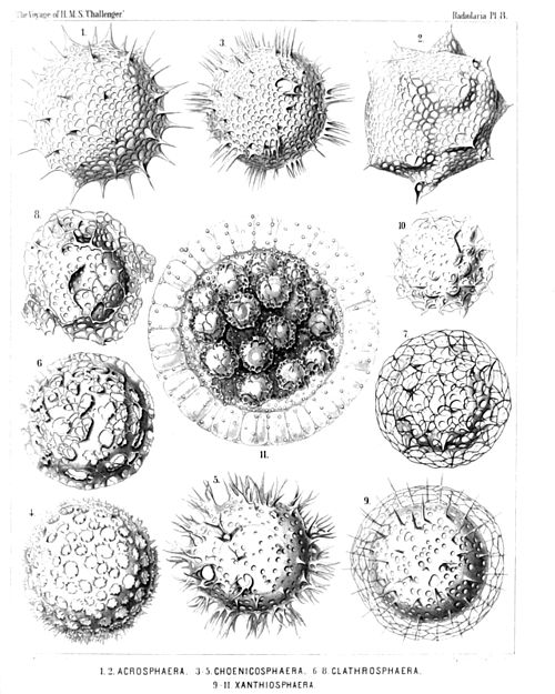
Fig. 1. Acrosphæra echinoides, n. sp., × 400 100
Fig. 2. Acrosphæra collina, n. sp., × 300 101
Fig. 3. Chænicosphæra nassiterna, n. sp., × 400 103
Fig. 4. Chænicosphæra murrayana, n. sp., × 300 102
Fig. 5. Chænicosphæra flammabunda, n. sp., × 300 103
Fig. 6. Clathrosphæra circumtexta, n. sp., × 400 118
Fig. 7. Clathrosphæra arachnoides, n. sp., × 300 119
Fig. 8. Clathrosphæra lamellosa, n. sp., × 300 119
Fig. 9. Xanthiosphæra erinacea, n. sp., × 400 120
Fig. 10. Xanthiosphæra lappacea, n. sp., × 300 120
Fig. 11. Xanthiosphæra lappacea, n. sp., × 100 120
A complete spherical cœnobium. The associated central capsules (each with a double shell) are densely crowded in the central part of the calymma, whilst its peripheral part is occupied by a layer of large alveoles. Numerous xanthellæ or yellow cells an scattered in the calymma.
Plate 8.
Plate 8.


PLATE 9.

Legion SPUMELLARIA.

Order LARCOIDEA.

Family Pylonida.


PLATE 9.
Pylonida.
Diam. Page.
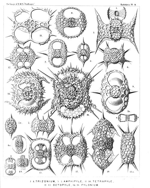
Fig. 1. Monozonium alatum, n. sp., × 300 633
Dorsal view.
Fig. 1a. Apical view.
Fig. 2. Dizonium pleuracanthum, n. sp., × 400 636
Fig. 3. Dizonium stauracanthum, n. sp., × 300 636
Fig. 4. Trizonium tricinctum, n. sp., × 300 637
Dorsal view.
Fig. 4a. Lateral view.
Fig. 4b. Apical view.
Fig. 5. Amphipyle tetraceros, n. sp., × 400 642
Dorsal view.
Fig. 6. Amphipyle callizona, n. sp., × 300 644
Dorsal view.
Fig. 7. Amphipyle amphiptera, n. sp., × 300 642
Dorsal view.
Fig. 7a. Lateral view.
Fig. 8. Tetrapyle circularis, n. sp., × 300 645
Dorsal view.
Fig. 9. Tetrapyle pleuracantha, n. sp., × 400 646
Dorsal view. The lentelliptical central capsule is visible between medullary and cortical shell.
Fig. 10. Tetrapyle turrita, n. sp., × 400 649
Oblique view, half dorsal, half lateral.
Fig. 11. Octopyle stenozona, n. sp., × 400 652
Dorsal view.
Fig. 12. Octopyle sexangulata, n. sp., × 300 653
Dorsal view.
Fig. 13. Octopyle decastyle, n. sp., × 300 654
Dorsal view.
Fig. 13a. Lateral view.
Fig. 14. Pylonium quadricorne, n. sp., × 400 655
Dorsal view.
Fig. 15. Tetrapylonium quadrangulare, n. sp., × 300 658
Dorsal view.
Fig. 16. Pylozonium octacanthum, n. sp., × 300 660
Dorsal view.
Plate 9.
Plate 9.


PLATE 10.

Legion SPUMELLARIA.

Order LARCOIDEA.

Family Tholonida.


PLATE 10.
Tholonida.
Diam. Page.
Fig. 1. Tholartus tricolus, n. sp., × 200 664
Fig. 2. Tholodes cupula, n. sp., × 500 665
Fig. 3. Amphitholus artiscus, n. sp., × 400 666
Fig. 4. Amphitholus panicium, n. sp., × 500 668
Fig. 5. Amphitholus acanthometra, n. sp., × 300 667
Fig. 6. Amphitholus acanthometra, n. sp., × 300 667
Frontal section of the shell.
Fig. 7. Amphitholonium tricolonium, n. sp., × 300 669
Fig. 8. Staurotholus tetrastylus, n. sp., × 300 673
Fig. 9. Staurotholus dodecastylus, n. sp., × 400 674
Fig. 10. Tholoma quadrigeminum, n. sp., × 200 672
Fig. 11. Staurotholonium octodoronium, n. sp., × 300 676
Fig. 12. Tholocubus tessellatus, n. sp., × 200 677
Fig. 13. Tholoma metallasson, n. sp., × 300 672
Fig. 14. Cubotholus regularis, n. sp., × 200 680
Fig. 15. Cubotholonium ellipsoides, n. sp., × 300 682
Fig. 16. Tholocubus tesseralis, n. sp., × 400 678
Fig. 17. Tholonium hexonium, × 400 679
Plate 10.
Plate 10.