-
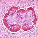
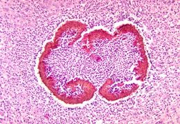
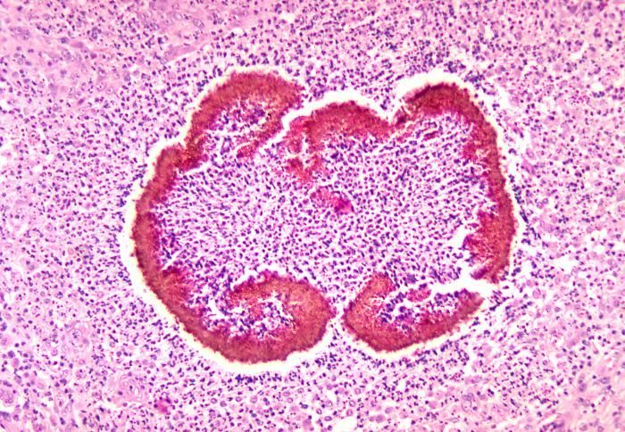
This micrograph depicts the histopathologic changes seen in black grain mycetoma due to Exophiala salmonis.Created: 1971
-
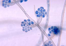
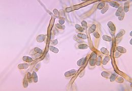
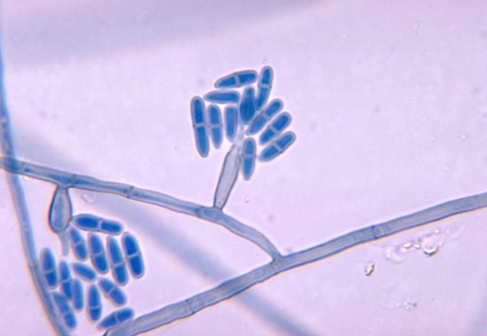
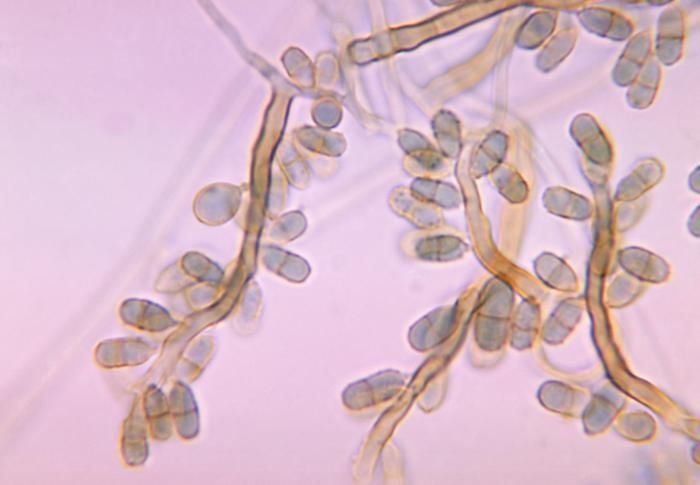
This micrograph depicts the conidia-laden conidiophores of the fungal organism Exophiala salmonis.Created: 1970
-
This micrograph depicts the conidia-laden conidiophores of the fungal organism Exophiala salmonis.Created: 1970
-
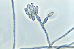
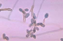
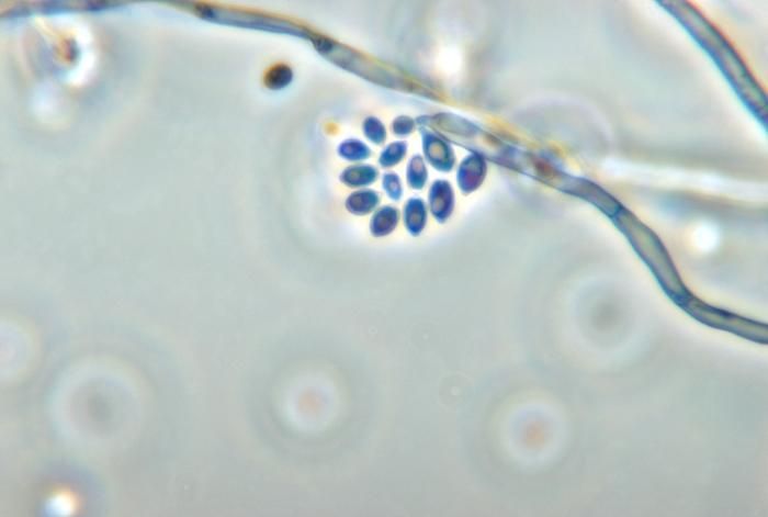
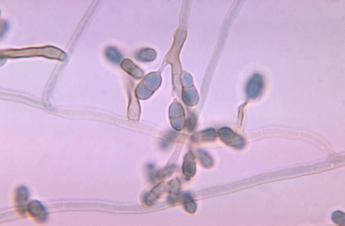
This micrograph depicts the conidia-laden conidiophore of the fungal organism Exophiala salmonis.Created: 1970
-
This micrograph depicts the histopathologic changes seen in black grain mycetoma due to Exophiala salmonis.Created: 1971
-
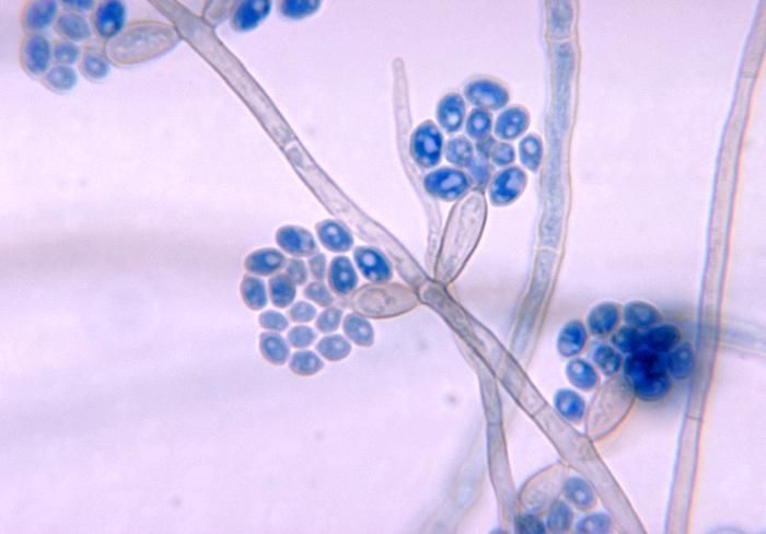
This micrograph depicts the conidia-laden conidiophores of the fungal organism Exophiala salmonis.Created: 1970
-
This micrograph depicts the conidia-laden conidiophores of the fungal organism Exophiala salmonis.Created: 1970
-
This micrograph depicts the conidia-laden conidiophore of the fungal organism Exophiala salmonis.Created: 1970
-
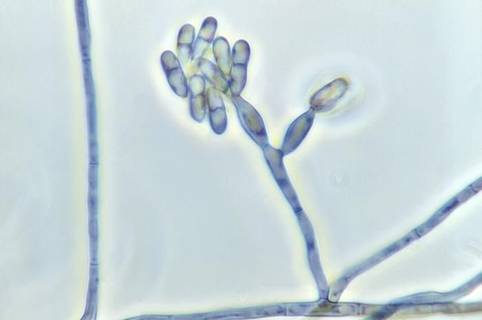
Magnified 1125X, this photomicrograph shows a conidia-laden condiophore of the fungus Exophiala salmonis.Created: 1973
-
This photomicrograph shows conidia-laden conidiophores of the fungus Exophiala salmonis.Created: 1970
-
This photomicrograph shows a condiophore with conidia of the fungus Exophiala salmonis.Created: 1970
-
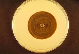
This is a plate culture growing a colony of the fungus Exophiala salmonis.Created: 1970