Eureka, its time for yet another post about Brettanomyces. And today, we are looking at some Brettanomyces lambicus cells. You might remember, that I did some plating of the two Brettanomyces strains from Wyeast: Brettanomyces bruxellensis and Brettanomyces lambicus on Sabouraud agar. And I already posted the results from the microscopy observation of a B. bruxellensis sample. Now is the time to talk about the microscopy observation of the B. lambicus cells from Wyeast.
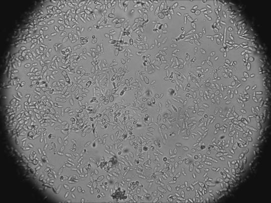
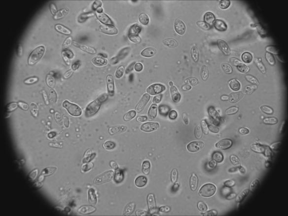
Lets begin with a few pictures now to get an overview. I will get into further details in the pictures below.
My first impression is that B. lambicus, much like B. bruxellensis, appears in very different shapes. There are boat-shaped cells, elongated cells, and even cells that attach to each other. But very few of the cells are circular like normal brewers yeast. Maybe the circular cells here might be brewers yeast impurities in the Brettanomyces sample. To summarize, B. lambicus seems to look quite different from brewers yeast (Saccharomyces cerevisiae). Lets go into more details at a higher magnification.
The cells seem to have a kind of vacuole (Fig 4). The vacuole can be observed as a kind of compartment in the cell itself. It is much easier to observe in the next picture (Fig 5).
Interestingly, there is something circular in the vacuole. I will come back to this observation later on.
It can be observed now, that the cells have again, like B. bruxellensis, one or two dark spots (Fig 6). I am still figuring out what these dark spots might be. But now back to the most interesting observation from my point of view. I already mentioned, that I could observe some kind of circular structure in the cells vacuole (see Fig 5). One disadvantage of pictures is their snapshot nature. The pictures just represent a short state of a cell. Just have a look the following video I took to see what the cells really looked like for real. Observe what happens inside the vacuole. I have to apologize for the bad quality of the video.
Have you noticed that the circular structure moved in the cells vacuole? Well, I have never observed something like that before. In my opinion there are several possible explanations for such a movement.
– One would be that there is a kind of dynamic movement in the vacuole itself. Much like mixing the vacuoles content in some way. And the circular structure then could be maybe a vesicle with some sort of nutrients in it and swirl around due to the dynamic movement in the vacuole.
– Another explanation, there is a kind of other cell in there. But this seems to be very unlikely to me. I have several reasons to come to such a conclusion. One would be how these cells would have gotten there in the first place.
– Sporulation. Could these structures be spores?
This is very exciting in my opinion. A lot of questions. I have several further investigations in mind to get further information about that circular structure in there. If someone out there has any idea what these structures could be, please comment below. Please comment as well if you have observed such a movement yourself.
What have I learned from the microscopy observations so far?
The observations helped me to understand the different morphologies of the Brettanomyces yeast cells. This can be observed if you look at different microscopy pictures as well. But looking at them yourself is very different. And this helped me to identify several yeasts I harvested from different sources to be Brettanomyces. On the other hand, colonies on the agar plates of the two strains looked very similar and I could not distinct between them. I addition, the colonies of Saccharomyces cerevisiae looked very similar to the ones from the Brettanomyces as well. I could not even distinct between Saccharomyces and Brettanomyces by just looking at the colonies on the Sabouraud agar. Only a microscopy observation can help to make such a distinction. It is therefore inevitable to look at the cells to get an idea what kind of yeast you have.
That’s all so far about the two Brettanomyces strains from Wyeast. Unfortunately, I can’t get my hands on the White Labs strains here. So no further pure strain observations will follow. What I have planned is to write some posts about Brettanomyces in general. And the first post about “Brettology” will be a post about the taxonomy of Brettanomyces. So stay tuned and thank you for your comments!


Hi Sam, I see the same thing, the “dancing” of the little particle in the vacuole. I’ll record a video to compare as well, if you’d like. I do not know what it is either. I have searched a lot online but cannot find anything. Sometimes I see the particles in the media as well, with the same “dancing” movement, the tumbling. The first time I saw them I was like oh shit, I got a contamination. But then I re-streaked, picked colonies, grew in media with ampicillin, and saw still the same thing. Especially prevalent in London Ale III from Wyeast. Great work you are doing! -Jasper
Hi Jasper, thats great. Now I know that I don’t just see things 😉 I did a lot of research as well and could not find any source talking about that matter. I thought about a contamination as well but in my case, it was a pure culture and the particle there is a bit big for a bacteria. You say, that you observed the same in colonies grown on ampicillin. That would exclude the possibility that the particle is a bacteria (although it could be an ampicillin resistance bacteria after all…). Not to mention again that it would be a rather big bacteria cell after all. You mention to observe the same thing in London Ale III as well. That makes it even more interesting to find out what this particle is. I would be very interested to see a picture of the particle in the London Ale yeast.
Unfortunately, I have no access to a lab at the moment but I will try to investigate those particles in the upcoming Fall. One test I am interested in is to do a DAPI stain, and I will search for a test to stain spores.
Thank you for your comment, really appreciated.
Cheers, Sam
Really nice post! Kinda missed it with exams and lab… Anyway, have you tried staining them just to make sure it really is a vacuole?
Hi there, the vacuole is just a guess. Unfortunately, I have no stains and such available at home. I hope to do some experiments in September… Cheers
I think its the vacuole. Especially in Fig. 5 this is pretty clear. But indeed to be sure a stain would be needed. The particle might be a “volutin granule” (see http://en.wikipedia.org/wiki/Volutin_granules, http://sols-fin.asu.edu/ugrad/solur/pdf/f08_marshall.pdf,
For some reason the final link did not work paste in, let me try again: http://books.google.com/books?id=DvNhR0xfHtMC&pg=PA102&lpg=PA102&dq=volutin+particles+vacuole+yeast&source=bl&ots=Be5qjjduoh&sig=zsUE8_f8zjZZzMI_IJpT4NnPsf8&hl=en&sa=X&ei=8umlT93FBIno9ATM8PmuAw&ved=0CF8Q6AEwBg#v=onepage&q=volutin%20particles%20vacuole%20yeast&f=false
I’m sorry the link is huge – but they talk about phosphate storage in the form of these granules, especially during slow growth as reserve-build up.
Thanks for the two links. I bet there is a stain out there used to stain phosphates… It makes a lot of sense to me that this particle there is a kind of granule. Whatever is in there. I should have a look at active growing Brettanomyces and hope to see none of these particles. The Bretts in the picture there came straight out of the Wyeast package. So not a lot of active metabolism I guess. Thanks for all the information.