
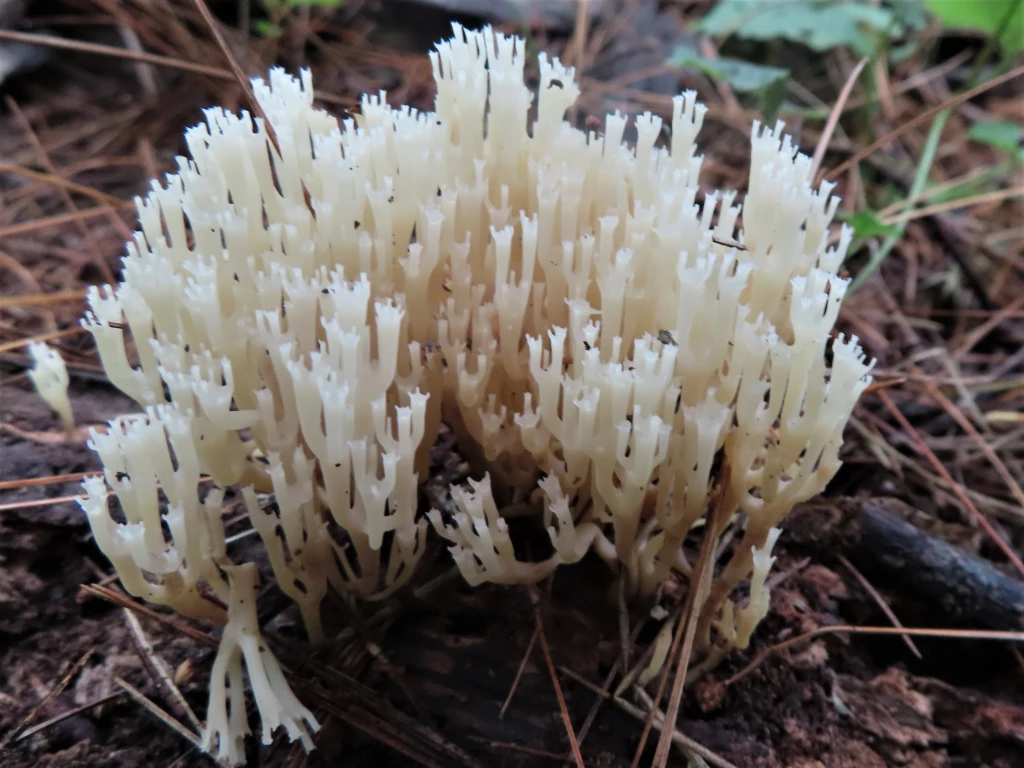
Common Name: Coral Fungus – The branching of the fungal thallus resembles the calcium carbonate structure of ocean corals. Other common names are applied to differentiated shapes, such as worm, club, or tube fungi for those lacking side branches and antler fungi for those with wider, flange-like appendages. An extreme is cauliflower fungus which looks nothing like coral but is usually included in the coral-like category in field guides. The common Crown-tipped coral is depicted; the ends of the coral segments have tines like miniature crowns.
Scientific Name: Clavariaceae – The family name for the coral fungi is derived from clava, the Latin word for “club;” the type-genus is Clavaria. The coral fungus above was originally Clavaria pyxidata, became Clavicorona pyxidata, and is now Artomyces pyxidatus. Pyx is from the Greek word pyxos meaning “box tree” from which boxes were made (and the etymology of the word box – a pyx is a container for Eucharist wafers). The implication for its use as a name for this species is “box-like.”
Potpourri: Coral fungi look like coral. The verisimilar likeness can be so convincing that it seems plausible that they were uprooted from a seabed reef and planted in the woods for decoration. The delicate ivory and cream-colored branches rising in dense clusters from a brown-black dead log are one of the wonders of the wooded paths sought by those who wander there. There is an abiding benefit to have some knowledge of the things that nature has created and coral fungi is a good collective mnemonic to apply to the group that surely must be closely related. And so it is for the traditionalists steeped in the lore of musty mushroom field guides who are referred to collectively as the “lumpers.” The new world order of DNA has taken the science of biology on a wild ride with many hairpin turns and dead ends; in the case of mycology, the train has left the tracks more than once. Coevolution … that which created a marsupial mouse in Australia unrelated to the placental house mouse everywhere else … globally demonstrates Darwin’s vision. Fungi that branch is a natural evolutionary path for two individual organisms that started at different places and times. The diaspora of species from one genus to another in search of a home on the genetic tree of life has exploded the coral fungi into fragments. This is the realm of the “splitters,” the subdividers for whom a bar code will become the only true arbiter of species. There is of course a hybrid middle ground, acknowledging the latter but practicing the former, the province of most mushroom hunters.
Like all epigeal fruiting bodies extending upward above the ground from the main body of a fungus, which is hypogeal or below ground, the branching arms of coral fungi function to support and project the spore bearing reproductive components called basidia. Gilled or pored mushrooms maximize the number of spores they can disperse by creating as much surface area as possible in the limited space beneath the cap or pileus. Similarly, coral fungi branch again and again or extend myriad singular shafts to get as many fingers of spore bearing surface into the air as possible. [1] The topology of using multiple extensions into a fluid medium is one of the recurring themes of evolution ― coevolution. In this case, it has nothing to do with fungi per se. They look like coral because real coral is doing essentially the same thing; the namesake polyps secrete a type of calcium carbonate called aragonite to form protective exoskeletons in reefs that extend outward into the water where their food floats by. To extend the analogy to the rest of biology is a matter of observation. Trees send branches covered with photosynthesizing leaves toward the sun and roots toward the water and minerals of the earth where they encounter the branching mycelia of fungi.
Fungi have evolved to distribute reproductive spores with different mechanisms that could only have been naturally selected by the variations in form and function of random mutation. Among the more creative methods are the puffing of puffball spores out a hole in the top by the impact force of raindrops, the odorous spore-laden goo of stinkhorns that attracts insects seeking nutrients, and the redolence of truffles sought by burrowing or digging animals as food digested, their spores excreted intact. The coral fungi are among the most primitive of all basidiomycete fungi in having their club-shaped spore bearing basidia positioned along the upper reaches of each prong so that they can be carried away by either wind or water. [2] Having more fruiting bodies with more branches creates more spores, which is why coral fungi are frequently found growing saprophytically in dense clusters on dead tree logs or growing in mycorrhizal clusters on the ground. Simply sticking indistinguishable club shapes into the air with a bunch of short rods with spores attached to the end is the most straightforward way to disperse them for germination.
The phylogenic diversity of the coral fungi belies their similar ramified appearance. Historically, structure was thought to be the basis for taxonomic classification, an assumption that works reasonably well with plants and animals but not with fungi. The delicate and colorful appearance of the coral fungi brought them to the attention of the earliest naturalists, who grouped them according to shapes. Since fungi were then considered members of the Plant Kingdom (Subkingdom Thallophyta), this was consistent with practice. The French botanist Chevallier placed them in the order Clavariées in 1826 with only two genera, Clavaria and Merisma noting that “se distingue du premier coup d’oeil” – they can be identified with a fleeting glance in having “la forme d’une petit massue” – the form of a little club. [3] The assignment of fungi to families according to form lasted for over a hundred years until the nuances in microstructure and spore appearance initiated cracks in the biological foundation. Toward the end of the last century the fungi were recast as one of five different kingdoms, the foundational genus Clavaria was dissected into six genera with derivative names like Clavulina (little club) and Clavariadelphis (brother of Clavaria), which is how they appear in the most popular fungi field guides. [4]
In spite of the distinctive shape that suggests a unique origin, coral fungi are agarics, the historical group name for almost all gilled fungi. What is now the order Agaricales is comprised of over 9,000 species, containing over half of all known mushroom forming macrofungi assigned to one of 26 families with about 350 genera that range from Amanita to Xerula. Carl Linnaeas, who established the first taxonomic structure in biology with the publication of Systema Naturae in the 18th century, placed all gilled mushrooms in a single genus that he named Agaricus. One hundred years later, Elias Fries published Systema Mycologicum, which separated the agarics into twelve genera based on macroscopic features such as the structure of the spore bearing surface or hymenium (e.g. gills, pores, teeth, ridges, vase-shaped) and spore color (white, pink, brown, purple-brown, or black). Six groups of basidiomycetes were recognized based on the shape of the sporocarp or fruiting body ― “coral-like fungi” was one of them. While there was some expansion of genera over the ensuing decades, the so-called Friesian approach to gilled mushroom identification has persisted and is what is still generally in use, spore print color and all. The use of field characteristics is crucial to the practical application of mycology that serves the community of foragers looking for edible species and other aficionados who enjoy their company. [5,6]
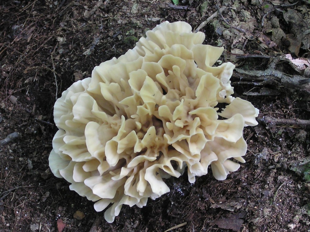
Over the last several decades, the use of DNA to map out the true phylogenetic relationships has upended the traditional taxonomy based on macroscopic structure and spore color. Unravelling the complex weave of evolutionary threads from one species to its predecessor is a monumental task that is just now gaining momentum. The goal is to determine the real or cladistic family tree so that a clade, the term adopted to refer to all species with a common ancestor, can be established with certainty. In one analysis, the agarics fell into six major clades, or single-ancestor groupings named Agaricoid, Tricholomatoid, Marasmioid, Pluteoid, Hygrophoroid and Plicaturopsidoid. The coral fungi are in the latter, which diverged from all the other agarics at the earliest evolutionary branching in the Cretaceous Era some 125 million years ago. It is not unreasonable to conclude from this analysis that the coral fungi evolved a reliable and efficient method of spore dispersal early on and have thrived ever since, branching out to form new species all using the same technique. It is now equally evident that the shape of a fungus does not necessarily establish its proper branch in the family tree. The agarics, now the Eugarics Clade, not only has fungi shaped like mushrooms and coral, but also puffballs like Calvatia and Lycoperdon. Likewise, shapes extend across multiple clades. For example, coral-shaped fungi also appear in the Russuloid Clade (Russulas) as Artomyces as pictured above and Sparassis as pictured below in the Polyporoid Clade (Polypores). This is then the dichotomy between the taxonomists of the old school steeped in the Linnaean traditions of field identification and the DNA systematists of the new school for which only the laboratory will do. [5,6]
The new biological life history of coral fungi is still subject to the findings of the most recent research paper devoted to the group and it may be decades before a settled taxonomy emerges. As a brief and incomplete history, in 1999 “four lineages containing cantharelloid and clavarioid fungi were identified,” with the clavarioid containing most of the corals, but also noting that “Clavicorona is closely related to Auriscalpium, which is toothed, and Lentinellus, which is gilled.” [7] In 2006, it was acknowledged that coral shaped fungi must have evolved at least five times over the millennia and that the “evolutionary significance of this morphology is difficult to interpret because the phylogenetic positions of many clavarioid fungi are still unknown.” The new genus Alloclavaria was added to accommodate the unique fungus Clavaria purpurea “not related to Clavaria but derived within the hymenochaetoid clade,” which consists mostly of bracket fungi. [8] Seven years later, the coral fungus family was found to consist of four major clades: Mucronella, Ramariopsis-Clavulinopsis, Hyphodontiella, and Clavaria-Camarophyllopsis-Clavicorona. This thorough phylogenic analysis of 47 sporocarp sequences merged with 243 environmental sequences concluded that “126 molecular operational taxonomic units can be recognized in the Clavariaceae … an estimate that exceeds the known number of species in the family.” [9] Phylogenic studies are continuing.
Returning to the more mundane walk through the woods looking for coral fungi, the two most pressing questions concern edibility and toxicity. Neither of these subjects is broached in the scientific literature, and, like most fungi, data points are empirical, relying on random trial and error anecdote. For coral fungi, this is complicated by the fact that most are small and delicate and therefore rarely sampled by those seeking massive brackets of Chicken-of-the-woods and yellow clusters of chanterelles. Edibility has been a question ever since Chevalier first singled them out in 1826, noting that “Presque tout les clavaires fournissent a l’homme une nouriteure saine, on mange ordinarements les plus grosses” – almost all are good to eat but only pick the big ones, and “Elles n’ont aucune qualité vénéneuses; quelques-une ont une saveur amère” – none are poisonous but some are bitter. [10] This sweeping assurance cannot have been the result of a thorough assessment, as there are good and bad corals. Modern guides are more circumspect, offering a range of information about edibility from choice to poisonous with caveats about having a laxative effect on some people and causing gastrointestinal distress in others. Many are of unknown edibility and likely to remain so. There is one standout worth noting that has the hallmarks of broad acceptability. The Cauliflower Mushroom (Sparassis americana – formerly crispa) is large, unusual, and common. It neither tastes nor looks much like a cauliflower. The “Elizabethan ruff of a mushroom” [11] is hard to miss and there is no doppelganger to fool the hapless hunter.
References:
1. Aurora, D. Mushrooms Demystified, Ten Speed Press, Berkeley, California, 1986 pp 630-658
2. Schaechter, E. In the Company of Mushrooms, Harvard University Press, Cambridge, Massachusetts, 1997, p.49
3. Chevallier F. Flore Générale des Environs de Paris, Ferra Jeune, Paris, France, 1826 p. 102.
4. Lincoff, G. National Audubon Society Field Guide to North American Mushrooms, Alfred A. Knopf, New York, 1981, pp 398-414.
5. Matheny P. et al “Major clades of Agaricales: a multilocus phylogenetic overview” Mycologia August 2006, Volume 98 Number 6 pp 982–995.
6. https://www.mykoweb.com/articles/Homobasidiomycete_clades.html
7. Pine E. et al “Phylogenetic relationships of cantharelloid and clavarioid Homobasidiomycetes based on mitochondrial and nuclear rDNA sequences”. Mycologia. 1999. Volume 91 Number 6 pp 944–963.
8. Dentinger B and McLaughlin D. “Reconstructing the Clavariaceae using nuclear large subunit rDNA sequences and a new genus segregated from Clavaria”. Mycologia. Volume 98 Number 5 September 2006 pp 746–762.
9. Birkebak J et al. “A systematic, morphological and ecological overview of the Clavariaceae (Agaricales)” Mycologia. Volume 105 Number 4, February 2013, pp 896–911.
10. Chevallier, op cit. p.104
11. Lincoff, op cit. p. 412.
