Microbiology Case Study: 76 Year Old Female with Upper Back Pain

Case History:
A 76 year old female presents with a two year history of worsening upper back pain. Imaging revealed compression fractures of the first three thoracic vertebrae (T1-T3). Fine needle aspiration and a core biopsy of the T3 vertebral body were examined in surgical pathology. There was acute and chronic granulomatous inflammation with fungal organisms observed on histologic examination. Surgery for decompression and fusion of C5-T6 vertebrae was performed and tissue was sent for fungal culture.

Potato flake agar shows a tan-brown fungus.
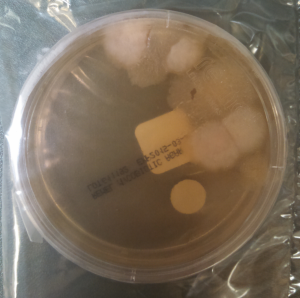
Mycosel agar shows beige-white fungal growth.
Scotch tape prep shows septate hyphae with unbranched conidiophores and single, terminal, "lollipop" conidia.
Scotch tape prep shows septate hyphae with unbranched conidiophores and single, terminal, “lollipop” conidia.
Silver stain of involved bone with fungal organisms exhibiting broad-based budding.

Laboratory Identification:
The workup revealed a thermally dimorphic fungus with a mold form growing in the laboratory at 25°C and a yeast form present in the surgical pathology specimen. The mold form is moderately slow growing and has septate hyphae with small, round, terminal conidia often described as “lollipops.” The yeast form is large (8-15 microns) with broad based buds and double contoured cell walls. The immune system reacts to the presence of the fungus by forming granulomas and leads to acute and chronic inflammation within the involved tissue. The organisms can occasionally be seen within giant cells in histologic sections. The silver stain, as seen above, highlights the organisms.

Discussion:
The fungus described above exhibits the features of Blastomyces dermatitidis. This organism resides in soil and decaying plant matter and is endemic to eastern North America including the Mississippi and Ohio River Valleys as well as areas surrounding the Great Lakes and St. Lawrence River. The most common primary sites of involvement for Blastomyces are cutaneous and pulmonary. Following a primary infection, the disease can progress to disseminated blastomycosis which involves other sites such as bone.

The primary site of infection in this case is unknown. There was no history of cutaneous ulcers and chest imaging was unremarkable. The patient did have a remote history of bloody sputum production which she had attributed to “dental difficulties” that she was experiencing and has since resolved. This may have been evidence of a primary pulmonary infection preceding the vertebral involvement; however it is difficult to say with certainty.

The classic double contoured cell walls are not evident on the silver stain of the surgical pathology specimen in this case. This may be due to the fact that the bone required decalcification before histologic sections could be taken. The decalcification process may have caused an artifactual loss of the double contour. Despite the fact that this classic finding was not seen, the macroscopic and microscopic morphology is most consistent with Blastomyces.

The patient is being treated with long-term itraconazole and is currently doing well.

-Britni Bryant, MD is a 2nd year anatomic and clinical pathology resident at the University of Vermont Medical Center.

 Wojewoda-small

-Christi Wojewoda, MD, is the Director of Clinical Microbiology at the University of Vermont Medical Center and an Assistant Professor at the University of Vermont.

Microbiology Case Study: 51 Year Old Woman with Fever and Cough

A 51 year old woman with a significant smoking history presented with 8-9 weeks of fever and cough. Shortly after the beginning of her illness, she developed pleuritic left-sided chest pain and hemoptysis. She was treated with amoxicillin and then prednisone without improvement. She had progressively worse pain and hemoptysis as well as fevers and night sweats, with weight loss. A chest x-ray and CT scan showed a left upper lobe mass- like infiltrate suspicious for a carcinoma. She underwent transbronchial fine needle aspiration biopsy of the lesion which showed the following morphology.

Bronchoalveolar lavage fluid (Pap stain).

The specimen was also sent for fungal culture.

Colony morphology on fungal media.
Organism morphology with lactophenol cotton blue scotch tape prep.


Laboratory diagnosis:

Blastomyces dermatidis was identified by microscopy and colony morphology. Septate, delicate hyphae with single, circular-to-pyriform condia on short conidiophores (lollipops) were seen on a scotch tape prep. The colonies appeared waxy and wrinkled, with a cream-tan color. Large, thick-walled yeast with buds attached by a broad base, 8-15 um with double-contoured walls, were demonstrated in tissue. Additionally, this patient had a positive urine antigen test for Blastomyces.

Discussion:

Blastomyces’ natural habitat is unknown but the organism is thought to reside in soil or wood, particularly in the Ohio, Mississippi, and Missouri River valley regions. It takes 2-30 days to grow in the lab. The infectious form is the conidia which are transmitted by inhalation. Common sites of infection include skin, lungs, and bone. The typical presentation in an immunocompetent individual is a pulmonary infection with associated acute or chronic suppurative and granulomatous lesions. Blastomyces infection may also cause osteomyelitis, prostatitis, urethritis, CNS infection, and disseminated infection. Immunocompromised patients may present with disseminated infection with involvement of skin, bone, and multiple organs. Infection may be confirmed by exoantigen testing or by nucleic acid probe testing.


-Lauren Pearson, D.O. is a 2nd year anatomic and clinical pathology resident at the University of Vermont Medical Center.

Wojewoda-small

-Christi Wojewoda, MD, is the Director of Clinical Microbiology at the University of Vermont Medical Center and an Assistant Professor at the University of Vermont.