|
||
|
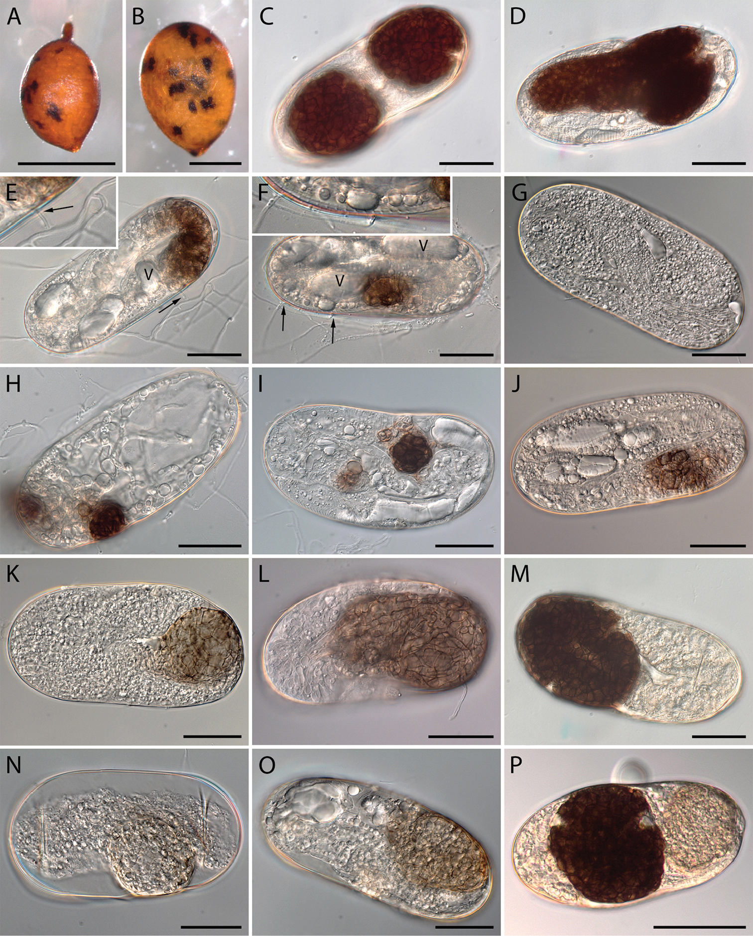
Cysts and eggs of Heterodera filipjevi infected by Monocillium gamsii exhibiting colonisation in vitro. A, B infected cysts rendered black-dotted due to fungal-colonised eggs containing microsclerotia C, D Eggs with mature sclerotia, extracted from symptomatic cysts E, F Individual hyphae penetrating eggshell (arrows indicate the individual hyphae; V indicates vacuole-like structures) G–M Fungal development inside the eggs G, H Earlier stages of infection in unembryonated eggs I, J Fungal development in the body cavity of developing juveniles where enlarged, thick-walled cells are formed and coalesce to initiate microsclerotia formation K–M Microsclerotia developing to full size and pigmentation N–P Pigmentation in microsclerotia from pale-olivaceous to darkly brown. Scale bars: 600 µm (A); 300 µm (B); 30 µm (C–O); 50 µm (P). |