

Abstract
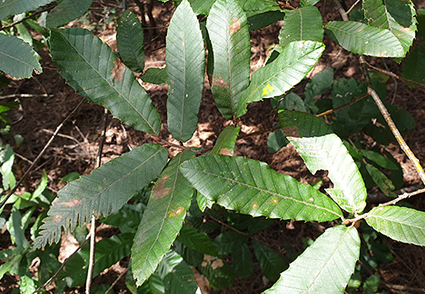
A routine survey to establish an inventory of fungal pathogens occurring on Quercus spp. in South Korea was conducted in the autumn of 2021 and 2022. Symptoms, including leaf spot, were frequently observed on Quercus variabilis leaves in a natural forest located in Sejong. A fungus with unique morphological characteristics, including pycnothyria, was found to be a major colonizer in the infected leaves of Q. variabilis. Based on the morphological characteristics of the fungus, it was identified as a member of Tubakia. Sequence comparisons of LSU, ITS, and TEF-1α gene regions revealed that the fungus was an undetermined species of Tubakia. This novel species is described as T. byeongjinii sp. nov., and this is the first report of Tubakia species on Q. variabilis in South Korea.
References
- Boroń, P. & Grad, B. (2016) The occurrence of Tubakia dryina in Poland—new hosts and ITS variation. Forest Pathology 47: 1–6. https://doi.org/10.1111/efp.12294
- Braun, U., Nakashima, C., Crous, P.W., Groenewald, J.Z., Moreno-Rico, O., Rooney-Latham, S., Blomquist, C.L., Haas, J. & Marmolejo, J. (2018) Phylogeny and taxonomy of the genus Tubakia s. lat. Fungal Systematics and Evolution 1: 41–99. https://doi.org/10.3114/fuse.2018.01.04
- Carbone, I. & Kohn, L.M. (1999) A method for designing primer sets for speciation studies in filamentous ascomycetes. Mycologia 91: 553–556. https://doi.org/10.2307/3761358
- Cohen, S.D. (1999) Technique for large scale isolation of Discula umbrinella and other foliar endophytic fungi from Quercus species. Mycologia 91: 917–922. https://doi.org/10.2307/3761547
- Gardes, M. & Bruns, T.D. (1993) ITS primers with enhanced specificity for basidiomycetes‐application to the identification of mycorrhizae and rusts. Molecular Ecology 2: 113–118. https://doi.org/10.1111/j.1365-294X.1993.tb00005.x
- Harrington, T.C. & McNew, D.L. (2016) Distribution and intensification of bur oak blight in Iowa and the Midwest. In: Porter, K.M. & Conkling, B.L. (Eds.) Forest health monitoring: national status, trends, and analysis 2015. Forest Service & Research Station, Southern Research Station, General Technical Report SRS-213: 105–110.
- Harrington, T.C. & McNew, D.L. (2018) A re-evaluation of Tubakia, including three new species on Quercus and six new combinations. Antonie van Leeuwenhoek 111: 1003–1022. https://doi.org/10.1007/s10482-017-1001-9
- Harrington, T.C., McNew, D. & Yun, H.Y. (2012) Bur oak blight, a new disease on Quercus macrocarpa caused by Tubakia iowensis sp. nov. Mycologia 104: 79–92. https://doi.org/10.3852/11-112
- Katoh, K., Misawa, K. & Kuma, K. (2002) MAFFT: a novel method for rapid multiple sequence alignment based on fast Fourier transform. Nucleic Acids Research 30: 3059–3066. https://doi.org/10.1093/nar/gkf436
- Kumar, S., Stecher, G. & Tamura, K. (2016) MEGA7: molecular evolutionary genetics analysis version 7.0 for bigger datasets. Molecular Biology and Evolution 33: 1870–1874. https://doi.org/10.1093/molbev/msw054
- Lee, D.H., Seo, S.T., Lee, S.K. & Lee, S.K. (2018) Leaf spot disease on seedlings of Quercus acutissima caused by Tubakia dryina in Korea. Australasian Plant Disease Notes 13: 14. https://doi.org/10.1007/s13314-018-0299-0
- O’Donnell, K., Kistler, H.C., Cigelnik, E. & Ploetz, R.C. (1998) Multiple evolutionary origins of the fungus causing Panama disease of banana: concordant evidence from nuclear and mitochondrial gene genealogies. Proceedings of the National Academy of Sciences 95: 2044–2049. https://doi.org/10.1073/pnas.95.5.2044
- Rehner, S.A. & Samuels, G.J. (1994) Taxonomy and phylogeny of Gliocladium analysed from nuclear large subunit ribosomal DNA sequences. Mycological Research 98: 625–634. https://doi.org/10.1016/S0953-7562(09)80409-7
- Saccardo, P.A. (1913) Notae mycologicae. Annales Mycologici 11: 312–325.
- Sieber, T.N. (2007) Endophytic fungi in forest trees: are they mutualists? Fungal Biology Reviews 21: 75–89. https://doi.org/10.1016/j.fbr.2007.05.004
- Stamatakis, A. (2006) RAxML-VI-HPC: maximum likelihood-based phylogenetic analyses with thousands of taxa and mixed models. Bioinformatics 22: 2688–2690. https://doi.org/10.1093/bioinformatics/btl446
- Stamatakis, A., Hoover, P. & Rougemont, J. (2008) A rapid bootstrap algorithm for the RAxML web servers. Systematic Biology 57: 758–771. https://doi.org/10.1080/10635150802429642
- Sutton, B.C. (1973) Tubakia nom. nov. Transactions of the British Mycological Society 60: 164–165. https://doi.org/10.1016/S0007-1536(73)80077-4
- Vilgalys, R. & Hester, M. (1990) Rapid genetic identification and mapping of enzymatically amplified ribosomal DNA from several Cryptococcus species. Journal of Bacteriology 172: 4238–4246. https://doi.org/10.1128/jb.172.8.4238-4246.1990
- White, T.J., Bruns, T., Lee, S. & Taylor, J. (1990) Amplification and direct sequencing of fungal ribosomal RNA genes for phylogenetics. In: Innis, M.A., Gelfand, D.H., Sninsky, J.J. & White, T.J. (Eds.) PCR protocols: a guide to methods and application. Academic Press, San Diego, pp. 315–322. https://doi.org/10.1016/B978-0-12-372180-8.50042-1