Download presentation
Presentation is loading. Please wait.

1
M. Refai

2
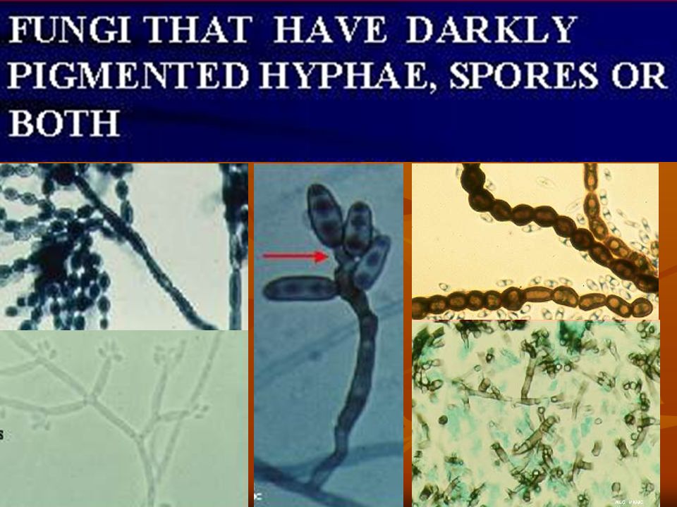
Dematiaceous Fungi The term "dematiaceous" refers to the characteristic dark appearance of this group of fungi as it grows on agar. Colonies are dark gray, brown, or black and, importantly, have a black reverse when the bottom of the agar plate is examined. Hyphae are dark yellow or brown when unstained or poorly stained This distinguishes the dematiaceous fungi from fungi with black conidia but an otherwise pale mycelium, such as A. niger.

5
A. flavus A. fumigatus A. niger

6
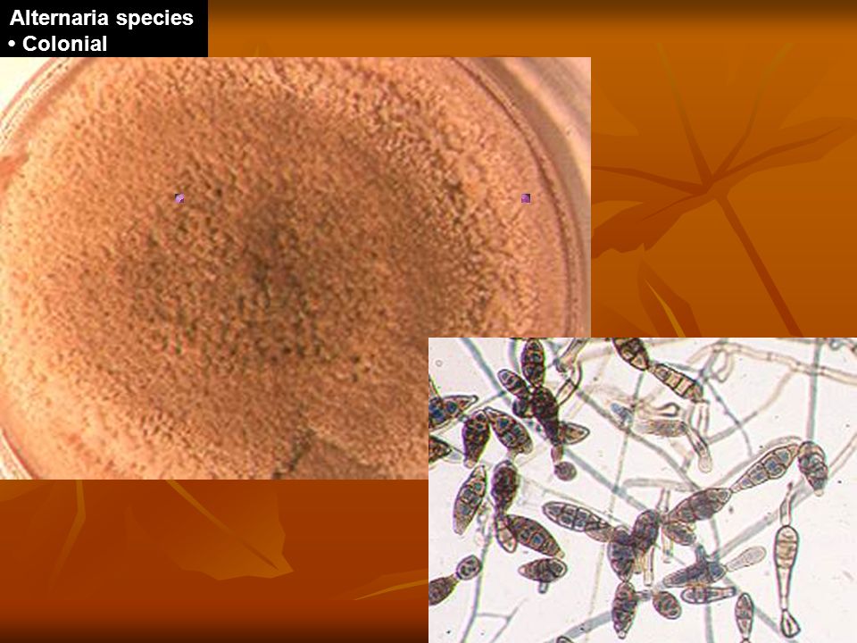
Dematiaceous Fungi with Muriform Macroconidia Alternaria Stemphylium
Ulocladium Epicoccum Note muriform septation in the macroconidia indicated by the arrow Conidia form chains or occur singly and are divided by transverse and longitudinal septations; conidia usually have a club-shaped configuration

7
Dematiaceous Fungi with Transversely Septated Macroconidia Curvularia
Drechslera characteristic boomerang shaped macroconidia.

8
Dematiaceous Fungi with Microconidia
Cladosporium Phialophora verrucosa Fonsecaea pedrosoi Wangiella dermatitidis

9
Dematiaceous Fungi that Defy the Above Grouping
Phaeoannellomyces werneckii Chaetomium species Note the 2 celled yeast forms. Perithecia with radially extending irregular brown filamentous setae

10
Alternaria tenuis Nees, 1817 Alternaria chartarum, Alternaria dianthicola, Alternaria geophilia, Alternaria infectoria, Alternaria stemphyloides, and Alternaria teunissima

11
Alternaria species • Colonial Morphology: Grows rapidly as downy to wooly, pale grey to brown on the surface and brown to black on reverse
12
Stemphylium botryosum
Wallroth, 1833 The genus Stemphylium contains several species, the most common one is Stemphylium sarcinaeforme

13
Ulocladium botrytis Preuss, 1851 The genus Ulocladium comprises 9 species

15
Curvularia species

16
Drechslera species

17
Bipolaris species

19
Pithomyces chartarum

20
Cladosporium Trejos, 1954 The genus comprises over 30 sp. Cladosporium elatum, Cladosporium herbarum, Cladosporium sphaerospermum, and Cladosporium cladosporioides. Many species have been classified in different other genera such as Exophiala, Fonseceae etc.

23
Phialophora verrucosa
Thaxter, 1915 The Phialophora species commonly involved P. verrucosa, P. hoffmanni, P. parasitica, P. repens and P. richardsiae.

24
Phialophora parasitica
• Colonial Morphology: Moderately slow growing, matures in 7 days. Initially cream colored and velvety, becoming olive grey to black with age. Reverse black.

25
Phialophora richardsiae
Conant, 1937

26
Cladophialora carioni

27
Cladophialophora bantiana (Xylohypha bantiana)
Lactophenol Aniline Blue • Magnification: x250 Magnification: x400

28
Rhinocladiella atrovirens
Rhinocladiella atrovirens

30
comprises species classified previously in the genera. Phialophora,
comprises species classified previously in the genera Phialophora, Hormodendrum Cladosporium F. compacta F. pedrosoi Fonsecaea. Powell, 1952

31
Fonsecaea pedrosoi

32
Fonsecaea compacta

34
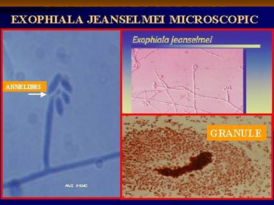
• Specimen: leg mass • Black yeast Exophiala jeanselmei
Exophiala castellanii, Exophiala moniliae, Exophila pisciphila, Exophiala salmonis, Exophiala spinifera. • Specimen: leg mass • Black yeast

35
• Specimen: Bronchial Washing
Exophiala spinifera • Specimen: Bronchial Washing •

37
Wangiella dermatitidis • Specimen: Bronchial Washing •

38
Hortae (Phaeoannellomyces) Phaeoannellomyces werneckii
(Cladosprium werneckii) .
 .")
39
Phaeococcomyces

40
Aureobasidium pullulans

45
Petriella setifera • Colonial Morphology: White, dark grey, brownish, becoming granular due to perithecium production • Stain: Lactophenol Aniline Blue x400 • Structure: Conidiophores arising from undifferentiated hyphae, producing single celled hyaline, ovoid conidia on the slimy heads.

46
Calcofluor White • Magnification: x400
Dactylaria gallopova

47
Chaetomium SP black perithecium

48
Phoma Sp.

49
Stachybotrys-atra

53
Chromoblastomycosis The ethiologic agents belong to the Fonsecaea, Phialophora, Rhinocladiella and Cladosporium genera [4, 5]. The causal agents are normally found as saprophytes in the soil, wood and vegetation [3]. They are more frequent in the tropics but have a worldwide distribution [7]. The vast majority of cases are attributed to Fonsecaea pedrosoi

54
Chromomycosis characterized by the formation of warty cutaneous nodules that develop very slowly, ultimately forming prominent papillomatous vegetations which may or may not ulcerate. Usually the lesions are confined to the feet and legs but may occur on the hands, face, ear, neck, chest or shoulders. The most common causes are Fonsecaea pedrosoi, F. compactum and Phialophora verrucosa.

59
Hand chromoblastomycosis

60
The dermis contains multiple black nodules with necrotic foci

61
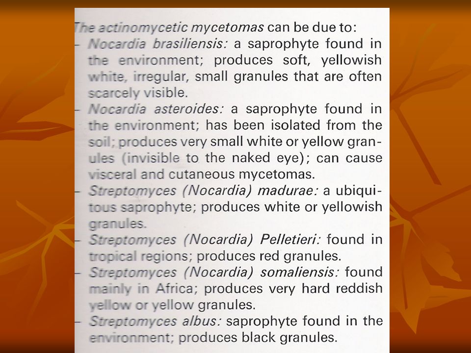
Mycetoma Madura foot Maduromycosis

62
is a chronic infection characterized by the development of tumifactions and sinuses.
The infection most often occurs in the feet but may appear on the hands or buttocks. The organism occasionally invades the body producing lesions in the brain, the meninges and other internal organs, including the bones. The disease occurs most frequently in tropical and subtropical zones.

63
The numerous fungi that have been isolated from cases of maduromycosis belong to several genera.
The dematiaceous fungi causing this disease fall in the genera Madurella, Phialophora , Curvularia , Pyrenochaeta and Leptosphaeria. These fungi produce black granules in the lesions. Other non-dematiaceous fungi that produce white to yellow granules include Cephalosporium, Petriellidium etc

68
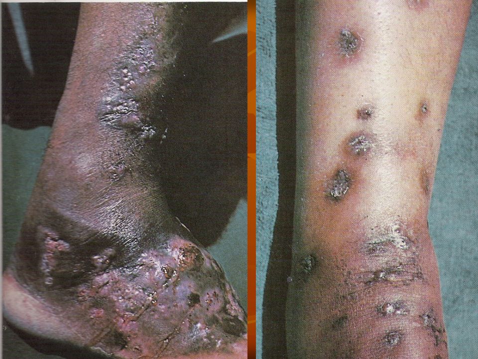
Mycetoma

70
Excision of fistule-forming inflammatory nodule at the ankle of a Japanese man
demarcated suppurative granuloma containing black grains

73
3. Phaeohyphomycosis: This term refers to infections, other than chromomycosis and maduromycosis, characterized by the presence of darkly pigmented, septate hyphae in tissues. Both cutaneous and systemic The clinical forms vary from solitary encapsulated cysts in the subcutaneous tissue to brain abscesses. The phaeomycotic cyst usually develops on the extremities and may enlarge to several centimeters

74
The most common causes of phaeohyphomycotic diseases are:
a. Phaeomycotic cyst: Exophiala jeanselmei, Wangiella dermatitidis, E. spinifera, Phialophora hoffmannii, P. parasitica, P. repens, P. richardsiae, Bipolaris spicifera. b. Subcutaneous phaeohyphomycosis: B. spicifera, Alternaria alternata, P. richardsiae, E. jeanselmei, Hermonema dermatitidis. c. Systemic phaeohyphomycosis: A. alternata, B. spicifera, Aureobasidium pullulans, Dactylaria constricta. d. Brain abscesses: Cladophialophora bantiana, Curvularia lunata.

76
Brain Biopsy • Stain: Gram Stain
Brain Biopsy • Stain: Gram Stain Structure: Long hyphae appearing pseudohyphae, observe septum. No indication of pigmentation.

77
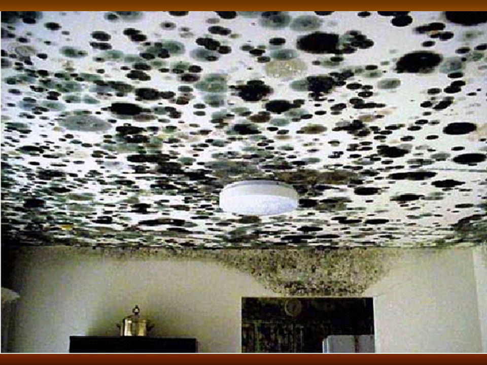
Other diseases and problems caused by dematiacious fungi
Allergy Mycotoxicosis Ocular infections in cats and a dog Color change and biodeterioration of antique marbles, walls, ceiling etc. Food spoilage - Black spots in frozen meat - Vegetable and fruit rot

80
Stachybotryotoxicosis
Stachybotryotoxicosis is a disease in animals caused by satratoxins produced by Stachybotrys alternans. The disease is manifested by 4 forms: a dermal form, generalized form, nervous form and abortion. In man it causes conjunctivitis, cough, rhinitis and skin inflammation.

81
Myrotheciotoxicosis Myrotheciotoxicosis is a disease of sheep, sometimes also cattle It is caused by the toxins verrucarin A and roridin A, produced by Myrothecium species. The disease is characterized by tympany and high mortality

82
Pithomycotoxicosis Pithomycotoxicosis is a disease of sheep and cattle associated with the consumption of plant stubs contaminated with sporidesmin produced by Pithomyces chartarum. It is characterized by diarrhoea and reduction of milk yield, photosensitization of the skin resulting in inflammation, oedema and serous exudation.

Similar presentations


>")


>")


