Sporothrix schenckii complex
It is now recognised that Sporothrix schenckii is a species complex of five distinct species: S. schenckii sensu strictu, S. brasiliensis, S. globosa, S. mexicana and S. luriei (Marimon et al. 2007, Romeo et al. 2011, Barros et al. 2011, Oliveira et al. 2014, Zhang et al. 2015b).
RG-2 organism.
Sporothrix schenckii complex is a dimorphic fungus and has a worldwide distribution, particularly in tropical and temperate regions. It is commonly found in soil and on decaying vegetation and is a well-known pathogen of humans and animals. Sporotrichosis is primarily a chronic mycotic infection of the cutaneous or subcutaneous tissues and adjacent lymphatics characterised by nodular lesions which may suppurate and ulcerate. Infections are caused by the traumatic implantation of the fungus into the skin, or very rarely, by inhalation into the lungs. Secondary spread to articular surfaces, bone and muscle is not infrequent, and the infection may also occasionally involve the central nervous system, lungs or genitourinary tract.
Sporothrix schenckii
Morphological description:
Colonies at 25C, are slow growing, moist and glabrous, with a wrinkled and folded surface. Some strains may produce short aerial hyphae and pigmentation may vary from white to cream to black. Conidiophores arise at right angles from thin septate hyphae and are usually solitary, erect and tapered toward the apex. Conidia are formed in clusters on tiny denticles by sympodial proliferation at the apex of the conidiophore, their arrangement often suggestive of a flower. As the culture ages, conidia are subsequently formed singly along the sides of both conidiophores and undifferentiated hyphae. Conidia are ovoid or elongated, 3-6 x 2-3 µm, hyaline, one-celled and smooth-walled. In some isolates, solitary, darkly-pigmented, thick-walled, one-celled, obovate to angular conidia may be observed along the hyphae. On brain heart infusion (BHI) agar containing blood at 37C, colonies are glabrous, white to greyish-yellow and yeast-like consisting of spherical or oval budding yeast cells.
Click images below to expand:

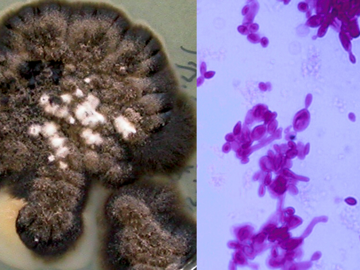
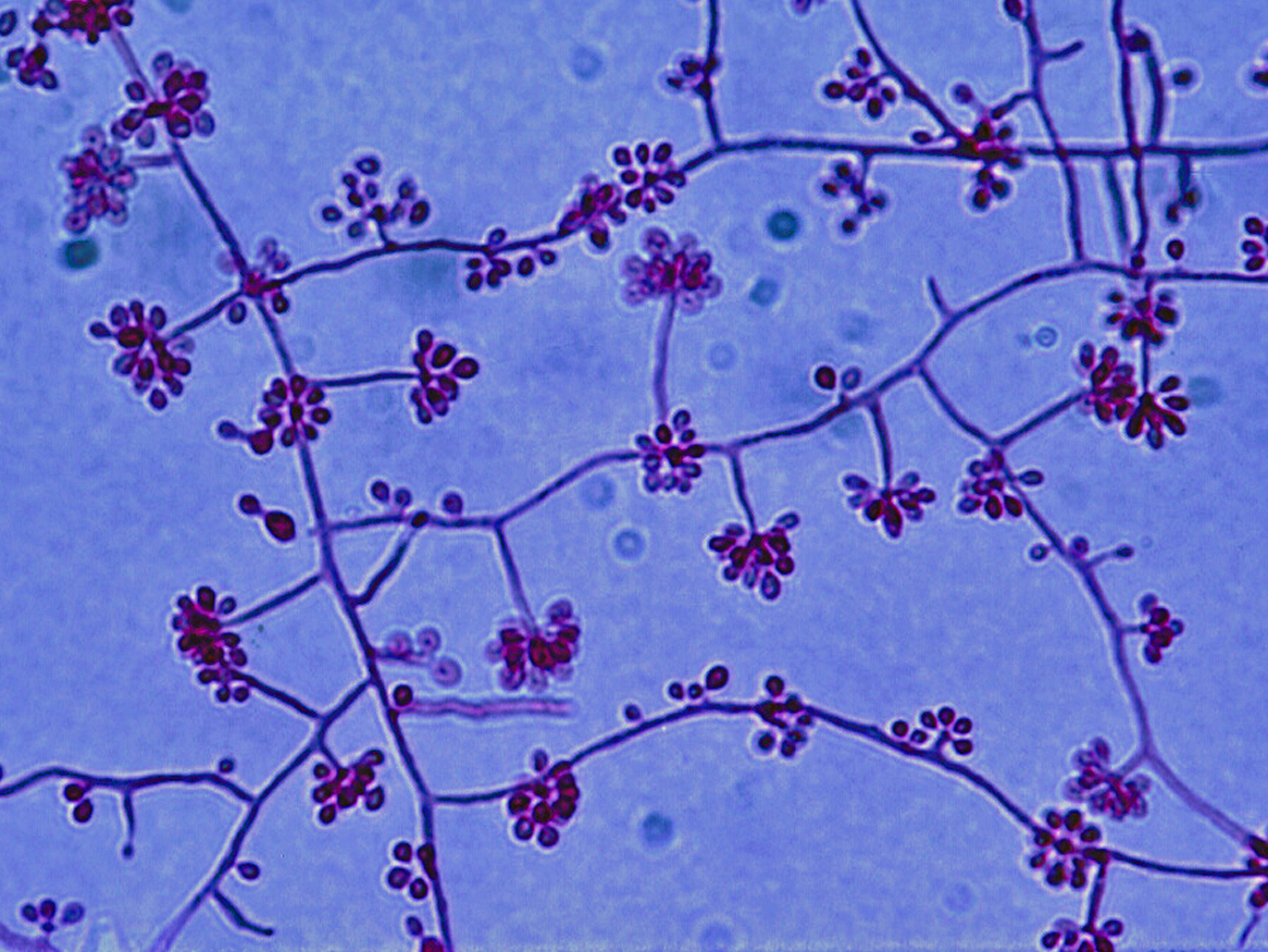
Photos show Sporothrix schenckii PAS stained tissue section showing budding yeast-like cells, culture at 25C and budding yeast cells in BHI at 37C, and conidiophores and conidia at 25C.
Molecular identification:
DNA sequencing using ITS, D1/D2, β-tubulin, calmodulin and chalcone synthase genes is recommended for species identification (Marimon et al. 2007, Romeo et al. 2011, Barros et al. 2011, Oliveira et al. 2014, Zhang et al. 2015b).
MALDI-TOF MS:
Oliverira et al. (2015) established a MALDI-TOF protocol and reference database for the identification of Sporothrix species.
Key features:
Hyphomycete characterised by thermal dimorphism and clusters of ovoid, denticulate conidia produced sympodially on short conidiophores.
| No | ≤0.016 | 0.03 | 0.06 | 0.125 | 0.25 | 0.5 | 1 | 2 | 4 | ≥8 | |
|---|---|---|---|---|---|---|---|---|---|---|---|
| AmB | 30 | 1 | 2 | 4 | 13 | 10 | |||||
| ISAV | 2 | 2 | |||||||||
| VORI | 30 | 1 | 2 | 8 | 9 | 6 | 4 | ||||
| POSA | 30 | 2 | 12 | 13 | 2 | 1 | |||||
| ITRA | 30 | 4 | 16 | 9 | 1 |
References: Sugiura and Hironaga (2010), Giraldo et al. (2014), Sandoval-Denis et al. (2014a), de Hoog et al. (2015).