Verruconis gallopava
Synonymy:
Ochroconis gallopava
Verruconis species are thermophilic, with Verruconis gallopava occurring in hot environments, such as thermal soils, broiler house litter, hot springs, and self-heated waste (Samerpitak et al. 2014).
Verruconis gallopava is neurotropic and is a recognised agent of human brain infections and is responsible for encephalitis in poultry and wild birds dogs and cats (Seyedmousavi et al. 2014). Occasional human pulmonary infections in immunocompetent hosts have also been reported (Samerpitak et al. 2014, Seyedmousavi et al. 2014, Giraldo et al. 2014).
RG-2 organism.
Morphological description:
Colonies are smooth to suede-like, dry, flat, tobacco-brown to brownish-black with a dark brown diffusible pigment. Hyphae are brown with relatively thick walls. Conidiophores are mostly cylindrical to acicular, sometimes poorly differentiated, bearing a few conidia at the tip. Conidia are two-celled, subhyaline to pale brown, smooth-walled to verrucose, cylindrical to clavate, constricted at the septum, 11-18 x 2.5-4.5 µm in size, with the apical cell wider than the basal cell. A remnant of a denticle may also be seen at the conidial base. Optimum growth at 35C, tolerant to 40C.
Click images below to expand:
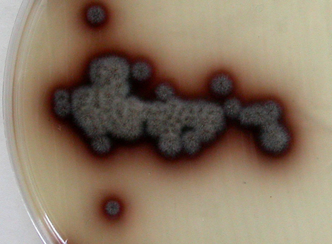
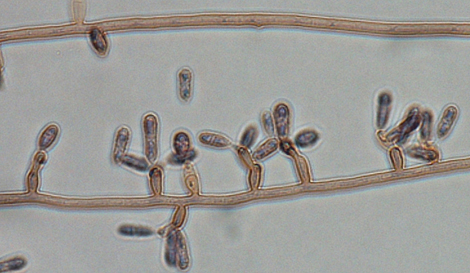
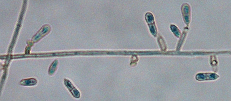
Photos of Verruconis gallopava culture and conidiophores and conidia.
Molecular identification:
ITS sequencing can identify species. Additional genes include β-tubulin, actin, and the D1/D2 region (Giraldo et al.2014, Seyedmousavi et al. 2014).
References:
Domsch et al. (1980), McGinnis (1980), de Hoog et al. (2000, 2015), Samerpitak et al. (2014), Seyedmousavi et al.(2014) and Giraldo et al. (2014).
| No | ≤0.016 | 0.03 | 0.06 | 0.125 | 0.25 | 0.5 | 1 | 2 | 4 | ≥8 | |
|---|---|---|---|---|---|---|---|---|---|---|---|
| AmB | 26 | 1 | 3 | 6 | 11 | 5 | |||||
| ISAV | 6 | 1 | 1 | 4 | |||||||
| VORI | 26 | 2 | 5 | 6 | 7 | 4 | 2 | ||||
| POSA | 25 | 5 | 3 | 8 | 8 | 1 | |||||
| ITRA | 26 | 4 | 4 | 14 | 4 |
References: Sugiura and Hironaga (2010), Giraldo et al. (2014), Sandoval-Denis et al. (2014a), de Hoog et al. (2015).