Coryneum modonium (Sacc.) Griffon & Maubl., (1910)
≡ Stilbospora modonia Sacc., (1884)
MycoBank number: MB 120927; Index Fungorum number: IF 120927; Facesoffngi number: FoF 11774; Figs. 19,20
Saprobic on a dead branch of Castanea sativa. Sexual morph: Pseudostromata 0.6–1.5 mm diam., solitary, scattered, circular, erumpent through the substrate, with perithecial bumps, containing 6–10 perithecia embedded in an entostroma. Ectostromatic disc 0.5–0.8 mm diam., distinct, circular, brownish. Central column and entostroma gray. Ostioles inconspicuous, invisible at surface of ectostromatic disc. Perithecia 300–500 μm × 400–600 μm (x̅ = 395 × 521, n = 20), globose to subglobose, uniloculate, black. Ostiole neck 350–450 µm long, 100–120 µm wide, central, cylindrical, blackish-brown. Peridium 45–60 µm wide, thick-walled, comprises of 8–11 layers, outermost layers pigmented, comprising dark brown to pale brown cells of textura angularis to flattened prismatica. Hamathecium comprises numerous, 5–7 µm wide, unbranched, hyaline, cellular paraphyses. Asci 180–230 × 15–25 μm (x̅ = 150 × 20 μm, n = 10), 8-spored, unitunicate, cylindrical, shortly rounded pedicellate, rounded at apex, with an ocular chamber. Ascospores 25–35 × 8.5–12 μm (x̅ = 29 × 10 µm, n = 40), uni-seriate, fusiform to ellipsoidal, rounded ends, 1-septate, constricted at septa, hyaline, guttulate, smooth-walled. Asexual morph: Coelomycetous. Conidiomata 850–1000 × 400–500 µm (x̅ = 900 × 460 µm, n = 5), stromatic, acervuli, solitary, erumpent on the substrate, immersed to semi immersed, surface tissues above slightly domed. Conidiomatal wall composed of thick-walled, dark brown cells. Conidiophores 30–60 × 6–9 μm (x̅ = 43 × 7.6 μm, n = 15), cylindrical, straight, septate, branched at the base, arising from basal stroma, smooth, hyaline to pale brown. Conidiogenous cells, holoblastic, indeterminate, cylindrical, annellidic, integrated, hyaline to pale brown. Conidia 30–70 × 10–20 µm (x̅ = 58 × 16.3 µm, n = 20), clavate to subcylindrical, club-shaped, rounded apex, hyaline at apical cells, truncate at the base, straight to slightly curved, hyaline or pale brown, becoming dark brown when mature, guttulate, 3–6-euseptate.
Material examined – Italy, Province of Forlì-Cesena [FC], Ridracoli – Bagno di Romagna, dead and fallen branch of Castanea sativa Mill. (Fagaceae), 03 April 2018, Erio Caporesi, IT 2866 (MFLU 18-1098; asexual morph), ibid., 15 October 2018, IT 4074 (MFLU 18-2312; sexual morph), ibid., 15 October 2018, IT 4074A, (MFLU 18-2498; sexual morph).
GenBank numbers – ITS: OM614591, OM614592; LSU: OM616562, OM616563.
Notes – Tulasne and Tulasne (1863) found both sexual and asexual morphs of Coryneum modonium (= Melanconis modonia) on chestnut wood. Fuckel (1869) reported another asexual morph as Melanconis modonia on dead Castanea vulgaris. Later, Saccardo (1883) reported the asexual morph of Coryneum modonium as Stilbospora modonia from Austria (Rhenogoviya) on the dead branches of Castanea. After several nomenclatural updates, currently Coryneum modonium Griffon & Maubl. is the accepted name (Species Fungorum 2022).
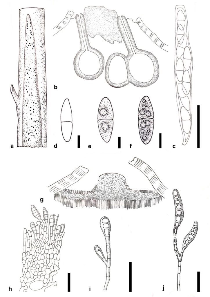
The updated redrawn line illustrations for sexual and asexual morphs of Melanconis modonia from Griffon and Maublanc (1910) are shown in Fig. 21. These morphologies are similar to our collection of Coryneum modonium, except for the absence of hyaline conidial tips. Sexual and asexual morphs of C. modonium show key characters with other Coryneum and Hyaloterminalis taxa (Senanayake et al. 2017b, Jiang et al. 2018, Rathnayaka et al. 2020). However, Talekpea morphology does not match the extant asexual taxa in Coryneaceae, including our strains, while it has a hyphomycetous asexual morph (Rathnayaka et al. 2020). Phylogenetically, Talekpea forms a distinct lineage within Coryneaceae similar to Rathnayaka et al. (2020). We provide two collections of Coryneum modonium from the Emilia–Romagna region based on morphology and multigene phylogeny (Figs. 19, 20).
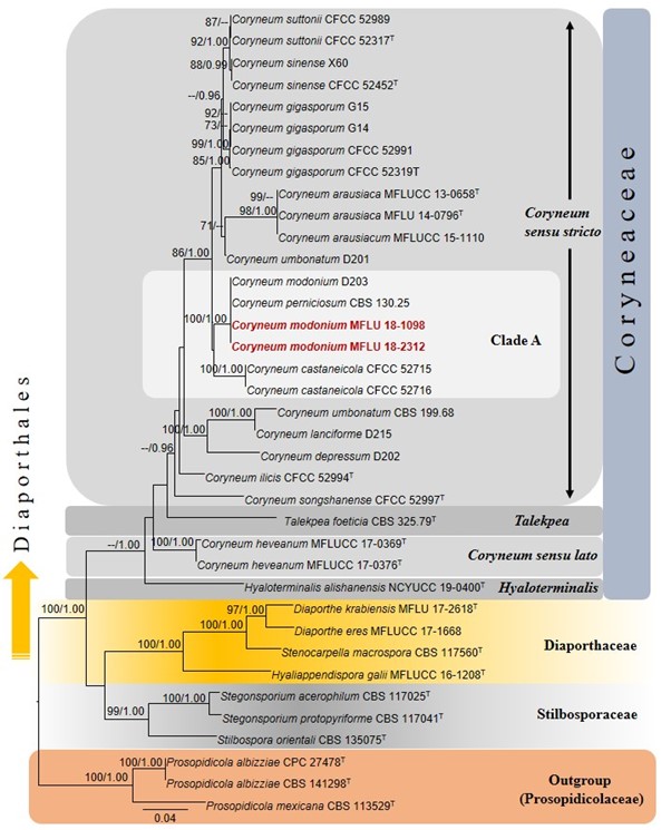
In the phylogenetic analysis of Coryneaceae, our new strains MFLU 18-2498 and MFLU 18-2312 grouped with Coryneum modonium (D203) and C. perniciosum (CBS 130.25) with 100% MLBS, 1.00 BYPP support (Fig. 22). Our strains of C. modonium (MFLU 18-1098 and MFLU 18-2312) group together within Coryneaceae in Clade A, including C. perniciosum (CBS 130.25) and sister to C. castaneicola (CFCC 52715 and CFCC 52716) (Fig. 22). Coryneum perniciosum was reported as a parasite of chestnut bark necrosis from Tuscany, Emilia, Lunigiana and Piedmont and Liguria regions in Italy (Briosi 1914). The base pair comparisons between C. modonium and C. perniciosum have revealed no differences in our two strains, MFLU 18-2498 and MFLU 18-2312 in ITS and LSU sequence data. Therefore, these four srains may be the same species. However, the holotype specimens or dry cultures, ex-type living cultures, and complete morphology for both C. modonium and C. perniciosum strains are lacking. Therefore, we keep C. perniciosum as the same and conclude our strains should be a new collection of C. modonium giving priority to the older name. We provided the first comprehensive descriptions, color illustrations, and multigene phylogenetic analyses with taxonomic discussion for Coryneum modonium. According to Sutton (1975), Coryneum species can be found on chestnut and oak trees, while our strains were recorded from Castanea sativa, generally named sweet chestnut and first record in Italy.

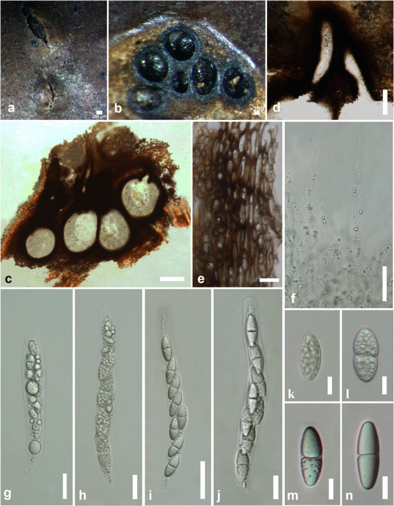
Figure 19 – Coryneum modonium (MFLU 18-2312). a. Appearance of ectostromatic discs on a twig of Castanea sativa. b, c. Longitudinal sections of perithecia. d. Ostioles. e. Peridium. f. Paraphyses. g–j. Asci (g–h mounted in water and i, j mounted in 10% KOH). k–n. Ascospores (k, l mounted in water and m, n mounted in 10% KOH). Scale bars: a–d = 200 µm, f, g–j = 20 µm, e, k–n = 10 µm.

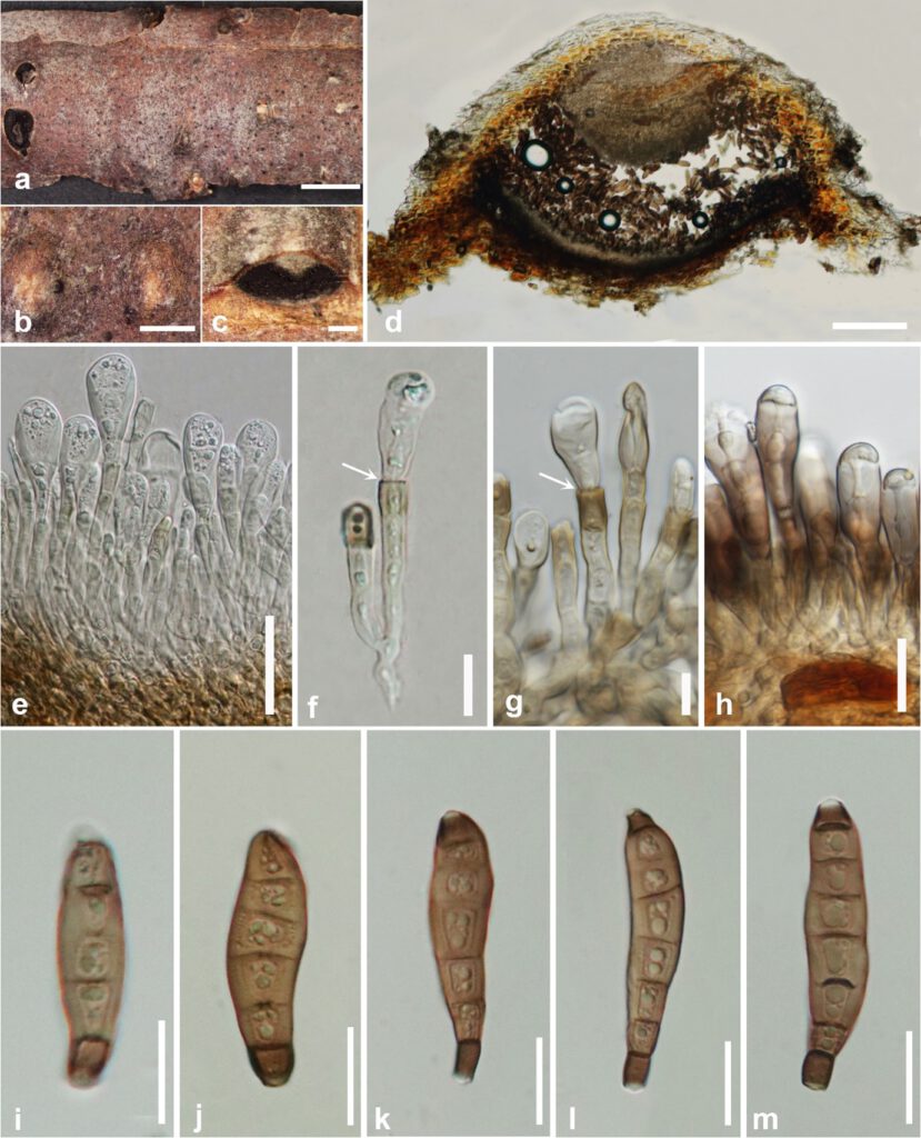
Figure 20 – Coryneum modonium (MFLU 18-1098). a–c. Conidiomata on a dead and hanging branch of Castanea sativa. d. Longitudinal section of conidiomata. e–h. Arrangement of conidiophores and conidiogenous cells (annellidic areas arrowed). i–m. Conidiospores. Scale bars: a = 2 mm, b–c = 500 µm, d = 200 µm, e–m = 20 µm.

Figure 21 – Coryneum modonium as Melanconis modonia Tul. Re-drawn from Griffon and Maublanc (1910). a. Appearance of fruiting bodies (spots) on chestnut branches. b. Longitudinal sections of stromata with ostiole. c. Ascus. d–f. Ascospores. g. Longitudinal sections of conidiomata. h. Fertile part with insertion of conidia. i–j. Arrangement of conidiophores, conidiogenous cells and conidiospores. Scale bars: c, h–j = 50 µm, d–f = 10 µm, g, a, b = NA.

Figure 22 – Phylogram generated from maximum likelihood analysis based on combined LSU, ITS and tef1-α sequenced data. Thirty seven strains were included in the combined sequence analyses, which comprised 3307 characters with gaps (ITS = 638, LSU = 849, tef1-α = 1271). Single gene analyses were also performed and topology and clade stability compared from combined gene analyses. Prosopidicola albizziae (CPC 27478, CBS 141298) and P. mexicana (CBS 113529) in Prosopidicolaceae were used as the outgroup taxa. Final ML optimization likelihood is -12145.448764. The matrix included 899 distinct alignment patterns, with 35.73 % undetermined characters or gaps. Estimated base frequencies were obtained as follows: A = 0.232426, C = 0.272338, G = 0.285542, T = 0.209694; substitution rates AC = 1.444912, AG = 1.869859, AT = 1.538490, CG = 1.206744, CT = 5.479664, GT = 1.0; gamma distribution. Bootstrap support values for ML (first set) equal to or greater than 70% and BYPP equal to or greater than 0.95 are given above the nodes. The strains from the current study are red bold and the type strains are indicated with T.
