Scolecofusarium L. Lombard, Sand.-Den. & Crous, gen. nov. Figs 8, 44.
MycoBank number: MB 838676; Index Fungorum number: IF 838676; Facesoffungi number: FoF 11006;
Etymology: From Greek skṓl¯ex, worm, in reference to the worm- like appearance of the macroconidia.
Type species: Scolecofusarium ciliatum (Link) L. Lombard, Sand.-Den. & Crous
Ascomata perithecial, solitary or gregarious, partially immersed on a stroma, smooth- and thin-walled, globose to broadly pyriform, red, with a broad, discoid apical region, turning darker in KOH, pigment dissolving in lactic acid to become yellow, lacking hairs and warts. Ascomatal wall of a single region composed of unevenly thickened cells of textura epidermoidea. Asci cylindrical, apex with an obscure refractive ring, 8-spored, ascospores uniseriate. Ascospores ellipsoidal to fusiform-ellipsoidal, 1- septate, not constricted at septum, yellow-brown, finely spinulose. Conidiophores mononematous (aerial) or grouped on sporodochia. Aerial conidiophores unbranched to loosely irregularly branched, bearing terminal phialides; conidiogenous cells monophialidic, subcylindrical, smooth- and thin-walled, with evident periclinal thickening and a non-flared collarette, producing only macroconidia. Sporodochia pink, orange to salmon coloured; sporodochial conidiophores irregularly and verticillately branched, consisting of short, often swollen, smooth- and thin- walled stipes bearing single terminal monophialides or apical whorls of 2– 3 monophialides; sporodochial conidiogenous cells monophialidic, cylindrical to subcylindrical, smooth- and thin- walled, with evident periclinal thickening. Macroconidia formed in pink to salmon slimy masses, subcylindrical, (0 –)3 – 7(– 10)- septate, straight or slightly curved, with blunt apical cell and obtuse to poorly developed, foot-shaped basal cell. Microconidia unknown. Chlamydospores unknown. [Description adapted from Samuels et al. (1991) & Gerlach & Nirenberg (1982)].
Diagnostic features: Red perithecia producing cylindrical asci containing ellipsoidal, 1-septate, finely spinulose ascospores and fusarioid asexual morph characterised by monophialides producing slender and delicate, almost cylindrical macroconidia from aerial conidiophores and pink to salmon coloured sporodochia, lacking microconidia as well as chlamydospores.
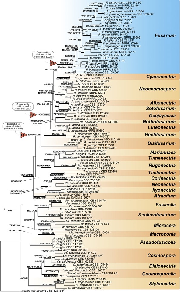
Fig. 7. Maximum-Likelihood (IQ-TREE-ML) consensus tree inferred from the combined ITS, LSU, rpb1, rpb2 and tef1 multiple sequence alignment of members of Nectriaceae. Numbers at the branches indicate support values (RAxML-BS / UFboot2-BS / BI-PP / gCF) above 70 % / 0.95 with thickened branches indicating full support (RAxML-BS / UFboot2-BS / gCF = 100 %; BI-PP = 1). The scale bar indicates expected changes per site. The tree is rooted to Nectria cinnabarina (CBS 125165). Arrows “F1”, “F2” and “F3” indicate the three alternative Fusarium hypotheses sensu Geiser et al. (2013). Ex-epitype, ex-isotype, ex-neotype and ex-type strains are indicated with ET, IT, NT, and T, respectively.
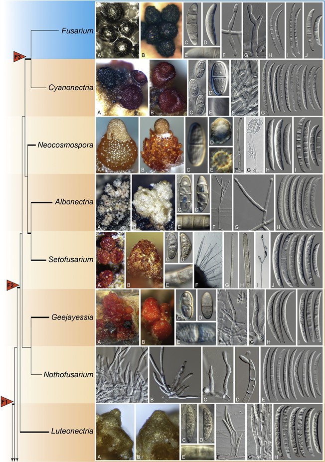
Fig. 8. (Continued).
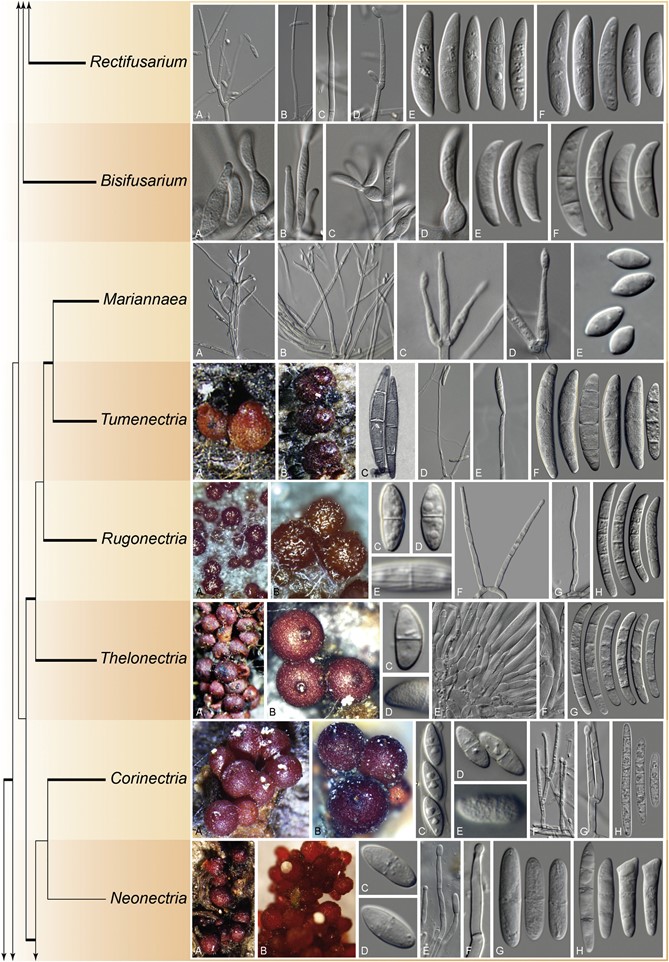
Fig. 8. (Continued).
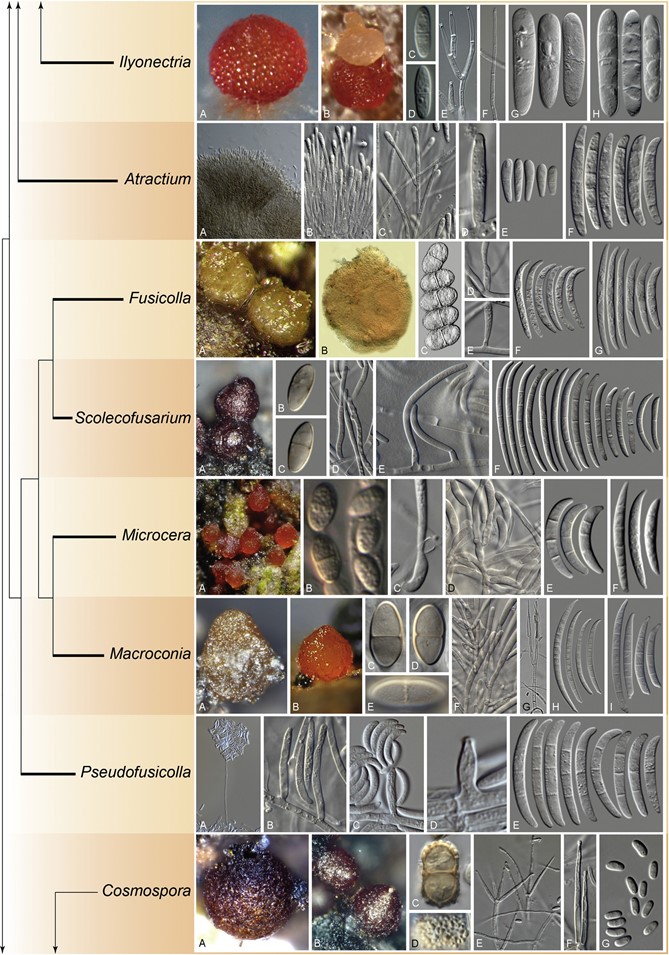
Fig. 8. (Continued).
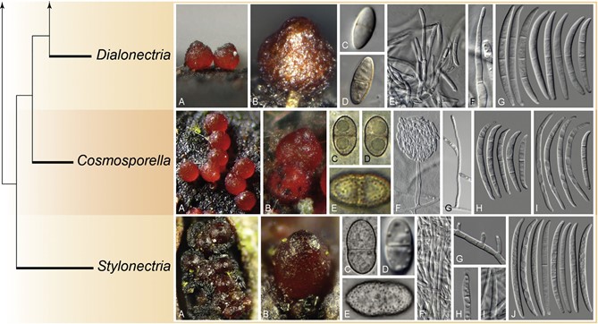
Fig. 8. Morphological features and phylogenetic affinities of fusarioid genera of Nectriaceae and close relatives. The tree was delineated based on the phylogeny presented in Fig. 7 and does not indicate phylogenetic distances. Fully supported branches are indicated in bold. The genus Fusarium is indicated in blue. Arrows “F1”, “F2” and “F3” indicate the three alternative Fusarium hypotheses sensu Geiser et al. (2013). Fusarium. A, B. Ascomata. C– E. Ascospores. F, G. Conidiogenous cells. H– J. Macroconidia. (B. Adapted from Schroers et al. 2011). Cyanonectria. A, B. Ascomata. C–E. Ascospores. F. Conidiogenous cells. G. Macroconidia. Neocosmospora. A, B. Ascomata. C–E. Ascospores. F, G. Conidiogenous cells. H, I. Macroconidia. [A. Adapted from Sandoval-Denis & Crous (2018). G. Adapted from Sandoval-Denis et al. (2019)]. Albonectria. A, B. Ascomata. C–E. Ascospores. F, G. Conidiophores and conidiogenous cells. H. Macroconidia. Setofusarium. A, B. Ascomata. C–E. Ascospores. F– H. Setae formed on sporodochia. I. Conidiophore. J. Conidia. Geejayessia. A, B. Ascomata. C–E. Ascospores. F, G. Conidiophores and conidiogenous cells. H, I. Macroconidia. [A. Adapted from Schroers et al. (2011)]. Nothofusarium. A–D. Conidiophores and conidiogenous cells. E. Conidia. Luteonectria. A, B. Ascomata. C– D. Ascospores. F, G. Conidiophores and conidiogenous cells. H. Conidia. Rectifusarium. A– D. Conidiophores and conidiogenous cells. E, F. Conidia. Bisifusarium. A– D. Conidiophores and conidiogenous cells. E, F. Conidia. Mariannaea. A, B. Conidiophores. C, D. Conidiogenous cells. E. Conidia. Tumenectria. A, B. Ascomata. C. Ascospores. D, E. Conidiophores and conidiogenous cells. F. Conidia. [A– C. Adapted from Salgado-Salazar et al. (2016)]. Rugonectria. A, B. Ascomata. C– E. Ascospores. F, G. Conidiophores and conidiogenous cells. H. Conidia. Thelonectria. A, B. Ascomata. C, D. Ascospores. E, F. Conidiophores and conidiogenous cells. G. Conidia. Corinectria. A, B. Ascomata. C– E. Ascospores. F, G. Conidiophores and conidiogenous cells. H. Conidia. (H. Picture by C. Gonz´alez). Neonectria. A, B. Ascomata. C, D. Ascospores. E, F. Conidiophores and conidiogenous cells. G, H. Conidia. [A. Adapted from Chaverri et al. (2011)]. Ilyonectria. A, B. Ascomata. C, D. Ascospores. E, F. Conidiophores and conidiogenous cells. G, H. Conidia. Atractium. A, B. Conidiophores. C, D. Conidiogenous cells. E, F. Conidia. Fusicolla. A, B. Ascomata. C. Ascospores. D, E. Conidiogenous cells. F, G. Conidia. (A–C. Pictures by C. Lechat). Scolecofusarium. A. Ascomata. B, C. Ascospores. D, E. Conidiophores and conidiogenous cells. F. Conidia. Microcera. A. Ascomata. B. Ascospores. C, D. Conidiogenous cells. E, F. Conidia. (A, B. Pictures by N. Aplin, Fungi of Great Britain and Ireland). Macroconia. A, B. Ascomata. C–E. Ascospores. F, G. Conidiophores and conidiogenous cells. H, I. Conidia. (B. Picture by P. Mlˇcoch). Pseudofusicolla. A, B. Conidiophores and conidiogenous cells. C, D. Conidia. [A– D. Adapted from Triest et al. (2016)]. Cosmospora. A, B. Ascomata. C, D. Ascospores. E, F. Conidiophores and conidiogenous cells. G. Conidia. Dialonectria. A, B. Ascomata. C– E. Ascospores. F, G. Conidiophores and conidiogenous cells. H. Conidia. (A. Picture by P. Mlˇcoch). Cosmosporella. A, B. Ascomata. C–E. Ascospores. F, G. Conidiophores and con- idiogenous cells. H, I. Conidia. (A–E. Pictures by P. Mlˇcoch). Stylonectria. A, B. Ascomata. C– E. Ascospores. F– I. Conidiophores and conidiogenous cells. J. Conidia. (A–C, E. Pictures by B. Wergen).
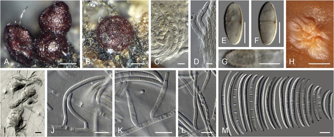
Fig. 44. Scolecofusarium ciliatum. A, B. Ascomata on natural substrate. C, D. Asci. E–G. Ascospores (G. Surface view). H. Pionnote on agar surface. I. Sporodochium. J–L. Conidiophores and conidiogenous cells. M. Macroconidia. A–H, J– L. CBS 146674. I. CBS 146676. M. CBS 144385. Scale bars: A, B = 100 μm; E–G = 5 μm; H = 1 mm; I = 20 μm; all others = 10 μm.
Species
