Ciliophora Pictures, Images and Stock Photos
Browse 1,200+ ciliophora stock photos and images available, or search for sporozoa or dinoflagellate to find more great stock photos and pictures.
Most popular
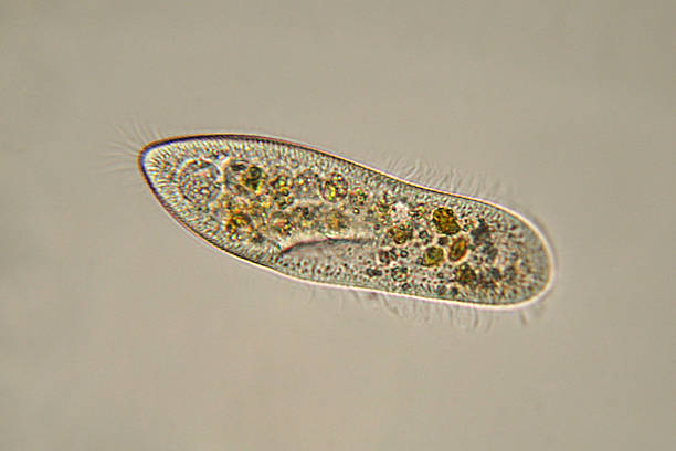
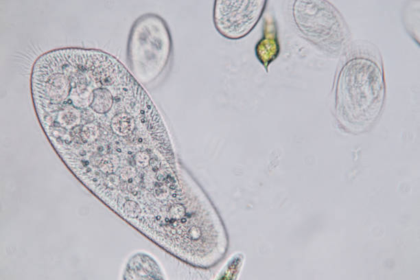
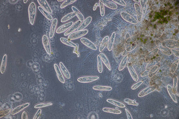
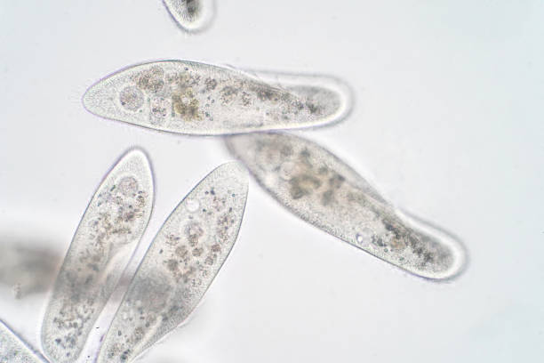
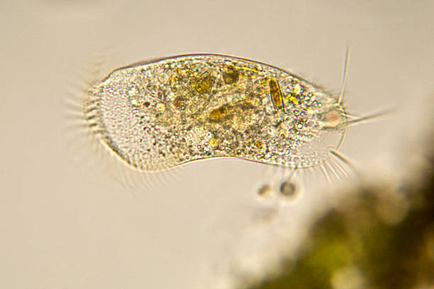
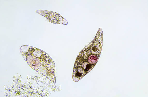
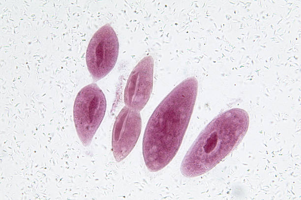
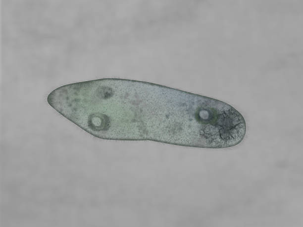
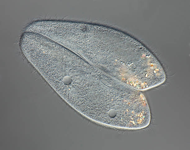
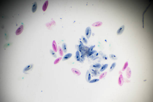
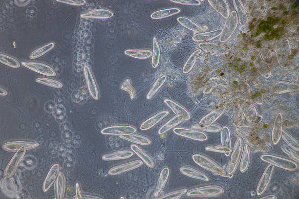
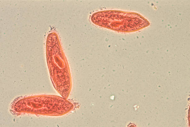
Photomicrograph of Paramecium caudatum. Consists of only one cell. Live specimen. Wet mount, 40X objective, transmitted brightfield illumination.
"contractile vacuole, differential interference contrast, Please keep in mind the special requirements of a micro photo. Motion blur of live specimen, very shallow depth of field, chromatic aberration and uneven focus are inherent in light microscopy."
Paramecium is a genus of unicellular ciliates, commonly studied as a representative of the ciliate group.
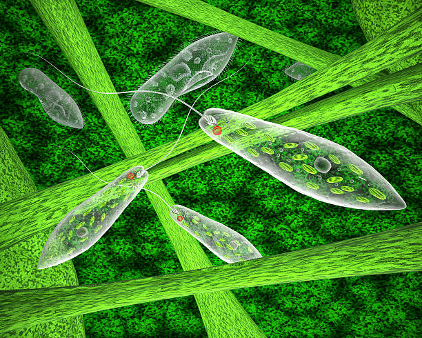
Ciliates and amoeba microorganisms in their natural habitat
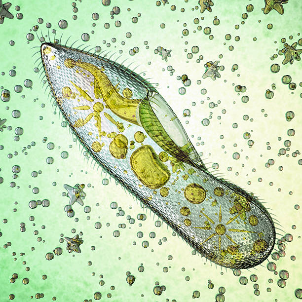
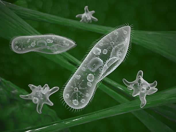
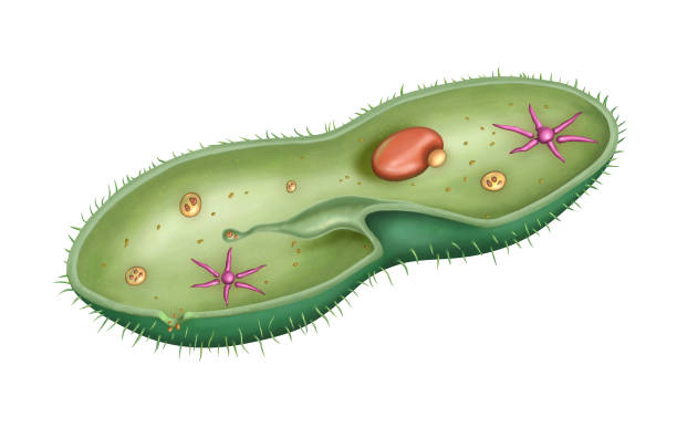
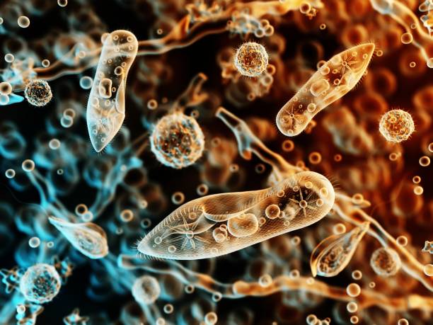
Biological micro organism paramecium caudatum. 3d image
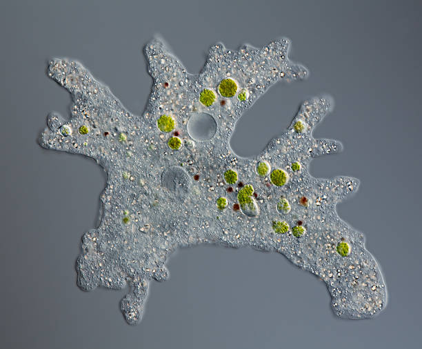
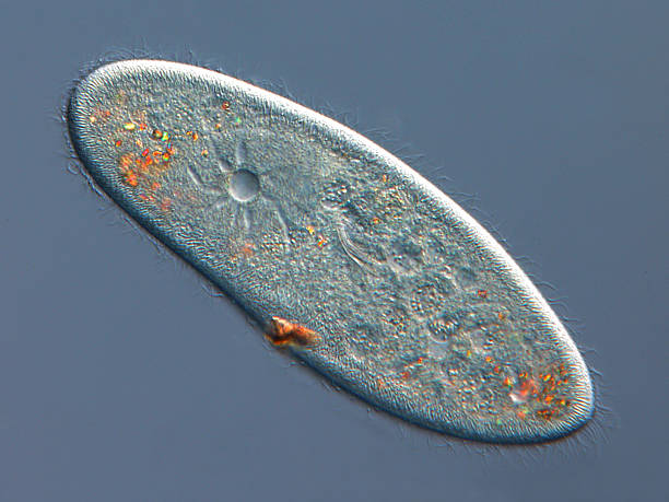
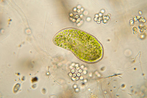
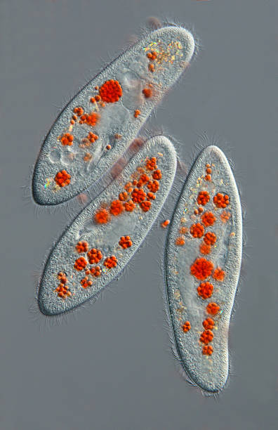
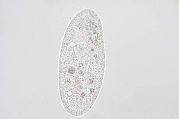
"focus to food vacuoles (with paramecium bursaria and haematococcus pluvialis), contractile vacuole, single nucleusdifferential interference contrastPlease keep in mind the special requirements of a micro photo. Motion blur of live specimen, very shallow depth of field, chromatic aberration and uneven focus are inherent in light microscopy."
Paramecium is a genus of unicellular ciliated protozoa, Paramecia are widespread in freshwater, brackish, and marine environments and are often very abundant in stagnant basins and ponds.
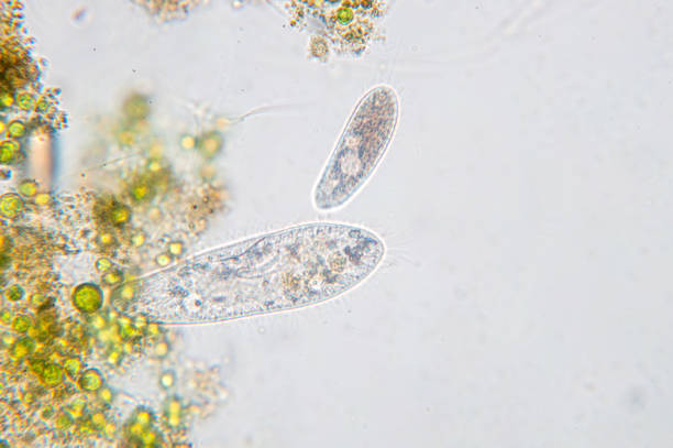
Paramecium caudatum is a genus of unicellular ciliated protozoan and Bacterium under the microscope
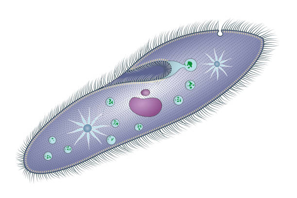
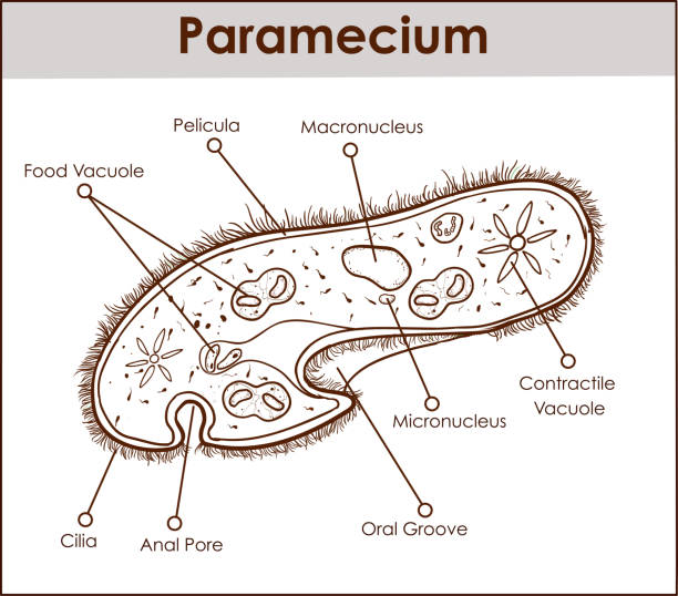
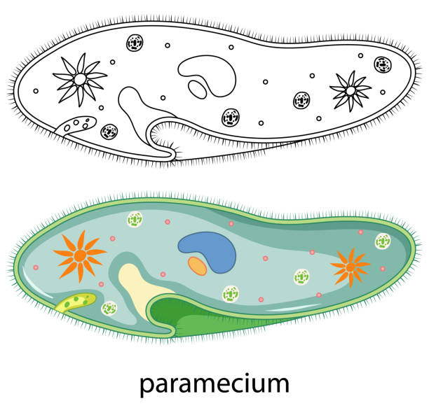
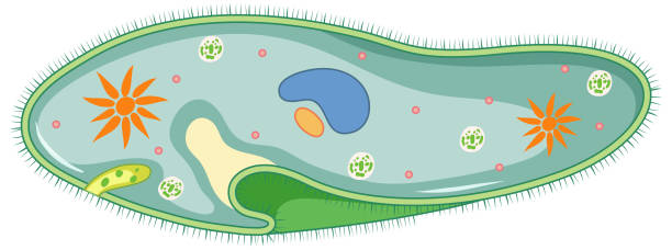
Cross-section diagram of a Paramecium caudatum, showing its internal structure. Digital illustration.
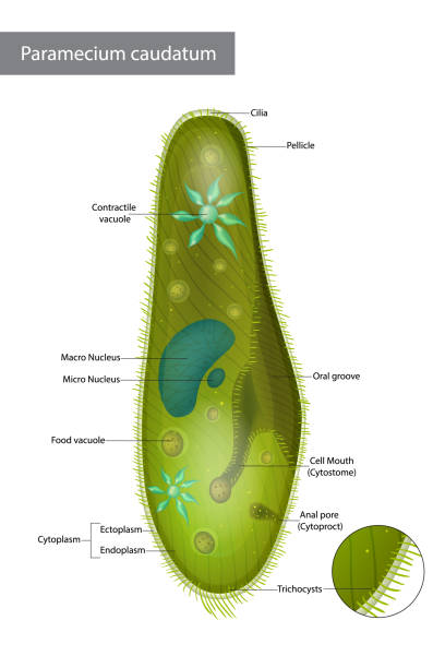
Structure Infusorian of the shoeshoe type or Paramecium caudatum. Paramecium caudatum is a species of unicellular protist in the phylum Ciliophora.
The structure of Paramecium saudatumThe structure of Paramecium saudatum
Photomicrograph of

Protozoa under a microscope. 3d illustration.
"differential interference contrast, Please keep in mind the special requirements of a micro photo. Motion blur of live specimen, very shallow depth of field, chromatic aberration and uneven focus are inherent in light microscopy."

Beauty girl love biology, looking at the microscope outdoors at sunny day
The structure of Paramecium saudatumThe structure of Paramecium saudatum
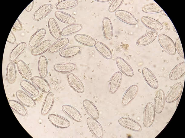
Enterobius vermicularis (EV) eggs. parasite in stool, image under light microscopy 40X objective at medical laboratory.
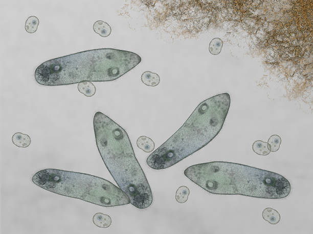
Microscopic of paramecium and amoeba

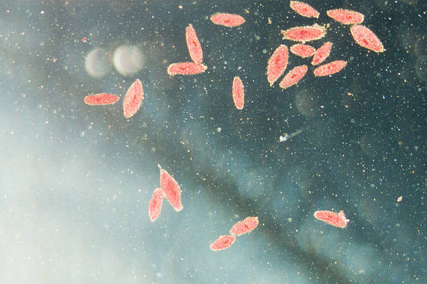
Photomicrograph of the ciliate Blepharisma americanum in various stages of development. Typical faint pink color. Each individual is only one cell. Live specimen. Wet mount, 10X objective, transmitted brightfield illumination.
microscopy micrograph animal, conjugation of Paramecium caudatum, magnification 50X
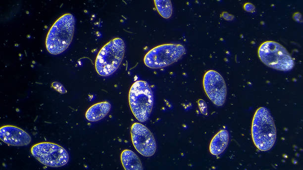
Colony of ciliates microorganisms floating in water, microscopic magnification 20x

model biological micro organism paramecium caudatum 3d render
"Microscopic photo of a professionally prepared slide demonstrating the cellular structure of the object.NOTE: Shallow DOF, uneven focus and chromatic aberration are inherent in microscopy, and what appears as dust is actually in the sample.See all my"

Seamless vector pattern of germs and bacteria. Beautiful abstract background. The concept of healthcare and medicine.
Protozoa, infusoria under a microscope. 3d illustration.
Paramecium in colour and doodle on white background illustration

Infusoria isolated on white. Vector illustration Paramecium. Infographics Ciliophora.
Microscopic of paramecium and amoeba
Paramecium is a genus of unicellular ciliated protozoa, commonly studied as a representative of the ciliate group. Paramecia are widespread in freshwater, brackish, and marine environments and are often very abundant in stagnant basins and ponds.
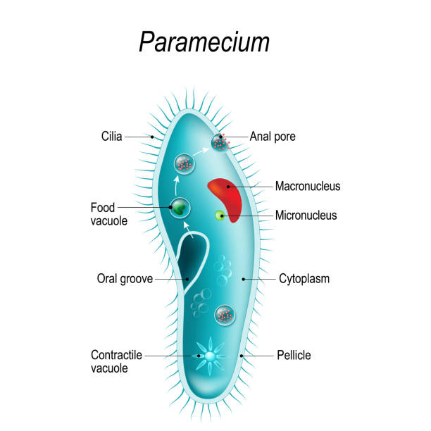
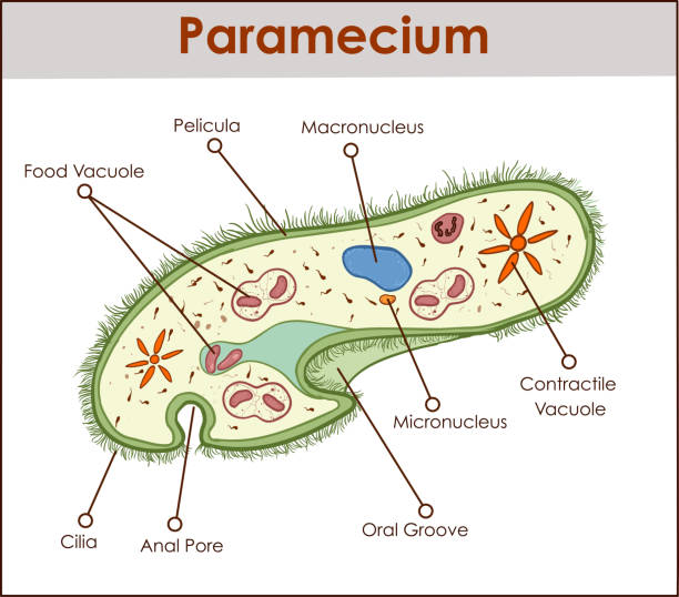
Anatomy of Paramecium caudatum. Vector diagram for educational, science, and biological use

Seamless vector pattern of germs and bacteria. Beautiful abstract background in violet tones. The concept of healthcare and medicine.
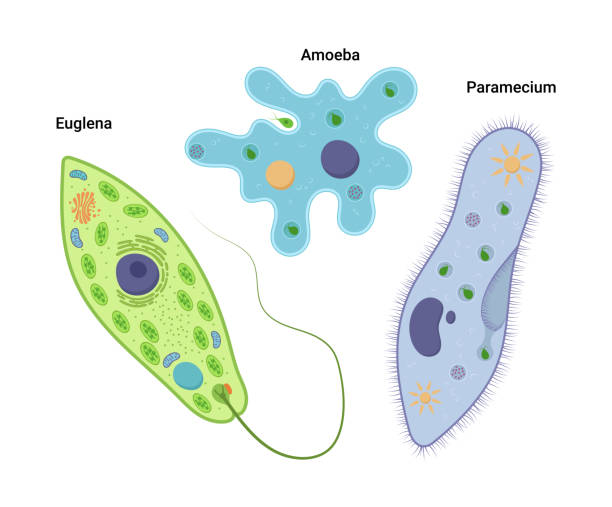
Euglena green and ciliate in the natural environment. 3d illustration
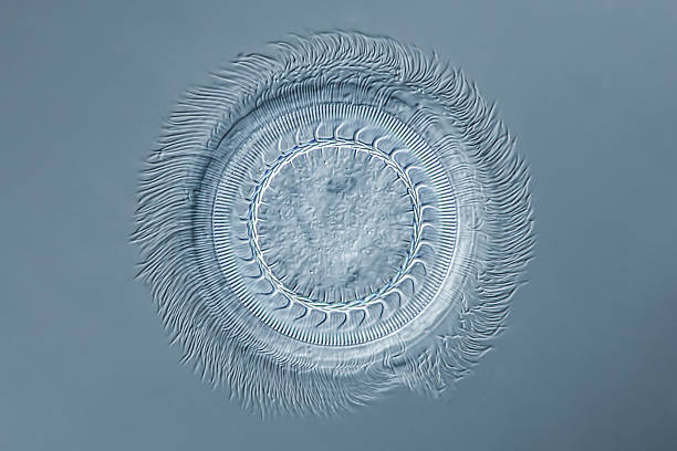
aboral ciliary wreath - 80µm - differential interference contrast
"focus to conjugationdifferential interference contrastPlease keep in mind the special requirements of a micro photo. Motion blur of live specimen, very shallow depth of field, chromatic aberration and uneven focus are inherent in light microscopy."

Creative hand drawn vector Biology background with doodle icons arranged in a circle.
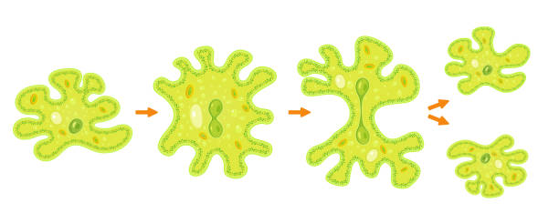
Amoeba binary fission infographic. Vector illustration of reproduction of simplest bacteria. Formation of unicellular organisms.

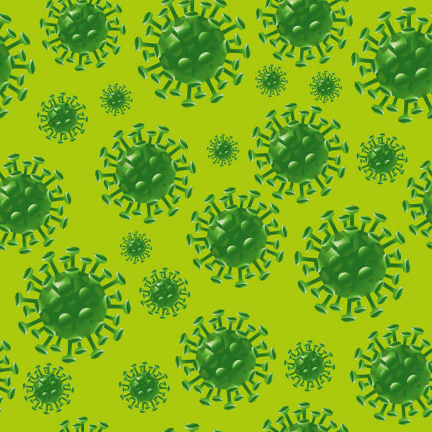
Vector Illustration of a Danger Virus Seamless Background. Coronavirus or other virus disease Background.
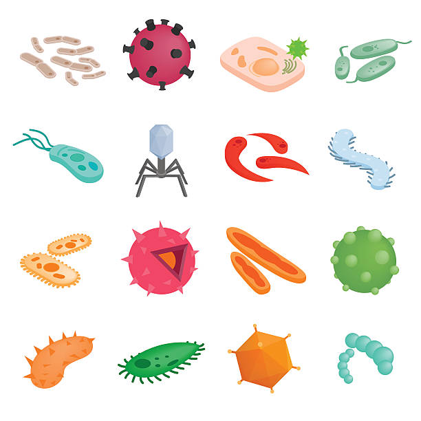
Protozoa isometric 3d illustrations. Color various microbes on a white background
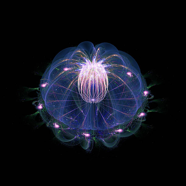
Abstract underwater background, isolated on black

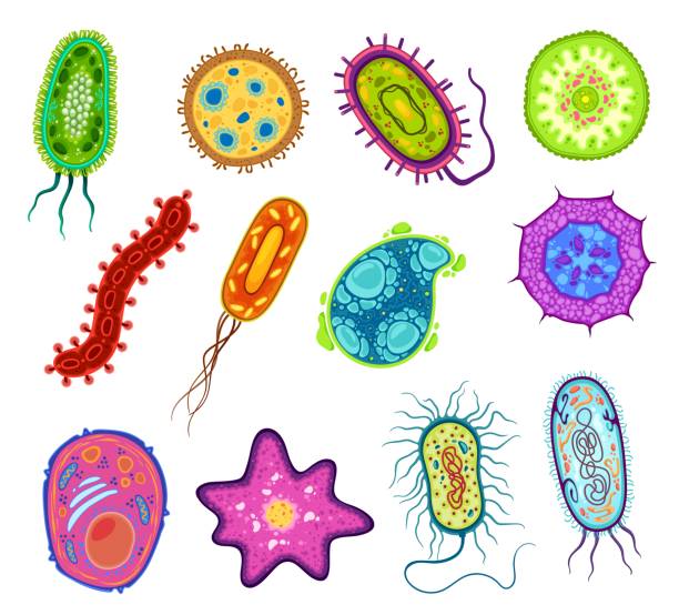
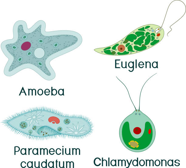
Protozoa, protista and amoeba microorganism cells, vector micro organism. Ameba and protist unicellular cells in lab microscope, protozoan eukaryotic organism types
A set of 15 simple blue and grey icons on white background for your designs and presentations.
Paramecium is a genus of unicellular ciliated protozoa, Paramecia are widespread in freshwater, brackish, and marine environments and are often very abundant in stagnant basins and ponds.

Biology hand drawn colorful vector illustration with doodle icons, biological images and objects, isolated on background.

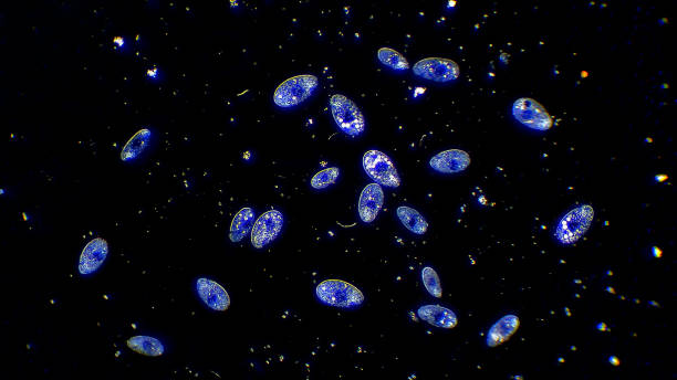
Colony of ciliates microorganisms floating in water, microscopic magnification 20x
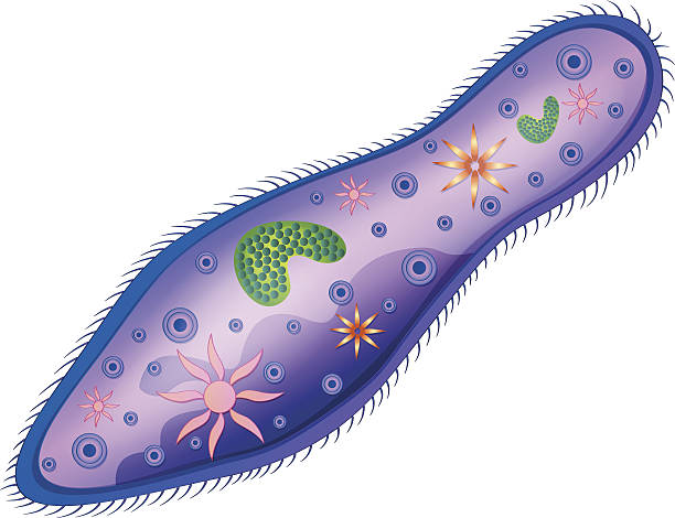
Illustration of the paramecium
Paramecium isolated on white background illustration
microscopy micrograph animal, conjugation of Paramecium caudatum, magnification 200X