
Exploring More on Dictyosporiaceae: The Species Geographical Distribution and Intriguing Novel Additions from Plant Litter

 ,
,  , ,
, ,  and
and
Abstract
:1. Introduction
2. Materials and Methods
2.1. Samples Collection, Fungal Isolation, and Examination
2.2. DNA Extraction, PCR Amplification, and Sequencing
2.3. Phylogenetic Analyses
3. Results
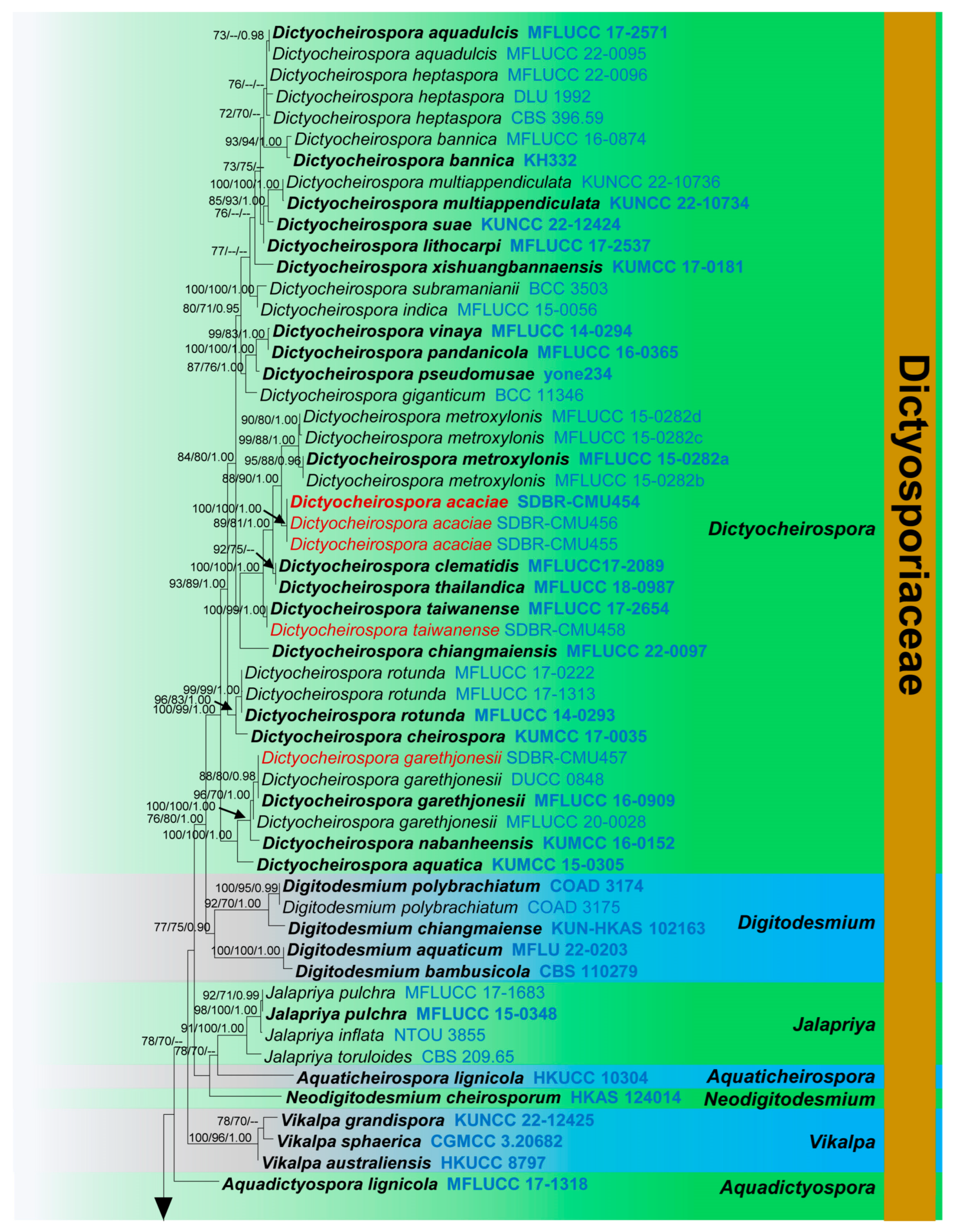
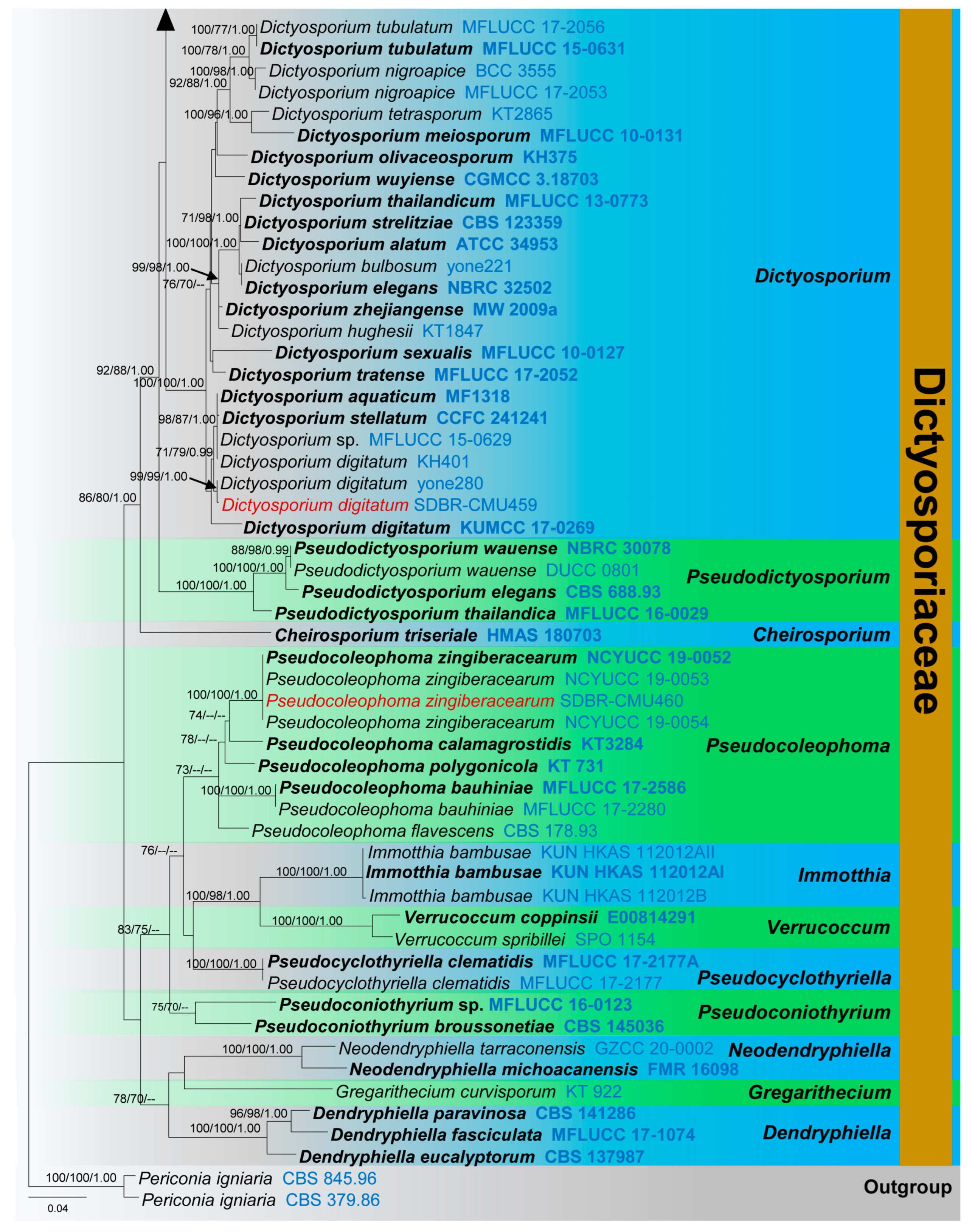
3.1. Phylogenetic Analyses
3.2. Taxonomy
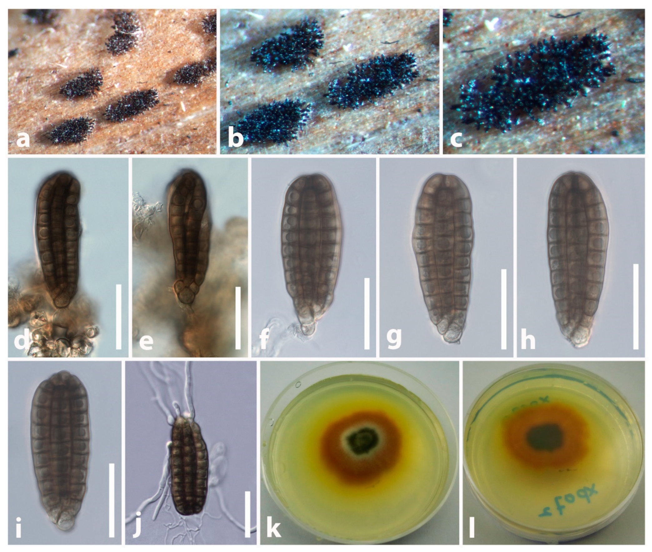
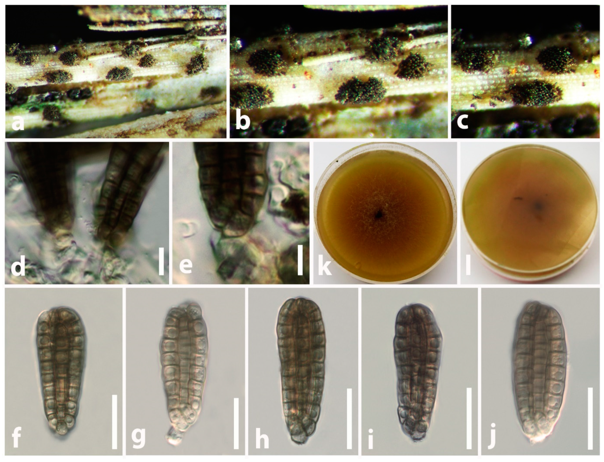
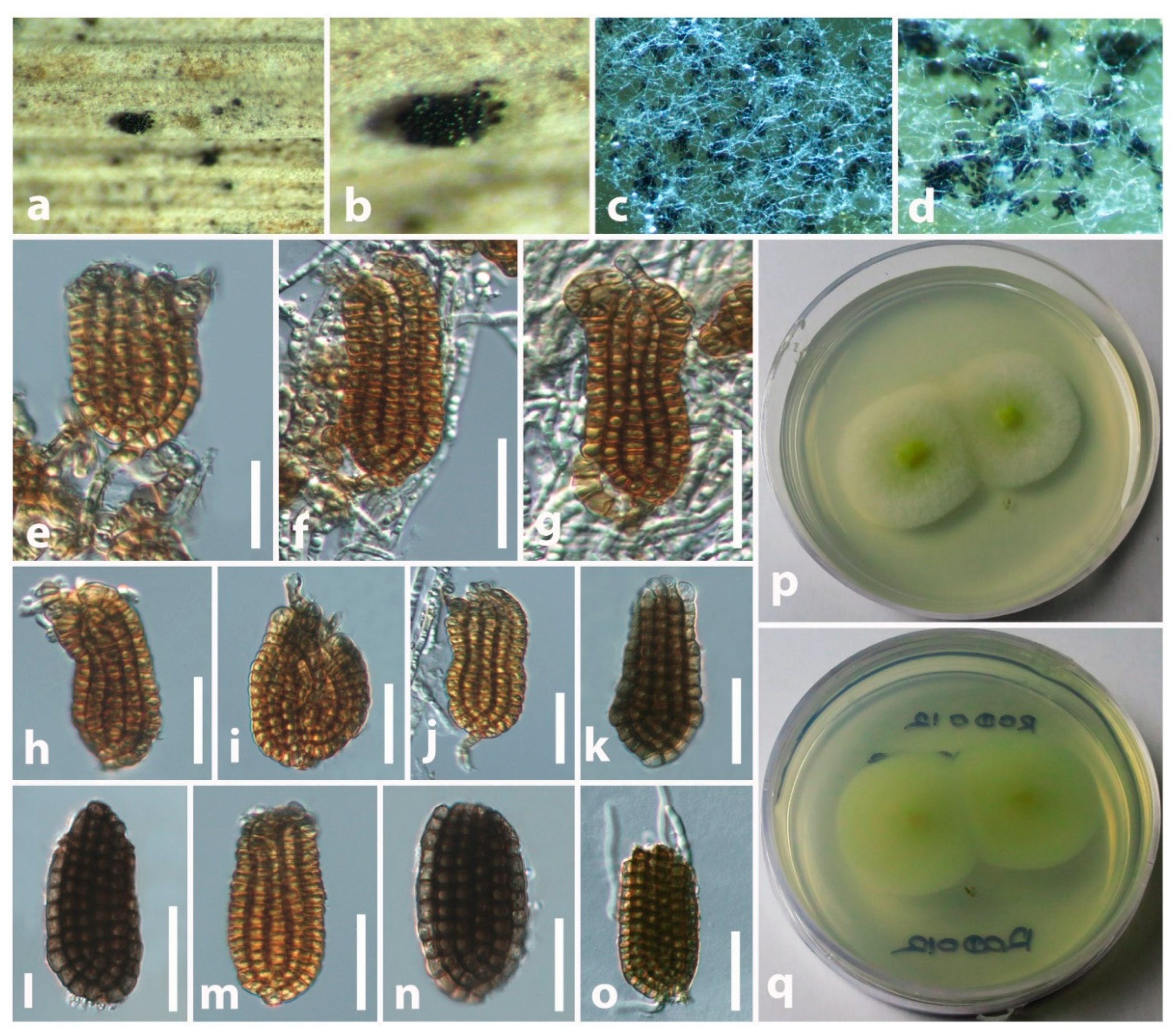
3.2.1. Dictyocheirospora M.J. D’souza, Boonmee, and K.D. Hyde, Fungal Divers. 80: 465 (2016)
3.2.2. Dictyosporium Corda, Weitenwe’er’s Beitr. Nat. 1: 87 (1837)
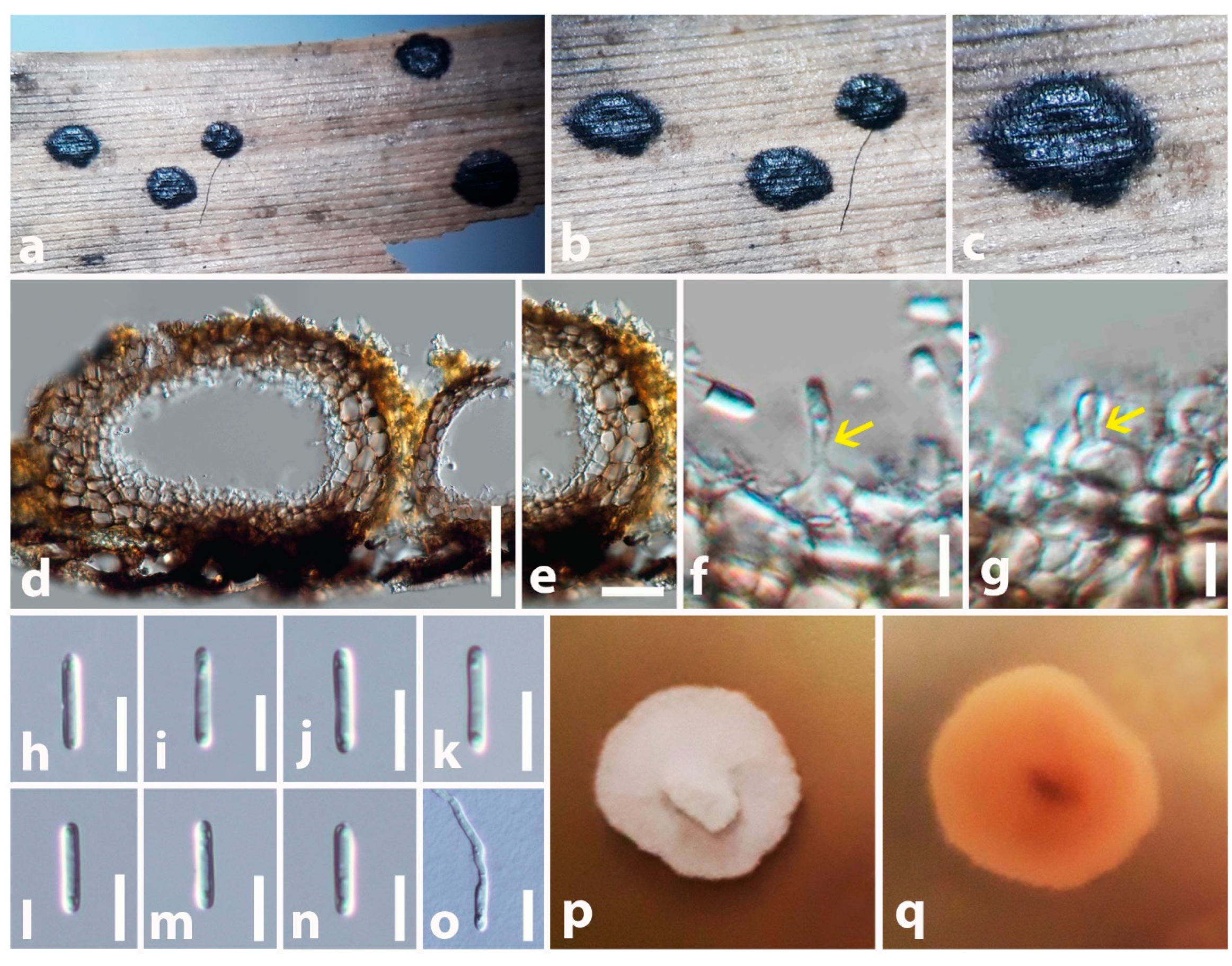
3.2.3. Pseudocoleophoma Kaz. Tanaka and K. Hiray., Stud. Mycol. 82: 89 (2015)
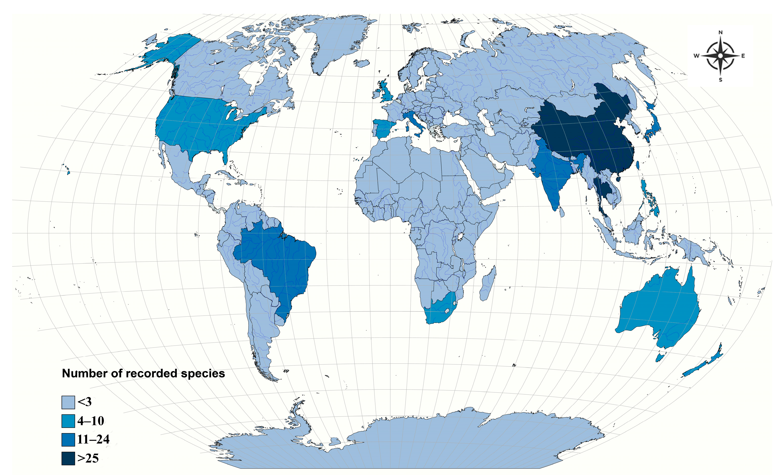
3.2.4. Geographical Distribution and Host Associations of Dictyosporiaceae Species

| Fungal Species | Host | Locality | Reference |
|---|---|---|---|
| Aquadictyospora clematidis Phukhams., D.J. Bhat, and K.D. Hyde | Clematis sikkimensis | Thailand | [44] |
| Aquadictyospora lignicola Z.L. Luo, W.L. Li, K.D. Hyde, and H.Y. Su | Unidentified submerged wood | China | [60] |
| Aquaticheirospora lignicola Kodsueb and W.H. Ho | Unidentified submerged wood | Thailand | [61] |
| Cheirosporium triseriale L. Cai and K.D. Hyde | Unidentified submerged wood | China | [62] |
| Cheirosporium vesiculare Abdel-Aziz | Unidentified decaying wood | Egypt | [14] |
| Dendryphiella aspera R.W. Barreto and J.C. David | Lantana camara | Brazil | [63] |
| Dendryphiella broussonetiae Y.L. Guo and Z.Y. Zhang | Broussonetia papyrifera | China | [64] |
| Dendryphiella dregeae A.N. Rai and Kamal | Dregea volubilis | India | [65] |
| Dendryphiella eucalypti Matsush. | Eucalyptus sp. | Chile and Taiwan | [53,66] |
| Dendryphiella eucalyptorum Crous and E. Rubio | Eucalyptus globulus | China, Spain, and South Africa | [53,67,68,69] |
| Dendryphiella fasciculata N.G. Liu, Z.Y. Liu, and K.D. Hyde | Unidentified decaying wood | Thailand | [70] |
| Dendryphiella indica V. Rao and Narania | Aloe sp. | India | [71] |
| Dendryphiella infuscans (Thüm.) M.B. Ellis | Cassia tora, Cucumis sativus, Curcuma aromatica, Desmodium strictum, Eichhornia crassipes, Lablab purpureus, Leucadendron sp., Merremia umbellata, and Vitis coignetiae | China, India, Japan, Taiwan, United States, and West Indies | [53] |
| Dendryphiella lycopersicifolia Bat. and Peres | Lycopersicon esculentum | Brazil | [72] |
| Dendryphiella paravinosa Crous and Guarnaccia | Citrus limon and Citrus sinensis | Italy | [53,73] |
| Dendryphiella phitsanulokensis N.G. Liu and K.D. Hyde | Unidentified decaying wood | Thailand | [59,74] |
| Dendryphiella stromaticola Cantillo, Gusmão, and Madrid | Unidentified wood | Brazil | [75] |
| Dendryphiella trisepta (J.J. Muchovej) B.W. Ferreira and R.W. Barreto | Glycine max | Brazil | [76] |
| Dendryphiella uniseptate Matsush. | Ficus sp. and Pistacia lentiscus | Italy and Papua New Guinea | [77,78] |
| Dendryphiella variabilis Iturrieta-González, Dania García, and Gené | Lauraceae sp. | Cuba | [15] |
| Dictyocheirospora acaciae Tennakoon and S. Lumyong | Acacia dealbata | Thailand | This study |
| Dictyocheirospora aquadulcis Sorvongxay, S. Boonmee, and K.D Hyde | Unidentified submerged wood | Thailand | [40,50] |
| Dictyocheirospora aquatica Z.L. Luo, Bhat, and K.D. Hyde | Unidentified submerged wood | China | [41] |
| Dictyocheirospora bannica Kaz. Tanaka, K. Hiray., Boonmee, and K.D. Hyde | Unidentified submerged wood | Thailand | [11,12] |
| Dictyocheirospora cheirospora S.K. Huang and f K.D. Hyde | Unidentified decaying wood | China | [43] |
| Dictyocheirospora chiangmaiensis H.W. Shen, Boonmee, and Z.L. Luo | Unidentified submerged wood | Thailand | [40] |
| Dictyocheirospora clematidis Phukhams., D.J. Bhat, and K.D. Hyde | Clematis sikkimensis | Thailand | [44] |
| Dictyocheirospora garethjonesii Z.L. Luo, Hong Y. Su, and K.D. Hyde | Unidentified submerged wood, Bismarkia nobilis, and Macaranga tanarius | China, and Thailand | [9,41] |
| Dictyocheirospora gigantica (Goh and K.D. Hyde) M.J. D’souza, Boonmee, and K.D. Hyde | Anisoptera oblonga | South Africa and Thailand | [45] |
| Dictyocheirospora heptaspora (Garov.) M.J. D’souza, Boonmee, and K.D. Hyde | Unidentified submerged wood | China and Thailand | [40] |
| Dictyocheirospora hydei (Prasher and R.K. Verma) J. Yang and K.D. Hyde | Tecoma stans | India | [46] |
| Dictyocheirospora indica (Prasher and R.K. Verma) J. Yang and K.D. Hyde | Phoenix rupicola | India and Thailand | [12,46] |
| Dictyocheirospora lithocarpi Jayasiri, E.B.G. Jones, and K.D. Hyde | Lithocarpus sp. | Thailand | [47] |
| Dictyocheirospora metroxyli Konta. and K.D. Hyde | Metroxylon sagu | Thailand | [42] |
| Dictyocheirospora multiappendiculata H.W. Shen and Z.L. Luo | Unidentified submerged wood | China | [40] |
| Dictyocheirospora musae (Photita) J. Yang, K.D. Hyde, and Z.Y. Liu | Musae acuminata | Thailand | [79] |
| Dictyocheirospora nabanheensis Tibpromma and K.D. Hyde | Pandanus sp. | China and Thailand | [40,48] |
| Dictyocheirospora pandanicola Tibpromma and K.D. Hyde | Pandanus sp. | Thailand | [11,48] |
| Dictyocheirospora pseudomusae (Kaz. Tanaka, G. Sato, and K. Hiray.) Kaz. Tanaka, K. Hiray., Boonmee, and K.D. Hyde | Unidentified decaying twigs | Japan | [49] |
| Dictyocheirospora rotunda M.J. D’souza, Bhat, and K.D. Hyde | Unidentified decaying wood | China and Thailand | [11,12,41,50,51] |
| Dictyocheirospora suae H.W. Shen and Z.L. Luo | Unidentified submerged wood | China | [40] |
| Dictyocheirospora subramanianii (B. Sutton) M.J. D’souza, Boonmee, and K.D. Hyde | Eucalyptus globulus | India | [52] |
| Dictyocheirospora taiwanense Tennakoon, C.H. Kuo, and K.D. Hyde | Ficus benjamina and Macaranga tanarius | Taiwan and Thailand | [50], this study |
| Dictyocheirospora tetraploides (L. Cai and K.D. Hyde) J. Yang and K.D. Hyde | Unidentified submerged wood | China | [12,80] |
| Dictyocheirospora thailandica X.D. Yu, W. Dong and H. Zhang | Unidentified submerged wood | Thailand | [51] |
| Dictyocheirospora vinaya M.J. D’souza, Bhat, and K.D. Hyde | Unidentified submerged wood | China and Thailand | [11,81] |
| Dictyocheirospora xishuangbannaensis Tibpromma and K.D. Hyde | Clematis sikkimensis and Pandanus sp. | Thailand | [44,48] |
| Dictyopalmispora palmae Pinruan and K.D. Hyde | Licuala longicalycata | Thailand | [11] |
| Dictyosporium acroinflatum Whitton, K.D. Hyde, and McKenzie | Freycinetia banksii | New Zealand | [56] |
| Dictyosporium alatum Emden | Machilus velutina, Pinus massoniana, and soil | China and Indonesia | [53,82,83,84] |
| Dictyosporium amoenum C.R. Silva, Gusmão, and R.F. Castañeda | Calophyllum brasiliense | Brazil | [85] |
| Dictyosporium appendiculatum Tibpromma and K.D. Hyde | Pandanus sp. | Thailand | [48] |
| Dictyosporium aquaticum Abdel-Aziz | Unidentified submerged wood | Egypt | [86] |
| Dictyosporium araucariae S.S. Silva, R.F. Castañeda, and Gusmão | Araucaria angustifolia | Brazil | [87] |
| Dictyosporium biseriale D.M. Hu, L. Cai, and K.D. Hyde | Unidentified submerged wood | China | [88] |
| Dictyosporium boydii A.L. Sm. and Ramsb. | Unidentified decaying wood | United Kingdom | [89] |
| Dictyosporium brahmaswaroopii M.D. Mehrotra | Leucaena leucocephala | India | [90] |
| Dictyosporium bulbosum Tzean and J.L. Chen | Bucida palustris, Clusia melchiori, Freycinetia banksia, and Pandanus sp. | Brazil, China, Japan, New Zealand, Spain, Taiwan, and West Indies | [45,49,53,91,92,93,94] |
| Dictyosporium campaniforme Matsush. | Trachycarpus fortune and Quercus myrsinaefolia | Japan and Switzerland | [53,95,96] |
| Dictyosporium canisporum L. Cai and K.D. Hyde | Unidentified submerged wood | China | [80] |
| Dictyosporium cocophylum Bat. | Butia yatay, Cocos nucifera, and Elaeis guineensis | Argentina, Brazil, and Ghana | [53,97,98] |
| Dictyosporium digitatum J.L. Chen, C.H. Hwang, and Tzean | Anisoptera oblonga, Archontophoenix alexandrae, Cyperus aggregatus, Licuala longicalycata, Machilus velutina, Pinus massoniana, Pandanus copelandii, P. furcatus, and Phoenix hanceana | Australia, Brunei, China, Japan, Mauritius, Philippines, Seychelles, Taiwan, and Thailand | [45,48,49,53,55,56], this study |
| Dictyosporium elegans Corda | Arenga engleri, Bamboo sp., Carpinus betulus, Chamaecyparis nootkatensis, Cistus sp., Cocos nucifera, Heritiera littoralis, Hordeum vulgare, Larix decidua, Livistona chinensis, Machilus velutina, Phillyrea angustifolia, Phoenix hanceana, Pinus massoniana, Pinus wallichiana, Pistacia lentiscus, Quercus ilex, Rhopalostylis sp., and Vitis sp. | Alaska, Australia, China, Germany, Italy, New Zealand, Pakistan, Poland, South Africa, and Taiwan | [53,99] |
| Dictyosporium foliicola P.M. Kirk | Ilex pernyi | Scotland and United Kingdom | [100] |
| Dictyosporium gauntii Bhat and B. Sutton | Unidentified decaying wood | Ethiopia | [101] |
| Dictyosporium guttulatum Tibpromma and K.D. Hyde | Pandanus sp. | Thailand | [48] |
| Dictyosporium hongkongensis Tibpromma and K.D. Hyde | Pandanus sp. | Hong Kong | [48] |
| Dictyosporium hughesii McKenzie | Rhopalostylis sapida and Stewartia monadelpha | Japan and New Zealand | [49,102] |
| Dictyosporium hymenaearum Bat. and J.L. Bezerra | Hymenaea sp. | Brazil | [103] |
| Dictyosporium krabiense Tibpromma and K.D. Hyde | Pandanus sp. | Thailand | [48] |
| Dictyosporium lakefuxianensis L. Cai, K.D. Hyde, and McKenzie | Freycinetia scandens | Australia | [104] |
| Dictyosporium manglietiae Kodsueb and McKenzie | Unidentified decaying wood | Thailand | [105] |
| Dictyosporium marinum Dayar. and E.B.G. Jones | Unidentified submerged wood | United Kingdom | [106] |
| Dictyosporium meiosporum Boonmee and K.D. Hyde | Unidentified decaying wood | Thailand | [86] |
| Dictyosporium minus Sacc. | Inga sp. | Brazil | [53] |
| Dictyosporium muriformis N.G. Liu, K.D. Hyde, and J.K. Liu | Unidentified decaying wood | China | [74] |
| Dictyosporium nigroapice Goh, W.H. Ho, and K.D. Hyde | Unidentified decaying wood and Machilus velutina | China and Thailand | [12,45] |
| Dictyosporium oblongum (Fuckel) S. Hughes | Phillyrea angustifolia, Phragmites australis, and Populus tremuloides | Canada, Italy, and Netherlands | [53] |
| Dictyosporium olivaceosporum Kaz. Tanaka, K. Hiray., Boonmee, and K.D. Hyde | Unidentified submerged wood | Japan | [11] |
| Dictyosporium palmae Abdel-Aziz | Licuala longicalycata and Phoenix dactylifera | Egypt and Thailand | [14,53] |
| Dictyosporium pandani Whitton, K.D. Hyde, and McKenzie | Pandanus spp. | Australia, Brunei, China, Nepal, and Philippines | [56] |
| Dictyosporium pandanicola Tibpromma and K.D. Hyde | Pandanus spp. | Thailand | [48] |
| Dictyosporium pelagicum (Linder) G.C. Hughes ex E.B.G. Jones | Unidentified decaying wood | Canada and United States | [53,107] |
| Dictyosporium prolificum Damon | Juncus sp. | United States | [108] |
| Dictyosporium rhopalostylidis McKenzie | Rhopalostylis sapida | New Zealand | [102] |
| Dictyosporium schizostachyfolium Bat. and M.L. Farr | Schizostachyum acutiflorum | Philippines | [109] |
| Dictyosporium sexualis Boonmee and K.D. Hyde | Decaying wood | Thailand | [11] |
| Dictyosporium sinense H.M. Liu and T.Y. Zhang | Soil | China | [53] |
| Dictyosporium solanii A.D. Sharma, Munjal, and Jandaik | Juglans regia and Trachycarpus fortunei | China and India | [53] |
| Dictyosporium splendidum Alves-Barb., Malosso, and R.F. Castañeda | Unidentified decaying leaves | Brazil | [110] |
| Dictyosporium stellatum G.P. White and Seifert | Unidentified decaying wood | United States | [111] |
| Dictyosporium strelitziae Crous and A.R. Wood | Strelitzia nicolai | South Africa | [112] |
| Dictyosporium taishanense G.Z. Zhao and T.Y. Zhang | Unidentified decaying wood | China | [113] |
| Dictyosporium tetraseriale Goh, Yanna, and K.D. Hyde | Livistona chinensis | Hong Kong | [45] |
| Dictyosporium tetrasporum L. Cai and K.D. Hyde | Unidentified submerged wood | China and Japan | [49,114] |
| Dictyosporium thailandicum M.J. D’souza, Bhat, and K.D. Hyde | Unidentified submerged wood | Thailand | [86] |
| Dictyosporium tratense J. Yang and K.D. Hyde | Unidentified decaying wood | Thailand | [12] |
| Dictyosporium triramosum Aramb., Cabello, and Cazau | Unidentified decaying wood | Argentina | [115] |
| Dictyosporium triseriale Matsush. | Phyllostachys sp. | China and Taiwan | [53,116] |
| Dictyosporium tubulatum J. Yang, K.D. Hyde, and Z.Y. Liu | Unidentified decaying wood | China and Thailand | [12,40] |
| Dictyosporium wuyiense Y. Zhang ter and G.Z. Zhao | Bamboo sp. | China | [117] |
| Dictyosporium yerbae Speg. | Ilex paraguariensis | Argentina | [118] |
| Dictyosporium yunnanense L. Cai, K.D. Hyde, and McKenzie | Unidentified submerged wood | China | [80] |
| Dictyosporium zeylanicum Petch | Unidentified decaying wood | Sri Lanka | [119] |
| Dictyosporium zhejiangense Wongs., H.K. Wang, K.D. Hyde, and F.C. Lin | Unidentified submerged wood | China | [120] |
| Digitodesmium aquaticum H.W. Shen, Boonmee, and Z.L. Luo | Unidentified submerged wood | Thailand | [40] |
| Digitodesmium bambusicola L. Cai, K.Q. Zhang, McKenzie, W.H. Ho, and K.D. Hyde | Bamboo sp. | Philippines | [121] |
| Digitodesmium chiangmaiense Q.J. Shang and K.D. Hyde | Unidentified decaying wood | Thailand | [50] |
| Digitodesmium elegans P.M. Kirk | Fagus sp. and Quercus sp. | United Kingdom | [122,123] |
| Digitodesmium heptasporum L. Cai and K.D. Hyde | Unidentified submerged wood | China | [80] |
| Digitodesmium intermedium J. Mena, Silvera, Gené, and Guarro | Plant debris | Spain | [124] |
| Digitodesmium macrosporum Silvera, Mercado, Gené, and Guarro | Soil | Spain | [124] |
| Digitodesmium polybrachiatum T.F. Nóbrega, B.W. Ferreira, and R.W. Barreto | Coffea canephora | Brazil | [125] |
| Digitodesmium recurvum W.H. Ho, K.D. Hyde, and Hodgkiss | Machilus velutina | Hong Kong | [53,126] |
| Digitodesmium tectonae (Rajeshk., Rajn. K. Verma, Boonmee, K.D. Hyde, Chandrasiri, and Wijayaw.) W.H. Tian and Maharachch. | Tectona grandis | India | [16,21] |
| Gregarithecium curvisporum Kaz. Tanaka and K. Hiray. | Sasa sp. | Japan | [49] |
| Immotthia atrograna (Cooke and Ellis) M.E. Barr | Aceri-Fraxinetum, Carya olivaeformis, Carya sp., Salix alba, Acer pseudoplatanus, and Fraxinus excelsior | United States | [127,128] |
| Immotthia atroseptata (Piroz.) M.E. Barr | Rhododendron maximum | United States | [128,129] |
| Immotthia bambusae H.B. Jiang and Phookamsak | Bamboo sp. | Thailand | [128] |
| Jalapriya apicalivaginata D.F. Bao, X. Fu, H.Y. Su, and Z.L. Luo | Unidentified submerged wood | China | [81] |
| Jalapriya aquatica D.F. Bao, X. Fu, H.Y. Su, and Z.L. Luo | Unidentified submerged wood | China | [81] |
| Jalapriya inflata (Matsush.) M.J. D’souza, Hong Y. Su, Z.L. Luo, and K.D. Hyde | Unidentified decaying wood | Canada | [11,66] |
| Jalapriya pulchra M.J. D’souza, Hong Y. Su, Z.L. Luo, and K.D. Hyde | Unidentified submerged wood | China | [11] |
| Jalapriya toruloides (Corda) M.J. D’souza, Hong Y. Su, Z.L. Luo, and K.D. Hyde | Laurus nobilis and Populus nigra | Pakistan and Spain | [53,130,131] |
| Neodendryphiella mali Iturrieta-González, Gené, and Dania García | Malus domestica | Italy | [15] |
| Neodendryphiella michoacanensis Iturrieta-González, Dania García, and Gené | Soil | Mexico | [15] |
| Neodendryphiella tarraconensis Iturrieta-González, Gené, and Dania García | Unidentified decaying wood and Soil | China and Spain | [15,69] |
| Neodigitodesmium cheirosporum W.H. Tian and Maharachch. | Unidentified submerged wood | China | [21] |
| Pseudocoleophoma bauhiniae Jayasiri, E.B.G. Jones, and K.D. Hyde | Bauhinia sp. | Thailand | [47] |
| Pseudocoleophoma calamagrostidis Kaz. Tanaka and K. Hiray. | Calamagrostis matsumurae | Japan | [49] |
| Pseudocoleophoma flavescens (Gruyter, Noordel. & Boerema) W.J. Li and K.D. Hyde | Soil | Netherlands | [132,133] |
| Pseudocoleophoma polygonicola Kaz. Tanaka and K. Hiray. | Polygonaceae sp. | Japan | [49] |
| Pseudocoleophoma puerensis L. Lu & Tibpromma | Coffea arabica | China | [134] |
| Pseudocoleophoma rhapidis Kular. and K.D. Hyde | Rhapis excelsa | China | [17] |
| Pseudocoleophoma rusci W.J. Li, Camporesi, and K.D. Hyde | Ruscus aculeatus | Italy | [133] |
| Pseudocoleophoma typhicola Kamolhan, Banmai, Boonmee, E.B.G. Jones, and K.D. Hyde | Typha latifolia | United Kingdom | [135] |
| Pseudocoleophoma yunnanensis L. Lu and Tibpromma | Coffea sp. | China | [134] |
| Pseudocoleophoma zingiberacearum Tennakoon, D.J. Bhat, C.H. Kuo, and K.D. Hyde | Hedychium coronarium and H. spicatum | Taiwan and Thailand | [29], this study |
| Pseudoconiothyrium broussonetiae Crous and R.K. Schumach. | Broussonetia papyrifera | Italy | [18] |
| Pseudocyclothyriella clematidis (Phukhams. and K.D. Hyde) Phukhams. and Phookamsak | Clematis vitalba | Italy | [44,128] |
| Pseudodictyosporium elegans (Tzean and J.L. Chen) R. Kirschner | Unidentified decaying wood | Taiwan | [136,137] |
| Pseudodictyosporium indicum (V.G. Rao and Subhedar) Boonmee and K.D. Hyde | Schleichera trijuga | India | [11,138] |
| Pseudodictyosporium thailandicum C.G. Lin, Yong Wang bis, and K.D. Hyde | Bamboo sp. | Thailand | [135] |
| Pseudodictyosporium wauense Matsush. | Bambusa vulgaris, Caesalpinia echinata, Phillyrea angustifolia, Pistacia lentiscus, and Quercus ilex | Brazil, China, Cuba, Italy, Papua New Guinea, and Venezuela | [53,60] |
| Sajamaea mycophile Flakus, Piątek, and Rodr. Flakus | Leptosphaeria polylepidis and Polylepis tarapacana | Bolivia | [19] |
| Verrucoccum coppinsii V. Atienza, D. Hawksw., and Pérez-Ort. | Lobaria pulmonaria | United Kingdom | [20] |
| Verrucoccum hymeniicola (Berk. & Broome) D. Hawksw., V. Atienza, and Pérez-Ort. | Sticta sp. | United States | [20] |
| Verrucoccum spribillei V. Atienza, D. Hawksw., and Pérez-Ort. | Lobaria linita | Alaska | [20] |
| Vikalpa australiensis (B. Sutton) M.J. D’souza, Boonmee, and K.D. Hyde | Eucalyptus sp. | Australia | [11,52] |
| Vikalpa freycinetiae (McKenzie) M.J. D’souza, Boonmee, and K.D. Hyde | Freycinetia banksii | New Zealand | [11,139] |
| Vikalpa grandispora H.W. Shen, Boonmee, and Z.L. Luo | Unidentified submerged wood | China | [40] |
| Vikalpa lignicola M.J. D’souza, Bhat, Hong Y. Su, and K.D. Hyde | Unidentified submerged wood | China | [11] |
| Vikalpa micronesiaca (Matsush.) M.J. D’souza, Bhat, and K.D. Hyde | Calophyllum inophyllum, Cocos nucifera, Drymophloeus pachycladus, and Theobroma cacao | Brazil, Cuba, United States, and Venezuela | [53] |
| Vikalpa sphaerica H.W. Shen and Z.L. Luo | Unidentified submerged wood | China | [40] |
4. Discussion
5. Conclusions
Author Contributions
Funding
Institutional Review Board Statement
Informed Consent Statement
Data Availability Statement
Acknowledgments
Conflicts of Interest
References
- Swift, M.J.; Heal, O.W.; Anderson, M.M. Decomposition in Terrestrial Ecosystems; Blackwell Scientific Publications: Oxford, UK, 1979. [Google Scholar]
- Berg, B.; McClaugherty, C. Plant Litter Decomposition, Humus Formation. In Carbon Sequestration, 2nd ed.; Springer: Berlin/Heidelberg, Germany, 2008. [Google Scholar]
- Graça, M.A. The role of invertebrates on leaf litter decomposition in streams–a review. Int. Rev. Hydrobiol. 2001, 86, 383–393. [Google Scholar] [CrossRef]
- Tennakoon, D.S.; Gentekaki, E.; Jeewon, R.; Kuo, C.H.; Promputtha, I.; Hyde, K.D. Life in leaf litter: Fungal community succession during decomposition. Mycosphere 2021, 12, 406–429. [Google Scholar] [CrossRef]
- Dechaine, J.; Ruan, H.; Sanchez de Leon, Y.; Zou, X. Correlation between earthworms and plant litter decomposition in a tropical wet forest of Puerto Rico. Pedobiologia 2005, 49, 601–607. [Google Scholar] [CrossRef]
- Krishna, M.P.; Mohan, M. Litter decomposition in forest ecosystems: A review. Energy Ecol. Environ. 2017, 2, 236–249. [Google Scholar] [CrossRef]
- Osono, T. Leaf litter decomposition of 12 tree species in a subtropical forest in Japan. Ecol. Res. 2017, 32, 413–422. [Google Scholar] [CrossRef]
- Dickinson, C.H.; Pugh, G.J.F. Biology of Plant Litter Decomposition; Academic Press: London, UK, 1974; Volume 2, pp. 245–775. [Google Scholar]
- Tennakoon, D.S.; Kuo, C.H.; Maharachchikumbura, S.S.N.; Thambugala, K.M.; Gentekaki, E.; Phillips, A.J.; Bhat, D.J.; Wanasinghe, D.N.; de Silva, N.I.; Promputtha, I.; et al. Taxonomic and phylogenetic contributions to Celtis formosana, Ficus ampelas, F. septica, Macaranga tanarius and Morus australis leaf litter inhabiting microfungi. Fungal Divers. 2021, 108, 1–215. [Google Scholar] [CrossRef]
- Holden, S.R.; Gutierrez, A.; Treseder, K.K. Changes in soil fungal communities, extracellular enzyme activities, and litter decomposition across a fire chronosequence in Alaskan boreal forests. Ecosystems 2013, 16, 34–46. [Google Scholar] [CrossRef] [Green Version]
- Boonmee, S.; Wanasinghe, D.N.; Calabon, M.S.; Huanraluek, N.; Chandrasiri, S.K.; Jones, G.E.; Rossi, W.; Leonardi, M.; Singh, S.K.; Rana, S.; et al. Fungal diversity notes 1387–1511: Taxonomic and phylogenetic contributions on genera and species of fungal taxa. Fungal Divers. 2021, 111, 1–335. [Google Scholar] [CrossRef]
- Yang, J.; Liu, J.K.; Hyde, K.D.; Jones, E.G.; Liu, Z.Y. New species in Dictyosporium, new combinations in Dictyocheirospora and an updated backbone tree for Dictyosporiaceae. MycoKeys 2018, 36, 83–105. [Google Scholar] [CrossRef]
- Hongsanan, S.; Hyde, K.D.; Phookamsak, R.; Wanasinghe, D.N.; McKenzie, E.H.C.; Sarma, V.V.; Lücking, R.; Boonmee, S.; Bhat, J.D.; Liu, N.-G.; et al. Refined families of Dothideomycetes: Orders and families incertae sedis in Dothideomycetes. Fungal Divers. 2020, 105, 17–318. [Google Scholar] [CrossRef]
- Abdel-Aziz, F.A. Two new cheirosporous asexual taxa (Dictyosporiaceae, Pleosporales, Dothideomycetes) from freshwater habitats in Egypt. Mycosphere 2016, 7, 448–457. [Google Scholar] [CrossRef]
- Iturrieta-González, I.; Gené, J.; Guarro, J.; Castañeda-Ruiz, R.F.; García, D. Neodendryphiella, a novel genus of the Dictyosporiaceae (Pleosporales). MycoKeys 2018, 37, 19–38. [Google Scholar] [CrossRef] [PubMed] [Green Version]
- Rajeshkumar, K.C.; Verma, R.K.; Boonmee, S.; Chandrasiri, S.; Hyde, K.D.; Ashtekar, N.I.K.H.I.L.; Lad, S.; Wijayawardene, N.N. Paradictyocheirospora tectonae, a novel genus in the family Dictyosporiaceae from India. Phytotaxa 2021, 509, 259–271. [Google Scholar] [CrossRef]
- Kularathnage, N.D.; Wanasinghe, D.N.; Senanayake, I.C.; Yang, Y.; Manawasinghe, I.S.; Phillips, A.J.; Hyde, K.D.; Dong, W.; Song, J. Microfungi associated with ornamental palms: Byssosphaeria phoenicis sp. nov. (Melanommataceae) and Pseudocoleophoma rhapidis sp. nov. (Dictyosporiaceae) from south China. Phytotaxa 2022, 568, 149–169. [Google Scholar] [CrossRef]
- Crous, P.W.; Schumacher, R.K.; Akulov, A.; Thangavel, R.; Hernández-Restrepo, M.; Carnegie, A.J.; Cheewangkoon, R.; Wingfield, M.J.; Summerell, B.A.; Quaedvlieg, W.; et al. New and Interesting fungi. 2. Fungal Syst. Evol. 2019, 3, 57–134. [Google Scholar] [CrossRef]
- Piątek, M.; Rodriguez-Flakus, P.; Domic, A.; Palabral-Aguilera, A.N.; Gómez, M.I.; Flakus, A. Phylogenetic placement of Leptosphaeria polylepidis, a pathogen of Andean endemic Polylepis tarapacana, and its newly discovered mycoparasite Sajamaea mycophila gen. et sp. nov. Mycol. Prog. 2020, 19, 1–14. [Google Scholar] [CrossRef] [Green Version]
- Atienza, V.; Hawksworth, D.L.; Pérez-Ortega, S. Verrucoccum (Dothideomycetes, Dictyosporiaceae), a new genus of lichenicolous fungi on Lobaria s. lat. for the Dothidea hymeniicola species complex. Mycologia 2021, 113, 1233–1252. [Google Scholar] [CrossRef]
- Tian, W.; Chen, Y.; Maharachchikumbura, S.S.N. Neodigitodesmium, a novel genus of family Dictyosporiaceae from Sichuan Province, China. Phytotaxa 2022, 559, 176–184. [Google Scholar] [CrossRef]
- Wijayawardene, N.; Hyde, K.; Dai, D.; Sánchez-García, M.; Goto, B.; Saxena, R.; Erdoğdu, M.; Selçuk, F.; Rajeshkumar, K.; Aptroot, A.; et al. Outline of Fungi and fungus-like taxa—2021. Mycosphere 2022, 13, 53–453. [Google Scholar] [CrossRef]
- Senanayake, I.; Rathnayaka, A.; Marasinghe, D.; Calabon, M.; Gentekaki, E.; Lee, H.; Hurdeal, V.; Pem, D.; Dissanayake, L.; Wijesinghe, S.; et al. Morphological approaches in studying fungi: Collection, examination, isolation, sporulation and preservation. Mycosphere 2020, 11, 2678–2754. [Google Scholar] [CrossRef]
- Jayasiri, S.C.; Hyde, K.D.; Ariyawansa, H.A.; Bhat, J.; Buyck, B.; Cai, L.; Dai, Y.C.; Abd-Elsalam, K.A.; Ertz, D.; Hidayat, I.; et al. The Faces of Fungi database: Fungal names linked with morphology, phylogeny and human impacts. Fungal Divers. 2015, 74, 3–18. [Google Scholar] [CrossRef]
- Index Fungorum. Available online: http://www.indexfungorum.org (accessed on 1 December 2022).
- Vilgalys, R.; Hester, M. Rapid genetic identification and mapping of enzymatically amplified ribosomal DNA from several Cryptococcus species. J. Bacteriol. 1990, 172, 4238–4246. [Google Scholar] [CrossRef] [PubMed] [Green Version]
- White, T.J.; Bruns, T.; Lee, S.; Taylor, J. Amplification and direct sequencing of fungal ribosomal RNA genes for phylogenetics. In PCR Protocols: A Guide to Methods and Applications; Innis, M.A., Gelfand, D.H., Sninsky, J.J., White, T.J., Eds.; Academic Press: New York, NY, USA, 1990; pp. 315–322. [Google Scholar]
- Rehner, S.A. Primers for Elongation Factor 1-Alpha (EF1-Alpha). 2001. Available online: https://www.docin.com/p-1613748809.html (accessed on 15 November 2022).
- Tennakoon, D.S.; Bhat, D.J.; Kuo, C.H.; Hyde, K.D. Leaf litter saprobic Dictyosporiaceae (Pleosporales, Dothideomycetes): Pseudocoleophoma zingiberacearum sp. nov. from Hedychium coronarium. Kavaka 2019, 53, 1–7. [Google Scholar] [CrossRef]
- Katoh, K.; Standley, D.M. MAFFT multiple sequence alignment software version 7: Improvements in performance and usability. Mol. Biol. Evol. 2013, 30, 772–780. [Google Scholar] [CrossRef] [Green Version]
- Hall, T. BioEdit: A user-friendly biological sequence alignment editor and analysis program for Windows 95/98/NT. In Proceedings of the Nucleic Acids Symposium Series, London, UK, 2–6 September 1999; pp. 95–98. [Google Scholar]
- Capella-Gutiérrez, S.; Silla-Martínez, J.M.; Gabaldón, T. TrimAl: A tool for automated alignment trimming in large-scale phylogenetic analyses. Bioinformatics 2009, 25, 1972–1973. [Google Scholar] [CrossRef] [PubMed] [Green Version]
- Miller, M.A.; Pfeiffer, W.; Schwartz, T. Creating the CIPRES science gateway for inference of large phylogenetic trees. In Proceedings of the 2010 Gateway Computing Environments Workshop (GCE), New Orleans, LA, USA, 14 November 2010; IEEE: New Orleans, LA, USA, 2010; pp. 1–8. [Google Scholar]
- Stamatakis, A.; Hoover, P.; Rougemont, J. A rapid bootstrap algorithm for the RAxML web servers. Syst. Biol. 2008, 57, 758–771. [Google Scholar] [CrossRef] [PubMed]
- Stamatakis, A. RAxML version 8: A tool for phylogenetic analysis and post-analysis of large phylogenies. Bioinformatics 2014, 30, 1312–1313. [Google Scholar] [CrossRef] [Green Version]
- Swofford, D.L. PAUP* Phylogenetic Analysis Using Parsimony * (and Other Methods), Version 4.0.; Sinauer Associates: Sunderland, UK, 2002. [Google Scholar]
- Ronquist, F.; Teslenko, M.; Van Der Mark, P.; Ayres, D.L.; Darling, A.; Höhna, S.; Larget, B.; Liu, L.; Suchard, M.A.; Huelsenbeck, J.P. MrBayes 3.2: Efficient Bayesian phylogenetic inference and model choice across a large model space. Syst. Biol. 2012, 61, 539–542. [Google Scholar] [CrossRef] [Green Version]
- Posada, D.; Crandall, K.A. Modeltest: Testing the model of DNA substitution. Bioinformatics 1998, 14, 817–818. [Google Scholar] [CrossRef] [Green Version]
- Rambaut, A.; Drummond, A.J. FigTree: Tree Figure Drawing Tool; Institute of Evolutionary Biology, University of Edinburgh: Edinburgh, Scotland, 2012. [Google Scholar]
- Shen, H.; Bao, D.; Wanasinghe, D.N.; Boonmee, S.; Liu, J.; Luo, Z. Novel Species and Records of Dictyosporiaceae from Freshwater Habitats in China and Thailand. J. Fungi 2022, 8, 1200. [Google Scholar] [CrossRef]
- Wang, R.X.; Luo, Z.L.; Hyde, K.D.; Bhat, D.J.; Su, X.J.; Su, H.Y. New species and records of Dictyocheirospora from submerged wood in north-western Yunnan, China. Mycosphere 2016, 7, 1357–1367. [Google Scholar] [CrossRef]
- Phookamsak, R.; Hyde, K.D.; Jeewon, R.; Bhat, D.J.; Jones, E.B.; Maharachchikumbura, S.S.N.; Raspé, O.; Karunarathna, S.C.; Wanasinghe, D.N.; Hongsanan, S.; et al. Fungal diversity notes 929–1035: Taxonomic and phylogenetic contributions on genera and species of fungi. Fungal Divers. 2019, 95, 1–273. [Google Scholar] [CrossRef] [Green Version]
- Hyde, K.D.; Norphanphoun, C.; Abreu, V.P.; Bazzicalupo, A.; Chethana, K.W.T.; Clericuzio, M.; Dayarathne, M.C.; Dissanayake, A.J.; Ekanayaka, A.H.; He, M.Q.; et al. Fungal diversity notes 603–708: Taxonomic and phylogenetic notes on genera and species. Fungal Divers. 2017, 87, 1–235. [Google Scholar] [CrossRef]
- Phukhamsakda, C.; McKenzie, E.H.; Phillips, A.J.; Jones, E.B.G.; Bhat, D.J.; Stadler, M.; Bhunjun, C.S.; Wanasinghe, D.N.; Thongbai, B.; Camporesi, E.; et al. Microfungi associated with Clematis (Ranunculaceae) with an integrated approach to delimiting species boundaries. Fungal Divers. 2020, 102, 1–203. [Google Scholar] [CrossRef]
- Goh, T.K.; Hyde, K.D.; Ho, W.H. A revision of the genus Dictyosporium, with descriptions of three new species. Fungal Divers. 1999, 2, 65–100. [Google Scholar]
- Prasher, I.B.; Verma, R.K. Two new species of Dictyosporium from India. Phytotaxa 2015, 204, 193–202. [Google Scholar]
- Jayasiri, S.C.; Hyde, K.D.; Jones, E.B.G.; McKenzie, E.H.C.; Jeewon, R.; Phillips, A.J.L.; Bhat, D.J.; Wanasinghe, D.N.; Liu, J.K.; Lu, Y.Z.; et al. Diversity, morphology and molecular phylogeny of Dothideomycetes on decaying wild seed pods and fruits. Mycosphere 2019, 10, 1–186. [Google Scholar] [CrossRef]
- Tibpromma, S.; Hyde, K.D.; Bhat, J.D.; Mortimer, P.E.; Xu, J.; Promputtha, I.; Doilom, M.; Yang, J.B.; Tang, A.M.; Karunarathna, S.C. Identification of endophytic fungi from leaves of Pandanaceae based on their morphotypes and DNA sequence data from southern Thailand. MycoKeys 2018, 33, 25–67. [Google Scholar] [CrossRef] [PubMed] [Green Version]
- Tanaka, K.; Hirayama, K.; Yonezawa, H.; Sato, G.; Toriyabe, A.; Kudo, H.; Hashimoto, A.; Matsumura, M.; Harada, Y.; Kurihara, Y.; et al. Revision of the Massarineae (Pleosporales, Dothideomycetes). Stud. Mycol. 2015, 82, 75–136. [Google Scholar] [CrossRef] [Green Version]
- Hyde, K.D.; Tennakoon, D.S.; Jeewon, R.; Bhat, D.J.; Maharachchikumbura, S.S.N.; Rossi, W.; Leonardi, M.; Lee, H.B.; Mun, H.Y.; Houbraken, J.; et al. Fungal diversity notes 1036–1150: Taxonomic and phylogenetic contributions on genera and species of fungal taxa. Fungal Divers. 2019, 96, 1–242. [Google Scholar] [CrossRef]
- Dong, W.; Wang, B.; Hyde, K.D.; McKenzie, E.H.; Raja, H.A.; Tanaka, K.; Abdel-Wahab, M.A.; Abdel-Aziz, F.A.; Doilom, M.; Phookamsak, R.; et al. Freshwater Dothideomycetes. Fungal Divers. 2020, 105, 319–575. [Google Scholar] [CrossRef]
- Sutton, B.C. Notes on some deuteromycete genera with cheiroid or digitate brown conidia. Proc. Indian Acad. Sci. Sect. 1985, 94, 229–244. [Google Scholar] [CrossRef]
- Farr, D.F.; Rossman, A.Y. Fungal Databases, Systematic Mycology and Microbiology Laboratory, ARS, USDA. Available online: http://nt.ars-grin.gov/fungaldatabases (accessed on 1 December 2022).
- Witenweber, W.R. Beiträge zur Gesammten Natur-Und Heilwissenschaften; Kronberger und Weber: Kronberg, Germany, 1836. [Google Scholar]
- Chen, J.L.; Hwang, C.H.; Tzean, S.S. Dictyosporium digitatum, a new hyphomycete from Taiwan. Mycol. Res. 1991, 95, 1145–1149. [Google Scholar] [CrossRef]
- Whitton, S.R.; McKenzie, E.H.C.; Hyde, K.D. Fungi Associated with Pandanaceae; Springer: Dordecht, The Netherlands, 2012; ISBN 978-94-007-4446-2. [Google Scholar]
- Whitton, S.R.; McKenzie, E.H.C.; Hyde, K.D. (Eds.) Fungi Associated with Pandanaceae; Fungal Diversity Research Series, 21; Springer: Dordrecht, The Netherlands, 2012; pp. 125–353. [Google Scholar]
- Species Fungorum. Available online: http://www.indexfungorum.org (accessed on 1 December 2022).
- Hyde, K.D.; Chaiwan, N.; Norphanphoun, C.; Boonmee, S.; Camporesi, E.; Chethana, K.W.T.; Dayarathne, M.C.; de Silva, N.I.; Dissanayake, A.J.; Ekanayaka, A.H.; et al. Mycosphere notes 169–224. Mycosphere 2018, 9, 271–430. [Google Scholar] [CrossRef]
- Li, W.L.; Luo, Z.L.; Liu, J.K.; Bhat, D.J.; Bao, D.F.; Su, H.Y.; Hyde, K.D. Lignicolous freshwater fungi from China I: Aquadictyospora lignicola gen. et sp. nov. and new record of Pseudodictyosporium wauense from northwestern Yunnan Province. Mycosphere 2017, 8, 1587–1597. [Google Scholar] [CrossRef]
- Kodsueb, R.; Lumyong, S.; Ho, W.H.; Hyde, K.D.; Mckenzie, E.H.; Jeewon, R. Morphological and molecular characterization of Aquaticheirospora and phylogenetics of Massarinaceae (Pleosporales). Bot. J. Linn. 2007, 155, 283–296. [Google Scholar] [CrossRef] [Green Version]
- Cai, L.; Guo, X.Y.; Hyde, K.D. Morphological and molecular characterization of a new anamorphic genus Cheirosporium, from freshwater in China. Persoonia 2008, 20, 53–58. [Google Scholar] [CrossRef] [Green Version]
- Barreto, R.W.; Evans, H.C.; Ellison, C.A. The mycobiota of the weed Lantana camera in Brazil, with particular reference to biological control. Mycol. Res. 1995, 99, 769–782. [Google Scholar] [CrossRef]
- Guo, Y.L.; Zhang, Z.Y. A new species of Dendryphiella. Mycosystema 1999, 18, 236–237. [Google Scholar]
- Rai, A.N.; Kamal, N. Fungi of Gorakhpur—XLII. Kavaka 1986, 14, 31–36. [Google Scholar]
- Matsushima, T. Matsushima Mycological Memoirs 3. Matsushima Mycol. Mem. 1983, 3, 1–89. [Google Scholar] [CrossRef]
- Crous, P.W.; Shivas, R.G.; Quaedvlieg, W.; van der Bank, M.; Zhang, Y.; Summerell, B.A.; Guarro, J.; Wingfield, M.J.; Wood, A.R.; Alfenas, A.C.; et al. Fungal Planet Description Sheets: 214–280. Persoonia 2014, 32, 184–306. [Google Scholar] [PubMed]
- Crous, P.W.; Wingfield, M.J.; Schumacher, R.K.; Akulov, A.; Bulgakov, T.S.; Carnegie, A.J.; Jurjević, Ž.; Decock, C.; Denman, S.; Lombard, L.; et al. New and Interesting Fungi. 3. Fungal Syst. Evol. 2020, 6, 157–231. [Google Scholar] [CrossRef]
- Hyde, K.D.; de Silva, N.I.; Jeewon, R.; Bhat, D.J.; Phookamsak, R.; Doilom, M.; Boonmee, S.; Jayawardena, R.S.; Maharachchikumbura, S.S.N.; Senanayake, I.C.; et al. AJOM new records and collections of fungi: 1–100. Asian J. Mycol. 2020, 3, 22–294. [Google Scholar]
- Liu, N.G.; Hongsanan, S.; Yang, J.; Lin, C.G.; Bhat, D.J.; Liu, J.K.; Jumpathong, J.; Boonmee, S.; Hyde, K.D.; Liu, Z.Y. Dendryphiella fasciculata sp. nov. and notes on other Dendryphiella species. Mycosphere 2017, 8, 1575–1586. [Google Scholar]
- Rao, V.G.; Narania, K. A new species of Dendryphiella. Curr. Sci. 1974, 43, 525–526. [Google Scholar]
- Batista, A.C.; Peres, G.E.P. Alguns Melanconiales e Moniliales. In Memórias da Sociedade Broteriana; Publicações do Instituto de Micologia da Universidade do Recife: Recife, Brazil, 1961; Volume 14, pp. 83–94. [Google Scholar]
- Crous, P.W.; Wingfield, M.J.; Richardson, D.M.; Le Roux, J.J.; Strasberg, D.; Edwards, J.; Roets, F.; Hubka, V.; Taylor, P.W.J.; Heykoop, M.; et al. Fungal Planet description sheets: 400–468. Persoonia 2016, 36, 316–458. [Google Scholar]
- Hyde, K.D.; Dong, Y.; Phookamsak, R.; Jeewon, R.; Bhat, D.J.; Jones, E.B.G.; Liu, N.G.; Abeywickrama, P.D.; Mapook, A.; Wei, D.; et al. Fungal diversity notes 1151–1276: Taxonomic and phylogenetic contributions on genera and species of fungal taxa. Fungal Divers. 2020, 100, 5–277. [Google Scholar]
- Crous, P.W.; Carnegie, A.J.; Wingfield, M.J.; Sharma, R.; Mughini, G.; Noordeloos, M.E.; Santini, A.; Shouche, Y.S.; Bezerra, J.D.; Dima, B.; et al. Fungal Planet description sheets: 868–950. Persoonia 2019, 42, 291–473. [Google Scholar] [CrossRef]
- Ferreira, B.W.; Barreto, R.W. Debunking Acroconidiella. Mycol. Prog. 2019, 18, 1303–1315. [Google Scholar]
- Matsushima, T. Microfungi of the Solomon Islands and Papua New Guinea; Matsushima: Kobe, Japan, 1971. [Google Scholar]
- Lunghini, D.; Granito, V.M.; Di Lonardo, D.P.; Maggi, O.; Persiani, A.M. Fungal diversity of saprotrophic litter fungi in a Mediterranean maquis environment. Mycologia 2013, 105, 1499–1515. [Google Scholar] [CrossRef] [PubMed]
- Photita, W.; Lumyong, P.; McKenzie, E.H.C.; Hyde, K.D.; Lumyong, S. A new Dictyosporium species from Musa acuminata in Thailand. Mycotaxon 2002, 82, 415–419. [Google Scholar]
- Cai, L.; Zhang, K.Q.; McKenzie, E.H.C.; Hyde, K.D. New species of Dictyosporium and Digitodesmium from submerged wood in Yunnan, China. Sydowia 2003, 55, 129–135. [Google Scholar]
- Fu, X.; Bao, D.F.; Luo, Z.L.; He, X.; Su, H.Y. Two new species of Jalapriya and a new record, Dictyocheirospora vinaya from freshwater habitats in China. Biodivers. Data J. 2021, 9, e74295. [Google Scholar] [CrossRef]
- van Emden, J.H. Three new fungi from Surinam soil. Acta Bot. Neerl. 1975, 24, 193–197. [Google Scholar] [CrossRef]
- Lu, B.S.; Hyde, K.D.; Ho, W.H.; Tsui, K.M.; Taylor, J.E.; Wong, K.M.; Yanna, Z.D.; Zhou, D.Q. Checklist of Hong Kong Fungi; Fungal Diversity Press: Hong Kong, China, 2000; p. 207. [Google Scholar]
- Zhuang, W.Y. (Ed.) Higher Fungi of Tropical China; Mycotaxon, Ltd.: Ithaca, NY, USA, 2001; 485p. [Google Scholar]
- Silva, C.R.; Gusmão, L.F.P.; Castañeda-Ruiz, R.F. Dictyosporium amoenum sp. nov. from Chapada Diamantina, Bahia, Brazil. Mycotaxon 2015, 130, 1125–1133. [Google Scholar] [CrossRef]
- Liu, J.K.; Hyde, K.D.; Jones, E.B.G.; Ariyawansa, H.A.; Bhat, D.J.; Boonmee, S.; Maharachchikumbura, S.S.N.; McKenzie, E.H.C.; Phookamsak, R.; Phukhamsakda, C.; et al. Fungal diversity notes 1–110: Taxonomic and phylogenetic contributions to fungal species. Fungal Divers. 2015, 72, 1–197. [Google Scholar] [CrossRef]
- Silva, S.S.; Castañeda Ruíz, R.F.; Gusmão, L.F.P. New species and records of Dictyosporium on Araucaria angustifolia (Brazilian pine) from Brazil. Nova Hedwig. 2015, 102, 523–530. [Google Scholar] [CrossRef]
- Hu, D.M.; Cai, L.; Chen, H.; Bahkali, A.H.; Hyde, K.D. Four new freshwater fungi associated with submerged wood from Southwest Asia. Sydowia 2010, 62, 191–203. [Google Scholar]
- Smith, A.L.; Ramsbottom, J. New or rare microfungi. Trans. Br. Mycol. Soc. 1914, 5, 156–168. [Google Scholar] [CrossRef]
- Mehrotra, M.D. Dictyosporium brahmaswaroopii sp. nov., from India. Mycol. Res. 1990, 94, 1149–1151. [Google Scholar] [CrossRef]
- Castañeda Ruiz, R.F.; Guarro, J.; Figueras, M.J.; Gene, J.; Cano, J. More conidial fungi from La Gomera, Canary Islands, Spain. Mycotaxon 1997, 65, 121–131. [Google Scholar]
- Tzean, S.S.; Chen, J.L. Two new species of Dictyosporium from Taiwan. Mycol. Res. 1989, 92, 497–502. [Google Scholar] [CrossRef]
- Minter, D.W.; Rodriguez Hernandez, M.; Mena Portales, J. Fungi of the Caribbean: An Annotated Checklist; PDMS Publishing: Isleworth, UK, 2001; 946p. [Google Scholar]
- Barbosa, F.R.; Gusmao, L.F.P.; Ruiz, R.F.C.; Marques, M.F.O.; Maia, L.C. Conidial fungi from the semi-arid Caatinga biome of Brazil. New species Deightoniella rugosa and Diplocladiella cornitumida with new records for the neotropics. Mycotaxon 2007, 102, 39–49. [Google Scholar]
- Matsushima, T. Icones Microfungorum a Matsushima Lectorum. Matsushima: Kobe, Japan, 1975; pp. 1–209. [Google Scholar]
- Taylor, J.E.; Hyde, K.D. Microfungi of Tropical and Temperate Palms; Fungal Diversity Press: Hong Kong, China, 2003; 459p. [Google Scholar]
- Batista, A.C. Notes on “Dictyosporium coccophilum” n. sp. and revision of the genus “Dictyosporium” (Speira). In Boletim da Secretaria de Agricultura Indústria e Comércio do Estado de Pernambuco; Instituto de Micologia da Universidade do Recife: Recife, Brazil, 1951; Volume 18, pp. 1–6. [Google Scholar]
- Capdeet, M.; Romero, A.I. Fungi from palms in Argentina. 1. Mycotaxon 2010, 112, 339–355. [Google Scholar] [CrossRef]
- Corda, A.K.J. Icones Fungorum Hucusque Cognitorum. Ann. Mag. Nat. Hist. 1837, 7, 61–63. [Google Scholar]
- Kirk, P.M.; Spooner, B.M. An account of the fungi of Arran, Gigha and Kintyre. Kew Bull. 1984, 38, 503–597. [Google Scholar] [CrossRef]
- Bhat, D.J.; Sutton, B.C. New and interesting hyphomycetes from Ethiopia. Trans. Br. Mycol. Soc. 1985, 85, 107–122. [Google Scholar] [CrossRef]
- McKenzie, E.H.C. Two new dictyosporous hyphomycetes on Rhopalostylis sapida (Arecaceae) in New Zealand. Mycotaxon 2010, 111, 155–160. [Google Scholar] [CrossRef]
- Batista, A.C.; Bezerra, J.L.; Siqueira, M.W.; Peres, G.E.P. Beneckea n. Gen. e Outros Fungos Imperfeitos; Publicações do Instituto de Micologia da Universidade do Recife: Recife, Brazil, 1960; Volume 299, pp. 1–42. [Google Scholar]
- Cai, L.; Zhang, K.Q.; McKenzie, E.H.C.; Lumyong, S.; Hyde, K.D. New species of Canalisporium and Dictyosporium from China and a note on the differences between these genera. Cryptogam. Mycol. 2003, 24, 3–11. [Google Scholar]
- Kodsueb, R.; Lumyong, S.; Hyde, K.D.; Lumyong, P.; Mckenzie, E.H. Acrodictys micheliae and Dictyosporium manglietiae, two new anamorphic fungi from woody litter of Magnoliaceae in northern Thailand. Cryptogam. Mycol. 2006, 27, 111–119. [Google Scholar]
- Dayarathne, M.C.; Jones, E.B.G.; Maharachchikumbura, S.S.N.; Devadatha, B.; Sarma, V.V.; Khongphinitbunjong, K.; Chomnunti, P.; Hyde, K.D. Morpho-molecular characterization of microfungi associated with marine based habitats. Mycosphere 2020, 11, 1–188. [Google Scholar] [CrossRef]
- Jones, E.B.G. Marine fungi. II. Ascomycetes and Deuteromycetes from submerged wood and drift Spartina. Trans. Br. Mycol. Soc. 1963, 46, 135–144. [Google Scholar] [CrossRef]
- Damon, S.C. Type studies in Dictyosporium, Speira and Cattanea. Lloydia 1952, 15, 110–124. [Google Scholar]
- Batista, A.C.; Farr, M.L. Algumas espécies de Dictyosporium e Podosporium. Saccardoa 1960, 1, 103–109. [Google Scholar]
- Alves-Barbosa, M.; Costa, P.M.O.; Malosso, E.; Castañeda Ruíz, R.F. Two new species of Dictyosporium and Helminthosporium from the Brazilian Atlantic Forest. Nova Hedwig. 2017, 105, 65–73. [Google Scholar] [CrossRef]
- Crous, P.W.; Groenewald, J.Z.; Shivas, R.G.; Edwards, J.; Seifert, K.A.; Alfenas, A.C.; Alfenas, R.F.; Burgess, T.I.; Carnegie, A.J.; Hardy, G.E.S.J.; et al. Fungal Planet Description Sheets: 69–91. Persoonia 2011, 26, 108–156. [Google Scholar] [CrossRef]
- Crous, P.W.; Braun, U.; Wingfield, M.J.; Wood, A.R.; Shin, H.D.; Summerell, B.A.; Alfenas, A.C.; Cumagun, C.J.R.; Groenewald, J.Z. Phylogeny and taxonomy of obscure genera of microfungi. Persoonia 2009, 22, 139–161. [Google Scholar] [CrossRef] [Green Version]
- Zhao, G.Z.; Zhang, T.Y. Notes on dictyosporic hyphomycetes from China 1. The genus Dictyosporium. Mycosystema 2003, 22, 19–22. [Google Scholar]
- Cai, L.; Hyde, K.D. Anamorphic fungi from freshwater habitats in China: Dictyosporium tetrasporum and Exserticlava yunnanensis spp. nov., and two new records for Pseudofuscophialis lignicola and Pseudobotrytis terrestris. Mycoscience 2007, 48, 290–296. [Google Scholar] [CrossRef]
- Arambarri, A.M. Dictyosporium triramosum, a new hyphomycete from Argentina. Mycotaxon 2001, 78, 185–189. [Google Scholar]
- Matsushima, T. Saprophytic microfungi from Taiwan, part 1. Hyphomycetes. Matsushima Mycol. Mem. 1980, 1, 8. [Google Scholar]
- Zhang, Y.; Cai, C.S.; Zhao, G.Z. Dictyosporium wuyiense sp. nov. from Wuyi Mountain China. Phytotaxa 2017, 314, 251–258. [Google Scholar] [CrossRef]
- Spegazzini, C. Hongos de la Yerba Mate; Anales del Museo Nacional de Historia Natural Buenos Aires: Buenos Aires, Argentina, 1908; Volume 17, pp. 111–141. [Google Scholar]
- Petch, T. Additions to Ceylon fungi. Ann. R. Bot. Gard. Perad. 1917, 6, 195–256. [Google Scholar]
- Wongsawas, M.; Wang, H.K.; Hyde, K.D.; Lin, F.C. Dictyosporium zhejiangense sp. nov., a new freshwater anamorphic fungus from China. Cryptogam. Mycol. 2009, 30, 355–362. [Google Scholar]
- Cai, L.; Zhang, K.; Mc Kenzie, E.H.; Hyde, K.D. Acrodictys liputii sp. nov. and Digitodesmium bambusicola sp. nov. from bamboo submerged in the Liput River in the Philippines. Nova Hedwig. 2002, 75, 525–532. [Google Scholar] [CrossRef]
- Kirk, P.M. New or interesting microfungi II. Dematiaceous Hyphomycetes from Ester Common, Surrey. Trans. Brit. Mycol. Soc. 1981, 77, 279–297. [Google Scholar] [CrossRef]
- Dennis, R.W.G. Fungi of the Hebrides; Royal Botanic Gardens, Kew: Richmond, UK, 1986; pp. 1–383. [Google Scholar]
- Silvera-Simon, C.; Mercado-Sierra, A.; Mena-Portales, J.; Gene, J.; Guarro, J. Two new species of Digitodesmium from Euskadi (Spain). Cryptogam. Mycol. 2010, 31, 211–218. [Google Scholar]
- Nóbrega, T.F.; Ferreira, B.W.; Barreto, R.W. Digitodesmium polybrachiatum sp. nov., a new species of Dictyosporiaceae from Brazil. Mycol. Prog. 2021, 20, 1135–1144. [Google Scholar] [CrossRef]
- Ho, W.H.; Hyde, K.D.; Hodgkiss, I.J. Digitodesmium recurvum, a new species of chirosporous hyphomycete from Hong Kong. Mycologia 1999, 91, 900–904. [Google Scholar] [CrossRef] [Green Version]
- Jaklitsch, W.; Scheuer, C.; Voglmayr, H. Notes on the genus Immotthia (Pleosporales, Ascomycetes), including some type studies. Österr. Z. Pilzk. 2002, 11, 93–106. [Google Scholar]
- Jiang, H.B.; Jeewon, R.; Karunarathna, S.C.; Phukhamsakda, C.; Doilom, M.; Kakumyan, P.; Suwannarach, N.; Phookamsak, R.; Lumyong, S. Reappraisal of Immotthia in Dictyosporiaceae, Pleosporales: Introducing Immotthia bambusae sp. nov. and Pseudocyclothyriella clematidis comb. et gen. nov. based on Morphology and Phylogeny. Front. Microbiol. 2021, 12, 656235. [Google Scholar] [CrossRef] [PubMed]
- Pirozynski, K.A. Three hyperparasites of ascomycetes. Mycologia 1973, 65, 761–767. [Google Scholar] [CrossRef]
- Gonzalez Fragoso, R. Algunos Demaciaceos de la Flora espanola. Bol. Real Soc. Esp. Hist. Nat. 1921, 21, 93–99. [Google Scholar]
- Ahmad, S.; Iqbal, S.H.; Khalid, A.N. Fungi of Pakistan; Sultan Ahmad Mycological Society of Pakistan: Lahore, Pakistan, 1997; p. 248. [Google Scholar]
- Boerema, G.H.; de Gruyter, J.; Noordeloos, M.E. New names in Phoma. Persoonia 1995, 16, 131. [Google Scholar]
- Li, W.J.; McKenzie, E.H.; Liu, J.K.J.; Bhat, D.J.; Dai, D.Q.; Camporesi, E.; Tian, Q.; Maharachchikumbura, S.S.N.; Luo, Z.L.; Shang, Q.J.; et al. Taxonomy and phylogeny of hyaline-spored coelomycetes. Fungal Divers. 2020, 100, 279–801. [Google Scholar] [CrossRef]
- Lu, L.; Karunarathna, S.C.; Dai, D.Q.; Xiong, Y.R.; Suwannarach, N.; Stephenson, S.L.; Elgorban, A.M.; Al-Rejaie, S.; Jayawardena, R.S.; Tibpromma, S. Description of Four Novel Species in Pleosporales Associated with Coffee in Yunnan, China. J. Fungi 2022, 8, 1113. [Google Scholar] [CrossRef]
- Hyde, K.D.; Hongsanan, S.; Jeewon, R.; Bhat, D.J.; McKenzie, E.H.C.; Jones, E.B.G.; Phookamsak, R.; Ariyawansa, H.A.; Boonmee, S.; Zhao, Q.; et al. Fungal diversity notes 367–490: Taxonomic and phylogenetic contributions to fungal taxa. Fungal Divers. 2016, 80, 1–270. [Google Scholar] [CrossRef]
- Tzean, S.S.; Chen, J.L. Cheiromoniliophora elegans gen. et sp. nov. (hyphomycetes). Mycol. Res. 1990, 94, 424–427. [Google Scholar] [CrossRef]
- Kirschner, R.; Pang, K.L.; Jones, E.B. Two cheirosporous hyphomycetes reassessed based on morphological and molecular examination. Mycol. Prog. 2013, 12, 29–36. [Google Scholar] [CrossRef]
- Rao, V.G.; Subhedar, A.W. Kamatia, a new genus of hyphomycetes. Trans. Br. Mycol. Soc. 1976, 66, 539–541. [Google Scholar] [CrossRef]
- McKenzie, E.H.C. Two new dictyosporous hyphomycetes on Pandanaceae. Mycotaxon 2008, 104, 23–28. [Google Scholar]
- Marod, D.; Kutintara, U. Biodiversity Observation and Monitoring in Thailand. In The Biodiversity Observation Network in the Asia-Pacific Region; Nakano, S., Ed.; Springer: Tokyo, Japan, 2012; pp. 53–63. [Google Scholar]
- Phengsintham, P.; Braun, U.; McKenzie, E.H.C.; Chukeatirote, E.; Cai, L.; Hyde, K.D. Monograph of cercosporoid fungi from Thailand. Plant Pathol. Quar. J. 2013, 3, 67–138. [Google Scholar] [CrossRef]
- Bimai, W. Status of Biodiversity in Thailand; Department of Biology, Faculty of Sciences, Mahidol University: Bangkok, Thailand, 1995. [Google Scholar]
- Vidthayanon, C. Aquatic Alien Species in Thailand (Part1). In Biodiversity. International Mechanisms for the Control and Responsible use of Alien Species in Aquatic Ecosystems; Report of an Ad Hoc expert Consultation; FAO: Rome, Italy, 2005; pp. 113–117. [Google Scholar]
- Pomoim, N.; Hughes, A.C.; Trisurat, Y.; Corlett, R.T. Thailand. Sci. Rep. 2022, 12, 5705. [Google Scholar] [CrossRef]
- Singh, M.; Griaud, C.; Collins, C.M. An evaluation of the effectiveness of protected areas in Thailand. Ecol. Indic. 2021, 125, 107536. [Google Scholar] [CrossRef]
- Photita, W.; Lumyong, S.; Lumyong, P.; Hyde, K.D. Endophytic fungi of wild banana (Musa acuminata) at doi Suthep Pui National Park, Thailand. Mycol. Res. 2001, 105, 1508–1513. [Google Scholar] [CrossRef]
- Pinnoi, A.; Lumyong, S.; Hyde, K.D.; Jones, E.G. Biodiversity of fungi on the palm Eleiodoxa conferta in Sirindhorn peat swamp forest, Narathiwat, Thailand. Fungal Divers. 2006, 22, 205–218. [Google Scholar]
- Thongkantha, S.; Jeewon, R.; Vijaykrishna, D.; Lumyong, S.; McKenzie, E.H.C.; Hyde, K.D. Molecular phylogeny of Magnaporthaceae (Sordariomycetes) with a new species Ophioceras chiangdaoense from Dracaena loureiroi in Thailand. Fungal Divers. 2009, 34, 157–173. [Google Scholar]
- Ali, I.; Kanhayuwa, L.; Rachdawong, S.; Rakshit, S.K. Identification, phylogenetic analysis and characterization of obligate halophilic fungi isolated from a man-made solar saltern in Phetchaburi province, Thailand. J. Microbiol. 2013, 63, 887–895. [Google Scholar] [CrossRef]
- Nguanhom, J.; Cheewangkoon, R.; Groenewald, J.Z.; Braun, U.; To-Anun, C.; Crous, P.W. Taxonomy and phylogeny of Cercospora spp. from Northern Thailand. Phytotaxa 2015, 233, 27–48. [Google Scholar] [CrossRef] [Green Version]
- Kuephadungphan, W.; Petcharad, B.; Tasanathai, K.; Thanakitpipattana, D.; Kobmoo, N.; Khonsanit, A.; Samson, R.A.; Luangsaard, J.J. Multi-locus phylogeny unmasks hidden species within the specialized spider-parasitic fungus, Gibellula (Hypocreales, Cordycipitaceae) in Thailand. Stud. Mycol. 2022, 101, 245–286. [Google Scholar] [CrossRef] [PubMed]
- Hyde, K.D.; Norphanphoun, C.; Chen, J.; Dissanayake, A.J.; Doilom, M.; Hongsanan, S.; Jayawardena, R.S.; Jeewon, R.; Perera, R.H.; Thongbai, B.; et al. Thailand’s amazing diversity: Up to 96% of fungi in northern Thailand may be novel. Fungal Divers. 2018, 93, 215–239. [Google Scholar] [CrossRef]
- Boonmee, S.; D’souza, M.J.; Luo, Z.; Pinruan, U.; Tanaka, K.; Su, H.; Bhat, D.J.; McKenzie, E.H.; Jones, E.B.; Taylor, J.E.; et al. Dictyosporiaceae fam. nov. Fungal Divers. 2016, 80, 457–482. [Google Scholar] [CrossRef]


| Fungal Species | Strain/Voucher No. | GenBank Accession Number | ||
|---|---|---|---|---|
| ITS | LSU | tef1-α | ||
| Aquadictyospora lignicola | MFLUCC 17-1318 T | MF948621 | MF948629 | MF953164 |
| Aquaticheirospora lignicola | HKUCC 10304 T | AY864770 | AY736378 | – |
| Cheirosporium triseriale | HMAS 180703 T | EU413953 | EU413954 | – |
| Dendryphiella eucalyptorum | CBS 137987 T | KJ869139 | KJ869196 | – |
| D. fasciculata | MFLUCC 17-1074 T | MF399213 | MF399214 | – |
| D. paravinosa | CBS 141286 T | KX228257 | KX228309 | – |
| Dictyocheirospora acaciae | SDBR-CMU454 T | OP965332 | OP965372 | OQ000838 |
| Di. acaciae | SDBR-CMU455 | OP965333 | OP965373 | OQ000839 |
| Di. acaciae | SDBR-CMU456C | OP965334 | OP965374 | OQ000840 |
| Di. aquadulcis | MFLUCC 17-2571 T | MK634545 | MK634542 | |
| Di. aquadulcis | MFLUCC 22-0095 | OP526634 | OP526644 | OP542236 |
| Di. aquatica | KUMCC 15-0305 T | KY320508 | KY320513 | – |
| Di. bannica | KH 332 T | LC014543 | AB807513 | AB808489 |
| Di. bannica | MFLUCC 16-0874 | MH381765 | MH381774 | – |
| Di. cheirospora | KUMCC 17-0035 T | MF177035 | – | – |
| Di. chiangmaiensis | MFLUCC 22-0097 T | OP526630 | OP526640 | OP542232 |
| Di. clematidis | MFLUCC 17-2089 T | MT310593 | MT214546 | MT394728 |
| Di. garethjonesii | MFLUCC 16-0909 T | KY320509 | KY320514 | – |
| Di. garethjonesii | DLUCC 0848 | MF948623 | MF948631 | MF953166 |
| Di. garethjonesii | MFLUCC 20-0028 | MW063152 | MW063213 | – |
| Di. garethjonesii | SDBR-CMU457 | OP965335 | OP965375 | – |
| Di. giganticum | BCC 11346 | DQ018095 | – | – |
| Di. heptaspora | CBS 396.59 | DQ018090 | – | – |
| Di. heptaspora | DLUCC 1992 | MT756244 | MT756243 | MT776563 |
| Di. heptaspora | MFLUCC 22-0096 | OP526635 | OP526645 | OP542237 |
| Di. indica | MFLUCC 15-0056 | MH381763 | MH381772 | MH388817 |
| Di. lithocarpi | MFLUCC 17-2537 T | MK347781 | MK347999 | – |
| Di. metroxylonis | MFLUCC 15-0282a T | MH742321 | MH742313 | – |
| Di. metroxylonis | MFLUCC 15-0282b | MH742322 | MH742314 | MH764303 |
| Di. metroxylonis | MFLUCC 15-0282c | MH742323 | MH742315 | MH764302 |
| Di. metroxylonis | MFLUCC 15-0282d | MH742324 | MH742316 | MH764301 |
| Di. multiappendiculata | KUNCC 22-10734 T | OP526632 | OP526642 | OP542234 |
| Di. multiappendiculata | KUNCC 22-10736 | OP526633 | OP526643 | OP542235 |
| Di. nabanheensis | KUMCC 16-0152 T | MH388340 | MH376712 | MH388375 |
| Di. pandanicola | MFLUCC 16-0365 T | MH388341 | MH376713 | MH388376 |
| Di. pseudomusae | yone 234 T | LC014550 | AB807520 | AB808496 |
| Di. rotunda | MFLUCC 14-0293 T | KU179099 | KU179100 | – |
| Di. rotunda | MFLUCC 17-0222 | MH381764 | MH381773 | MH388818 |
| Di. rotunda | MFLUCC 17-1313 | MF948625 | MF948633 | MF953168 |
| Di. suae | KUNCC 22-12424 T | OP526631 | OP526641 | OP542233 |
| Di. subramanianii | BCC 3503 | DQ018094 | – | – |
| Di. taiwanense | MFLUCC 17-2654 T | MK495821 | MK495820 | – |
| Di. taiwanense | SDBR-CMU458 | OP965336 | OP965376 | – |
| Di. thailandica | MFLUCC 18-0987 T | MT627734 | MN913743 | – |
| Di. vinaya | MFLUCC 14-0294 T | KU179102 | KU179103 | – |
| Di. xishuangbannaensis | KUMCC 17-0181 T | MH388342 | MH376714 | MH388377 |
| Dictyosporium sp. | MFLUCC 15-0629 | MH381766 | MH381775 | MH388819 |
| Dic. alatum | ATCC 34953 T | NR_077171 | DQ018101 | – |
| Dic. aquaticum | MF 1318 T | KM610236 | – | – |
| Dic. bulbosum | yone 221 | LC014544 | AB807511 | AB808487 |
| Dic. digitatum | KH 401 | LC014545 | AB807515 | AB808491 |
| Dic. digitatum | yone 280 | LC014547 | AB807512 | AB808488 |
| Dic. digitatum | KUMCC 17-0269 T | MH388344 | MH376716 | MH388378 |
| Dic. digitatum | SDBR-CMU459 | OP965337 | OP965377 | OQ000841 |
| Di. elegans | NBRC 32502 T | DQ018087 | DQ018100 | – |
| Dic. hughesii | KT 1847 | LC014548 | AB807517 | AB808493 |
| Dic. meiosporum | MFLUCC 10-0131 T | KP710944 | KP710945 | – |
| Dic. nigroapice | BCC 3555 | DQ018085 | – | – |
| Dic. nigroapice | MFLUCC 17-2053 | MH381768 | MH381777 | MH388821 |
| Dic. olivaceosporum | KH 375 T | LC014542 | AB807514 | AB808490 |
| Dic. sexualis | MFLUCC 10-0127 T | KU179105 | KU179106 | – |
| Dic. stellatum | CCFC 241241 T | NR_154608 | JF951177 | – |
| Dic. strelitziae | CBS 123359 T | NR_156216 | FJ839653 | – |
| Dic. tetrasporum | KT 2865 | LC014551 | AB807519 | AB808495 |
| Dic. thailandicum | MFLUCC 13-0773 T | KP716706 | KP716707 | – |
| Dic. tratense | MFLUCC 17-2052 T | MH381767 | MH381776 | MH388820 |
| Dic. tubulatum | MFLUCC 15-0631 T | MH381769 | MH381778 | MH388822 |
| Dic. tubulatum | MFLUCC 17-2056 | MH381770 | MH381779 | – |
| Dic. wuyiense | CGMCC 3.18703 T | KY072977 | – | – |
| Dic. zhejiangense | MW-2009a T | FJ456893 | – | – |
| Digitodesmium aquaticum | MFLU 22-0203 T | OP749872 | OP749877 | – |
| Dig. bambusicola | CBS 110279 T | DQ018091 | DQ018103 | – |
| Dig. chiangmaiense | KUN HKAS 102163 T | – | MK571766 | – |
| Dig. polybrachiatum | COAD 3174 T | MW879318 | MW879316 | – |
| Dig. polybrachiatum | COAD 3175 | MW879319 | MW879317 | – |
| Gregarithecium curvisporum | KT 922 | AB809644 | AB807547 | – |
| Immotthia bambusae | KUN-HKAS 112012AI T | MW489455 | MW489450 | MW504646 |
| I. bambusae | KUN-HKAS 112012AII | MW489456 | MW489451 | MW504647 |
| I. bambusae | KUN-HKAS 112012B | MW489457 | MW489452 | |
| Jalapriya inflata | NTOU 3855 | JQ267362 | JQ267363 | – |
| J. pulchra | MFLUCC 15-0348 T | KU179108 | KU179109 | – |
| J. pulchra | MFLUCC 17-1683 | MF948628 | MF948636 | MF953171 |
| J. toruloides | CBS 209.65 | DQ018093 | DQ018104 | – |
| Neodendryphiella michoacanensis | FMR 16098 T | LT906660 | LT906658 | – |
| N. tarraconensis | GZCC20-0002 | MN999922 | MN999927 | – |
| Neodigitodesmium cheirosporum | HKAS 124014 T | ON595714 | ON595713 | – |
| Periconia igniaria | CBS 379.86 | LC014585 | AB807566 | AB808542 |
| P. igniaria | CBS 845.96 | LC014586 | AB807567 | AB808543 |
| Pseudocoleophoma bauhiniae | MFLUCC 17-2280 | – | – | MK360075 |
| Ps. bauhiniae | MFLUCC 17-2586 T | – | – | MK360076 |
| Ps. calamagrostidis | KT 3284 T | LC014592 | LC014609 | LC014614 |
| Ps. flavescen | CBS 178.93 | – | GU238075 | – |
| Ps. polygonicola | KT 731 T | AB809634 | AB807546 | AB808522 |
| Ps. typhicola | MFLUCC 16-0123 | KX576655 | KX576656 | – |
| Ps. zingiberacearum | NCYUCC 19-0052 T | MN615939 | MN616753 | MN629281 |
| Ps. zingiberacearum | NCYUCC 19-0053 | MN615940 | MN616754 | MN629282 |
| Ps. zingiberacearum | NCYUCC 19-0054 | MN615941 | MN616755 | MN629283 |
| Ps. zingiberacearum | SDBR-CMU460 | OP965338 | OP965378 | OQ000841 |
| Pseudoconiothyrium broussonetiae | CBS 145036 T | MK442618 | MK442554 | MK442709 |
| Pseudocyclothyriella clematidis | MFLU 16-0280 | MT310596 | MT214549 | – |
| Pse. clematidis | MFLUCC 17-2177A T | MT310595 | MT214548 | MT394730 |
| Pseudodictyosporium elegans | CBS 688.93 T | DQ018099 | DQ018106 | – |
| Pseu. thailandica | MFLUCC 16-0029 T | KX259520 | KX259522 | KX259526 |
| Pseu. wauense | NBRC 30078 T | DQ018098 | DQ018105 | – |
| Pseu. wauense | DLUCC 0801 | MF948622 | MF948630 | MF953165 |
| Verrucoccum coppinsii | E 00814291 T | MT918785 | MT918770 | – |
| V. spribillei | SPO 1154 | MT918781 | MT918764 | – |
| Vikalpa australiensis | HKUCC 8797 T | DQ018092 | – | – |
| Vi. grandispora | KUNCC 22-12425 T | OP526638 | OP526648 | OP542240 |
| Vi. sphaerica | CGMCC3.20682 T | OP526639 | OP526649 | OP542241 |
| Dictyocheirospora Species | Conidiomata Size (μm diam) | Conidia | Reference | ||
|---|---|---|---|---|---|
| Size (μm) | No. of Rows | No. of Cells/Row | |||
| D. acaciae | 170–300 | 42–60 × 15–18 | 5–6 | 9–11 | This study |
| D. aquatica | 150–250 | 34–42 × 12.5–19.5 | 5–6 | 6–8 | [41] |
| D. bannica | 100–260 | 73–86 × 21–26 | (5–)7 | 17–19 | [11] |
| D. cheirospora | – | 54–63 × 15–26 | 5–7 | 8–12 | [43] |
| D. chiangmaiensis | – | 42–46 × 16–18 | 4–6 | 9–10 | [40] |
| D. clematidis | 200–340 | 42–60 × 15–30 | 6–7 | 10–12 | [44] |
| D. garethjonesii | 200–300 | 45.5–54.5 × 15.5–24.5 | 6–7 | 7–10 | [41] |
| D. gigantica | – | 105–121 × 25–32 | 7 | 19–22 | [45] |
| D. heptaspora | – | 50–80 × 20–30 | 7 | – | [45] |
| D. hydei | 120–240 | 30–33 × 14–17 | 7 | 5–6 | [46] |
| D. indica | 130–415 | 36–46 × 13–18 | 6–7 | 8–10 | [46] |
| D. lithocarpi | 225–248 | 35–40 × 12–18 | 6 | 10–16 | [47] |
| D. rotunda | 300–350 | 42–58 × 19–38 | 5–7 | 8–12 | [11] |
| D. suae | – | 72–79 × 20–25 | 5–7 | 12–15 | [40] |
| D. metroxyli | 100–200 | 45–69 × 15–29 | 4–6 | 9–14 | [42] |
| D. multiappendiculata | – | 55–62 × 19–22 | (5–)7 | 9–13 | [40] |
| D. pandanicola | – | 60–75 × 18.5–35.5 | 5–7 | 13–18 | [48] |
| D. pseudomusae | 170–490 | 61–78 × 19–29 | (6–)7 | 13–15 | [49] |
| D. taiwanense | 110–230 | 74–84 × 16–20 | 5 | 10–13 | [50] |
| D. thailandica | – | 42–65 × 20–45 | 6–7 | 9–12 | [51] |
| D. nabanheensis | – | 35–40 × 18–21 | 6 | 6–10 | [48] |
| D. subramanianii | – | 33–42 × 16–20 | 7 | 9–13 | [52] |
| D. vinaya | 200 | 58–67 × 15.5–26.5 | 6–7 | 9–13 | [11] |
| D. xishuangbannaensis | – | 35–50 × 17–25 | 6 | 6–12 | [48] |
Disclaimer/Publisher’s Note: The statements, opinions and data contained in all publications are solely those of the individual author(s) and contributor(s) and not of MDPI and/or the editor(s). MDPI and/or the editor(s) disclaim responsibility for any injury to people or property resulting from any ideas, methods, instructions or products referred to in the content. |
© 2023 by the authors. Licensee MDPI, Basel, Switzerland. This article is an open access article distributed under the terms and conditions of the Creative Commons Attribution (CC BY) license (https://creativecommons.org/licenses/by/4.0/).
Share and Cite
Tennakoon, D.S.; de Silva, N.I.; Maharachchikumbura, S.S.N.; Bhat, D.J.; Kumla, J.; Suwannarach, N.; Lumyong, S. Exploring More on Dictyosporiaceae: The Species Geographical Distribution and Intriguing Novel Additions from Plant Litter. Diversity 2023, 15, 410. https://doi.org/10.3390/d15030410
Tennakoon DS, de Silva NI, Maharachchikumbura SSN, Bhat DJ, Kumla J, Suwannarach N, Lumyong S. Exploring More on Dictyosporiaceae: The Species Geographical Distribution and Intriguing Novel Additions from Plant Litter. Diversity. 2023; 15(3):410. https://doi.org/10.3390/d15030410
Chicago/Turabian StyleTennakoon, Danushka S., Nimali I. de Silva, Sajeewa S. N. Maharachchikumbura, Darbhe J. Bhat, Jaturong Kumla, Nakarin Suwannarach, and Saisamorn Lumyong. 2023. "Exploring More on Dictyosporiaceae: The Species Geographical Distribution and Intriguing Novel Additions from Plant Litter" Diversity 15, no. 3: 410. https://doi.org/10.3390/d15030410