

Bacterial Diseases of Bioenergy Woody Plants in Ukraine
by
 , , and
Anatolyj Goychuk
1,
Ivanna Kulbanska
1,
Maryna Shvets
2,
Lidiia Pasichnyk
3,
Volodymyr Patyka
3,
Antonina Kalinichenko
4,5,* and
Larysa Degtyareva
5 1
Department of Forestry, National University of Life and Environmental Sciences of Ukraine, 03041 Kyiv, Ukraine
2
Department of Forestry, Forest Cultures and Forest Inventory, Polissia National University, 10008 Zhytomyr, Ukraine
3
Zabolotny Institute of Microbiology and Virology, National Academy of Sciences of Ukraine, 03143 Kyiv, Ukraine
4
Institute of Environmental Engineering and Biotechnology, University of Opole, 45-040 Opole, Poland
5
Information System and Technology Department, Poltava State Agrarian University, 36003 Poltava, Ukraine
*
Author to whom correspondence should be addressed.
Sustainability 2023, 15(5), 4189; https://doi.org/10.3390/su15054189
Submission received: 3 January 2023
/
Revised: 16 February 2023
/
Accepted: 23 February 2023
/
Published: 25 February 2023
(This article belongs to the Special Issue Clean Energy Management: Emerging Technologies and Mathematical Modeling)
Abstract
:In this study, the characterization of several bacterial diseases affecting silver birch (Betula pendula Roth.), common ash (Fraxinus excelsior L.), white poplar (Populus alba L.), and white willow (Salix alba L.) in Ukraine were described. The typical symptoms, features of pathogenesis, and characteristics of the causative agents of the most common bacterial diseases of these tree species were shown. The following types of bacterioses were noted to be especially dangerous, namely, bacterial wetwood, fire blight, bacterial canker, and tuberculosis. Bacterial necrosis of the bark was a less dangerous disease. At the same time, all of the listed types of bacterioses were registered within the forest areas of the investigated region. The study revealed that bacterial wetwood of birch and poplar was caused by Lelliottia nimipressuralis; the bacterial canker of poplar is Pseudomonas syringae (Pseudomonas syringae f. populi and Pseudomonas cerasi); the fire poplar blight is caused by Pseudomonas cerasi (P. syringae); the common ash tuberculosis is caused by Pseudomonas syringae pv. savastanoi; and the bacterial wilt of the willow is caused by Brenneria salicis. The phenomenon of the introduction of microorganisms of different functional orientations as well as the formation of conditions for their activity in the rhizosphere of plants have been studied. In the future, it will provide the development of effective methods for the rapid identification of causative agents of bacterioses and plant protection measures based on multi-functional microbiological preparations based on highly effective strains of microorganisms.
1. Introduction
The etiology of the dieback of woody plants, including bioenergy forest cultures, has not been sufficiently studied in Ukraine and worldwide. Currently, more than 150 hypotheses for forest extinction are known. The most significant of them are influential deviations from the average long-term values of meteorological factors (in particular, severe frosty winters and summer droughts), a sharp change in groundwater levels [1], environmental pollution, harmful entomofauna [2], and bacterial [3,4,5] and fungal pathogens [6].
Currently, the increased attention of scientists and practitioners worldwide is paid to the problem of the wide spread of diseases on forest woody plants’ bacterial etiology [4,5,7]. Attention is focused on the fact that several hundred bacterioses with different symptoms and degrees of harm are described on forest woody plants in the world. They are caused by bacteria from the genera Rhizobium, Pseudomonas, Erwinia, Enterobacter, Agrobacterium, Brenneria, Xanthomonas, and Xylella [3,5,8]. At the same time, the species composition of bacterial structures and their quantitative ratio are constantly changing. Bacteriosis equally affects forest woody plants in natural stands, forest cultures, forest protection strips, as well as in urban, park, and forest park stands [8].
The aggressiveness and pathogenicity of the causative agent and the virulence of the causative agent and the host plant determined by many morphological, physiological, biochemical, and genetic properties of the bacteria and their hosts (woody plants), as well as various environmental factors, are the main factors in the harmfulness of phytopathogenic bacteria. Usually, they are evolutionarily determined and controlled via the time factor [5,9].
Particularly dangerous types of bacterioses are bacterial wetwood (other names for the disease include flux slime, bacterial slime, bacterial dropsy, etc.), Lelliottia nimipressuralis (Carter 1945), found by Brady et al. 2013; fire blight, Erwinia amylovora, found by Burill Winslow et al.; bacterial canker, Rhizobium radiobacter (Beijerinck and van Delden, 1902); tuberculosis, Pseudomonas syringae pv. savastanoi (Smith 1908), found by Young et al., etc. These diseases are registered within the forest areas of Ukraine on many species of forest woody plants, in particular, the silver birch (Betula pendula Roth.) [5], common oak (Quercus robur L.) [7], common ash (Fraxinus excelsior L.) [10,11], silver fir (Abies alba Mill.) [12], and others.
The consequence of the spread of these diseases is a violation of the integrity and normal functioning of the forest biocenosis, as well as a decrease in the quality indicators of certain types of forest woody plants that can be used as sources of biomass, that is, bioenergy cultures.
As a fast-growing bioenergy wood, poplar attracts the attention of foresters and power engineers from alternative energy sources in many countries of the world to cover the growing shortage of commercial wood and as a source of biomass energy. The stock of poplar wood reaches 700 m3 per 1 ha of culture under favorable conditions in 25 years [13]. Poplar is often used in diverse industries, as an element of windbreaks or urban plantings.
However, their growing is associated with significant difficulties, since poplar is affected by diseases of bacterial etiology more often than other species [14].
Bacterial wetwood of poplar (Populus alba L.) [15] has been known in the territory of Ukraine since 1974. It was shown to be caused by Lelliottia nimipressuralis (Erwinia nimipressuralis). The wetwood-type poplar disease was described in the Czech Republic, Slovakia, Bulgaria, the USA, and other countries [14].
Bacterial canker of poplar (Populus alba L.) is a mixed bacterial disease caused by a range of pathogens depending on environmental conditions. The main pathogen in the coastal zone of Western Europe is Xanthomonas populi (Aplanobacterium populi), in Eastern Europe it is Pseudomonas syringae (P. cerasi), and in Central Europe it is Enterobacter cancerogenus (Erwinia cancerogena) [14,16,17].
Currently, the following causative agents of poplar canker have been identified: poplar canker by Xanthomonas populi (Aplanobacterium populi) and canker-ulcer disease by Pseudomonas syringae (Pseudomonas cerasi and Pseudomonas syringae f. populi) [14]. A bacterial disease of the fire poplar blight type was first mentioned in the 20th century. The fire poplar bark blight was noted by Hartley and Glenn in the USA (1920), but its causative agent was not isolated. The disease was only assumed to be caused by Erwinia amylovora, an etiological factor of fire blight of fruit trees [14]. Over time, diseases of the fire blight type were described in the Czech Republic and Slovakia, and then in Ukraine. However, Pseudomonas syringae appeared to be the causative agent. Bacterial species Pantoea agglomerans (Erwinia herbicola) and Cladosporium and Penicillium fungi were a constant companion of the pathogen. According to the report of Kwaśna [17], Li A. [18] also isolated virulent strains of P. syringae in this disease. Birch (Betula pendula Roth) is one of the main species of protective afforestation. Birch is a widespread species of great forestry importance in open forest areas. Like poplar, birch is characterized by rapid growth of biomass and can be considered as a renewable energy source [19]. However, both of these species are vulnerable to bacterial wetwood causing their rapid dieback [5].
Ash (Fraxinus excelsior L.) produces high-quality wood. The wood core is light brown, sometimes with a greenish tinge, and sapwood is white with a yellowish or pinkish tinge. Ash wood has a beautiful texture. It is strong, heavy, hard, elastic, viscous, flexible, machined, hard to prick, slightly grooved and cracked, and well-polished, but not resistant to rot. Therefore, the growing of common ash is known to be associated with significant difficulties, since ash is more often affected by infectious diseases than other species [10,11]. Forestry practitioners and scientists are alarmed by the deterioration of sanitary conditions and the dieback of Fraxinus excelsior in more than 30 European countries in recent years. This trend was first registered in Lithuania and north-eastern Poland in the early 1990s [20]. The disease then spread north to Latvia and Estonia [21]; Germany and Sweden [22]; the Czech Republic, Slovakia, Finland, and Denmark [23]; Austria [24]; Hungary, Slovenia, and Norway [25]; France [26]; Italy and Greece [27]; Belgium [28]; the Netherlands, England, and Ireland [10]; and Ukraine [11]. Researchers have isolated a significant species and the diversity of myco- and microorganisms. They are follows: the bacterium Pseudomonas syringae subsp. savastanoi (ex Smith) subsp. nov., nom. rev. pv. fraxini and the fungus Phoma riggenbachii Boerema and Janse [29]; P. syringae subsp. savastanoi rv. oleae and the fungus Nectria galligena [30]; micromycetes (Cytophoma pruinosa (Fries) von Höhnel [31], Gloeosporium aridum Ell. and Holw., Fusicoccum sp.); viruses; nematodes (Xiphinema americanum Cobb); and mycoplasma (“witch’s broom” was found on trunks in advanced stages of dieback of F. excelsior in New York [32]). The latest confirmed studies in Ukraine and the world indicate that the phytopathogenic bacterium Pseudomonas savastanoi (ex Smith 1908), found by Gardan et al. 1992, is the causative agent of tuberculosis of F. excelsior. In pathology, it is accompanied by certain micromycetes and other components of the microbiota [11].
Biofuels are the obvious direction to apply white willow (Salix alba L.) raw materials. Obtaining energy from willow brings a profit every three years. We can obtain from 10 to 20 tons of dry matter from 1 hectare of willow after every three years. In addition, due to their resilience, willows are used for protection from wind and sunlight, to form landscaping elements, fences, and to make furniture from wicker. Thus, it is quite understandable that white willow is a promising bioenergy culture. However, like other tree species, it is affected by bacterioses.
Today, the most serious environmental problem with bacterial tree diseases is observed in Ukraine. White willow very often suffers from verticillosis. It is a broad-spectrum disease caused by a fungus of Verticillium genus. Two forms of the course of the disease are distinguished: rapid death within a month and chronic, when the plant slowly becomes unproductive. There is also Erwinia salicis [33], causing the “watermark disease” of willow. It infects trees, clogging the vessels of the xylem, preventing blood circulation. The mass dieback of the Willow family in England in the early twenties of the last century contributed to a detailed study of this disease. It was studied in detail in the Netherlands, but its causative agent was named Pseudomonas saliciperda Lindeijer [33]. Such pathogenicity of willows is experimentally confirmed.
Since 1999, salicis species have been transferred to the new genus Brenneria, currently Brenneria salicis [34,35,36]. The pathogen is transmitted by insects. Wind and rain also contribute to its spread. The important thing is that willow trees lose their value even at the very beginning of the disease due to the change in the color of wood [15].
Each microorganism is a complex system of biochemical reactions, which changes its direction depending on the conditions of existence [3,37,38,39]. The mechanisms by which the properties of bacteria evolve are diverse and not fully understood. It is especially important in the process of transition of saprotrophic bacterial species to parasitism, their ability not only to initiate the development of the infectious process but also to maintain it persistently. Phytopathogenic bacteria in minor quantities have been experimentally proved to be an integral component of automicrobiota (normal microbiota) in healthy plants (organs). They are not only selected by the plant and accompany it at different stages of ontogenesis, but also perform a number of useful functions. However, these components can cause a pathological process (disease) of a woody plant under certain conditions. As a result, the approaches to primary plant infection, the incubation period, etc., are changing significantly. At present, these issues are relevant for both science and forestry practice to protect bioenergy plants from phytopathogenic bacteria [3].
The main aim of the work is to conduct an analytical review of literary sources on the topic of the work, as well as to study the features of the pathogenesis and etiology of potentially bioenergetic forest woody plants (white poplar, silver birch, common ash, white willow), which grow in the conditions of the Kyiv Polissia of Ukraine, which, in the future, will make it possible to carry out the early diagnosis of the manifestations of the pathological process and will allow for quick and rational decisions regarding the implementation of forest protection measures.
2. Materials and Methods
The study is based on the analysis of scientific papers, in particular, articles, conference materials, monographs, and other scientific publications, considering the influence of pathogens of bacterial diseases on certain species of forest woody plants (white poplar (Populus alba L.), silver birch (Betula pendula Roth.), common ash (Fraxinus excelsior L.), and white willow (Salix alba L.)), which are potentially suitable biomass energy sources, i.e., bioenergy cultures. The typical symptoms of the most dangerous types of bacterioses are noted. Detailed morphological and biological characteristics of their pathogens are given, and the harmfulness is described. The sampling was conducted in the Kyiv Polissia of Ukraine. For bacteriological analysis, samples of vegetative and generative organs of white poplar (shoots—30 pcs., leaves—30 pcs.), silver birch (shoots—30 pcs., leaves—30 pcs.), common ash (shoots—30 pcs., leaves—30 pcs., seeds—15 pcs.), and white willow (shoots—30 pcs., leaves—30 pcs., seeds—15 pcs.) trees with typical signs of bacterial etiology diseases were used. In particular, the material of the affected wood was selected (on the border with the apparently healthy tissue). A total of 287 samples were taken for micro- and mycobiological studies, and 142 cultures of fungi and bacterial isolates (in pure culture) were isolated. A total of 201 artificial lesions of organs (trunks and leaves) of the studied species of woody plants were carried out (in vitro and in vivo conditions), and strains of phytopathogenic bacteria were isolated.
Bacteriological analysis was carried out by homogenization of plant material followed by inoculation in Petri dishes under thermostatic conditions at 28 °C for 4–5 days. Inoculation of microorganisms was carried out in test tubes on agar media. Cultural, morphological, and biochemical properties of bacterial isolates were determined according to generally accepted methods [37,38,39,40], as well as using the API 20E test system and ErbaLachema, NEFERMtest24 MikroLaTEST®.
A standard collection of bacterial strains from the Department of Phytopathogenic Bacteria of the D.K. Zabolotny Institute of Microbiology and Virology of the National Academy of Sciences of Ukraine was used as test cultures. They were as follows: Pseudomonas fluorescens 8573, Pseudomonas syringae UCM B-1027T, Xanthomonas campestris 8003 b, Pectobacterium carotovorum 8982, Pseudomonas savastanoi 9174, and others.
To identify phytopathogenic bacteria, the cultural properties were studied not only on a solid nutrient medium (potato agar), but also in a liquid Omelyansky’s mineral medium with bromothymol blue indicator and the addition of 0.5% carbohydrates. For diagnosis, monosaccharides (mannose, arabinose, xylose), disaccharides (raffinose, sucrose, lactose, maltose), alcohols (mannitol, sorbitol, galactitol, glycerin, inositol), glucosides (salicin), organic acids (formic, acetic, malic), and amino acids (aspartic, glutamine) were used as sources of carbohydrates. The belonging of bacteria to aerobes or facultative anaerobes was determined by studying the enzymatic or oxidative pathways of glucose uptake. A cell suspension of bacterial isolates (1 × 10⁷ CFU/cm3 titer) was inoculated into 2 test tubes per 2 mL of liquid glucose medium. Restricting the access to air, one tube was filled with sterile vaseline oil (1 cm thick), and the other was left untreated.
The growth of bacteria, indicating the enzymatic type of glucose uptake, was revealed by the change in the color of the indicator from blue to yellowish, determining whether the isolates belonged to facultative anaerobes. The growth of bacteria in a test tube without oil indicated an oxidizing type of glucose uptake. Such isolates were classified as obligate aerobes. The proteolytic activity of bacteria (tissue maceration) was demonstrated by the decomposition of sterile potato pieces. They were washed with sterile water, soaked in ethanol, and ignited for sterilization. The sliced circles were peeled and laid out into Petri dishes. A two-day bacterial biomass grown on potato agar was applied to the central part of the potato pieces and incubated for 1–3 days in an incubator (at 28 °C).
3. Results and Discussion
3.1. The Bioenergetic Culture of Populus alba L. and the Pathogenesis of Its Bacterial Diseases
Bacterial dropsy of poplar (Populus alba L.). The disease is characterized by the following symptoms. In early spring, longitudinal cracks appear on the bark of tree trunks and branches. A colorless or brown exudate appears, darkening in the air. Abundant exudate may appear throughout the growing season at high humidity. The bark shows dieback, cracks, and hangs, which is a characteristic disease feature. The wood becomes reddish-brown. The affected wood and bark become wet. When it is pressed, liquid begins to ooze, sometimes in large quantities. Under the influence of bacterial activity, maceration of bast fibers and wood tissues occurs, and gas accumulates in the wood, causing considerable pressure inside (from 0.05 to 0.5 ATM). It consists of methane (up to 50%), nitrogen (up to 29%), and carbon dioxide (up to 18%). As a result of bark dieback, depressed cankers on the trunks are formed, covered with bark in some places. Cracks in the branches and trunk can sometimes be covered with smooth bark. In old trees, the disease can take on a chronic form. There is an abundant exudate on the trunks and dieback of individual branches on such trees from year to year.
Bacterial wetwood is ubiquitous, particularly in nurseries, forest stands, and stands in the green zone. The disease is especially dangerous for poplar aged 4–8 years.
The causative agent of poplar bacterial wetwood is polyphagous, affecting almost all shrub and tree species, although in varying degrees. They are fir, larch, spruce, pine, juniper, yew berry, all oaks, hornbeam, beech, alder, birch, Tatar maple, aspen, elms, Amur velvet, horse chestnut, walnut, cultural pear, linden, spindle, grapes, and ginseng. Bacteria Lelliottia nimipressuralis, isolated from poplar, infect the leaves of oak, beech, and hornbeam, and cause necrosis on the leaves of beans (variety “Second five-year plan”) and tomatoes (variety “Mayak”). Tobacco leaves, beans, sunflower cotyledons, leaves of linden, birch, fruit trees, and other plants are not affected by L. nimipressuralis. The resistance of Populus × canescens, Populus alba, Populus candicans, Populus × canadensis, and Populus bolleana to the causative agent of bacterial wetwood L. nimipressuralis was established using artificial infection under in vivo conditions. Other types of poplars are affected by the causative agent of bacterial wetwood.
Other functional and systematic groups of myco- and microorganisms also participate in the pathological process together with the causative agent of wetwood. Their participation may cause some specific symptoms of the disease: the formation of “stair-step wounds”, wood rotting, etc. The disease‘s causative agent L. nimipressuralis was isolated from the affected samples of poplar trees both in pure culture and in a mixture with yellow-pigmented bacteria, bacteria Pseudomonas sp., and fungi Fusarium sp., Penicillium sp., and Cladosporium sp.
Bacterial poplar canker. The first signs of the disease are well visible on the 1–2-year-old branches and coincide with the time of the spring pruning of trees. Tree trunks and branches are affected. This phenomenon takes place in early spring and early summer. Small blisters are formed at the base of the branches, at the level of the internodes, near the affected shoots. Later, they open with cracks, sometimes forming ulcers. Ulcers can be small (1–3 cm) and nodular, called “closed ulcers”. However, typically, they are large (from 1 to 15 cm) and elongated, called “open ulcers”. The wood under the affected area has a light red color. The bast fiber has a dark gray color. In cross-section, the cortex and part of the cambium are translucent. The rapid progression of the disease is visualized with the necrosis and the dieback of the branches. The slow one causes the formation of bark roller growing over the years. The most intensive development of tumors and ulcers is observed in May. In August, it is practically absent. The vital activity of buds weakens on the affected trees. They show dieback. Flower buds do not open. The affected ones become oily, dark brown, and fall off prematurely.
In the initial phase of the disease, cracks and ulcers are formed on the branches at the base of such buds. In July, opaque necrosis is developed on poplar leaves. It is irregular in shape, more intensely colored towards the edge, dark brown or gray-brown, sometimes with a chlorotic border. Bacteria P. syringae f. populi and P. cerasi were isolated from the affected plant leaves. Most of the spots were observed on the edges of the leaves and on their upper part. The necrosis covers most of the leaf blade in the first half of August. The necrotic parts crack and separate. In several variants, additional damage to the vein with the adjacent tissue of the leaf was noted.
The strains of both species were highly virulent, infecting trunks, branches, and leaves.
The development of large necrosis was observed on the branches after the artificial infection with P. syringae f. populi. Sometimes the necrosis completely covered them. In the case of P. cerasi infection, only localized necroses ulcers, callus boundaries, with cracks on the bark were formed on the branches. The symptoms were similar to the natural manifestations of the disease. During the winter period, the causative agent remains in the ulcers and resumes its activity in the spring. Both types of pathogens (P. syringae f. populi and P. serasi) have the same name—P. syringae. However, P. syringae f. populi is distinguished by the fact that it causes necrosis of leaf tissues adjacent to its vessels while moving through them.
Associated microbiota of poplar cankers includes bacteria of the genus Pseudomonas, Bacillus, Erwinia, Pseudomonas syringae f. populi, yellow-pigmented bacteria Corynebacterium, Chromobacterium bergonzini and Xanthomonas, fungi of the genera Fusarium, Cladosporium, Cytospora, Nectria, and Penicillium as well as yeast. Some of them cause only necrosis of branches (Erwinia), their thickening and partial death (Pseudomonas), or complete death (Pseudomonas, Xanthomonas). Associations of bacteria and fungi are formed in wood wounds. They include P. syringae; some fungi Chondroplea populea, Cytospora chrysosperma, and Phomopsis pulator; and some of the genus Fusarium and Hypoxylon, which are often the cause of poplar disease.
The harmfulness of the disease is not only in the rapid and large-scale dieback of trees, the liquefaction of the crown, the reduction in the number of leaves, and the instability to windbreaks, but also in the reduction of wood growth. Losses account for 22.9% of the total wood growth of healthy plants. The development of wood-destroying fungi is increasing, sharply reducing the quality of wood. In fact, it becomes unsuitable for appropriate industrial application. It causes a two-fold decrease in the value of wood. To compare with other diseases, this level does not exceed 2.5%.
Fire poplar blight. Under natural conditions, large necrotic yellowish-brown zones with a clearly separated brown border develop at the edge of the leaf, along which a narrow strip of chlorosis sometimes spreads. A small-sized necrosis of light brown color with a dark brown border is observed very rarely. In case of severe damage, the leaves curl, dry up, hang withered for a while, then fall off and the trees die. The disease begins to appear in July and then the pathogen P. syringae (P. cerasi) is released.
No lesions (ulcers, cracks, necrosis) were found on the branches and trunks. However, after artificial infection, P. syringae (P. cerasi) attacks leaf stakes, green branches, and buds. Being transmitted through the vessels, it causes necrosis of adjacent tissues on the leaf, but does not affect the lignified parts of the branch. The typical pattern of leaf bacteriosis in the experiment was manifested by spraying swollen buds of annual seedlings. Populus angulata and its hybrids with Populus canadensis, Populus nigra, rarely Populus pyramidalis, and some species of Populus trichocarpa were mainly affected.
An artificial infection of leaves through a wound of P. syringae (P. cerasi) infected Populus alba, Populus canescens, P. nigra, P. pyramidalis, P. tremula, and P. simonii. Populus laurifolia was resistant.
The disease is extremely harmful, since it leads to the rapid dieback of trees. However, the percentage of affected plants does not exceed 5% today. The quick removal of the dead poplar trees in settlements may be related to this phenomenon.
3.2. The Bioenergetic Culture of Betula pendula Roth. and the Pathogenesis of Its Bacterial Diseases
Bacterial wetwood of birch (Betula pendula Roth.). The liquefaction of the crown and the presence of dry branches near the tree are the external signs of the disease.
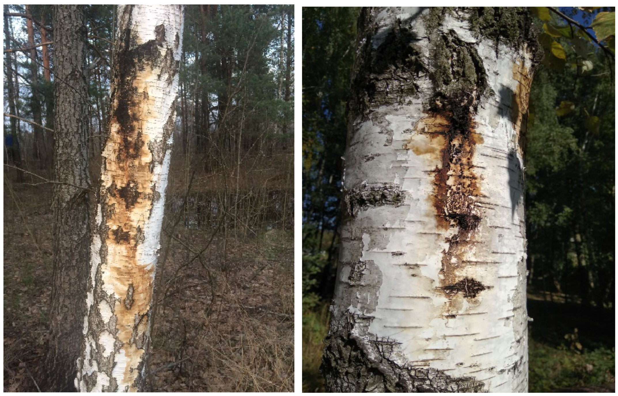
The symptoms of bacterial wetwood are primarily related to the moisture content in the birch trunk and include the formation of a weeping pathological rod, cracks and ulcers, as well as abundant exudation (Figure 1). Shoots on the affected tree trunks indicate a deep pathology. Chronic pathogenesis is accompanied by the death of the upper part of the plant and, in the future, the death of a weakened tree. The initial diagnostic sign of bacterial wetwood is the appearance of dieback in stands with low crown density. For example, traces of the vital activity of bacteria appear on trees affected by bacterial wetwood. They are weeping reddish spots. Signs of bacterial infection usually appear in the spring or early summer. Certain secretions accumulate under the bark, forming a swelling on the trunks that becomes visible. The bast and wood in the affected areas become wet, acquiring a dark brown color and a sour smell. Birches with coarsely fissured and longitudinal bark are distinguished by the peculiar fermentation odors and brown exudate emerging from cracks.
The birch (Betula pendula Roth.) with smooth bark usually ruptures under the pressure of gases released by bacteria, especially hydrogen sulfide. As a result, the exudate often becomes black. A short-term current is observed mainly in May and June, and then dries up. In autumn, exudate sometimes occurs again. Infected trees have much smaller leaves than healthy ones, and chlorotic conditions often appear. Often, numerous shoots appear below the crown, which die off after a year or two, and in some cases after a few months. About a third of birches die from bacterial swampiness in mature and overmature plantations.
The results of studies of bacterial dropsy of silver birch are presented. The phytopathogenic bacterium Lelliottia nimipressuralis (Enterobacter nimipressuralis) is its causative agent. It is indicated that the death of drooping birch when it is affected by bacterial wetwood occurs as a result of exfoliation of the integumentary parts with the formation of necrosis with a width of 10–20 cm and a length of 1 m or more. The pathogenicity of the causative agent of dropsy was proved in an experiment on indicator plants and on silver birch. Shoots and leaves of B. pendula Roch. were not sensitive to the pathogen. The infection of bacterial wetwood with the formation of persistent foci of drying was established to occur more intensively in birch stands of older age groups. In the lesions, different types of bacteria were isolated along with L. nimipressuralis, as well as fungi, which, obviously, are a concomitant microbiota in the pathology of bacterial wetwood.
In laboratory conditions, the causative agent of bacterial wetwood, L. nimipressuralis, was confirmed and data on cell sizes were clarified. They are small, straight, rod-shaped, located singly or in pairs, less often in chains or groups, rounded at the ends (somewhat elliptical in shape), polymorphic, ranging in size from 0.45 to 1.75 μm, and well stained. Bacteria are motile, do not form spores, are Gram-negative, and have long peritrichous flagella. L. nimipressuralis are facultative anaerobes, grow well on potato agar (PA), meat-potato agar (MPA), and meat-potato bouillon (MPB). On the MPA, they form white, shiny, smooth, rounded colonies with somewhat uneven edges. Slight turbidity with traces of a pellicle is formed on the MPB.
According to morphotypes, opaque white-cream, brilliant grayish-white, and yellow-pigmented colonies were identified from the pathological tissues of affected with bacterial wetwood. They were assigned to the genera Bacillus, Lelliottia, Xanthomonas, and Pantoea. Mycobiota in the pathogenesis of bacterial dropsy was represented by micromycetes of the genera Mucor (Zygomycota), Rhizopus, Acremonium (Ascomycota), and Penicillium. It was associated with the accompanying microbiota.
Bacterium L. nimipressuralis was pathogenic for B. pendula in the experiments. The pathogenic properties of X. campestris and P. agglomerans were variable. X. campestris showed weak pathogenic properties in only one variant. Isolated strains of bacteria were similar to those described in the literature according to the main characteristics. Definitely, the variability of some L. nimipressuralis isolates in the assimilation of carbohydrates and alcohols may be related to the specific conditions of bacteria existence. Associated with bacterial wetwood, P. agglomerans, which found variability in the case of artificial infection of birch, was also similar to that described in the literature according to the morphological, physiological, and biochemical characteristics.
To some extent, the species and form diversity of bacteria in the pathogenesis of bacterial wetwood may be due to the fact that the affected trunk is a kind of ecological niche for the growth of a complex of anaerobic saprotrophic mycobiota and microbiota. Bacteria isolated from waterlogged layers of wood and exudate are involved in butyric fermentation but may not be directly related to bacterial wetwood. In particular, some researchers have linked the formation of the exudate and its intensity to fungi, including the genus Torula (apparently referring to fungi associated with wetwood). As the integral components of the ecosystem and facultative symbiotrophs, enterobacteria were suggested to have the property to become parasitic when the host plant is weakened.
3.3. The Bioenergetic Culture of Fraxinus excelsior L. and the Pathogenesis of Its Bacterial Diseases
There is a difficult situation with the phytosanitary state of ash Fraxinus excelsior L., which must be urgently addressed in Ukraine. The consistent geographical deterioration of tree stands under the auspices of the State Forestry Agency of Ukraine, as well as ash trees in forest parks, is characteristic of infection. Infection of common ash begins at the age of two or three years at a certain height of the trunk. It is possible through both exogenous and endogenous ways. The “scab” is the first sign of tuberculosis. Visually, the manifestation of pathology is directly related to the presence of phytophagous insects. However, in fact, ash tuberculosis (phytopathogenic bacterium Pseudomonas syringae pv. savastanoi), affecting ash trunks, branches, shoots, and inflorescences, is of particular concern. Instead of a greenish-gray bark, small elliptical tumors form due to local swelling of the cortex and voids filling with a gray, sticky, odorless bacterial mass. The pathogen disrupts not only physiological processes in trees, but also fundamentally threatens seed generation, affecting the genitals (Figure 2).
Ash tuberculosis is one of the most dangerous diseases of common ash. It has reached epiphytoty in Ukraine, especially on overmature trees.
There are specific symptomatic differences at different stages of the manifestation of the disease. They provide the conditional distinction of several phases of the pathology. In our opinion, it provides the opportunity to timely diagnose an affected tree at any stage of the disease with a certain development of appropriate protective measures. A narrow, shallow, oblong, straight, and tortuous crack is formed in the center of the bulge. The exudate released through the cracks forms a thick or thin gray membrane after drying. It remains on the surface of the periderm for a long time. The rhytidome of the affected trees becomes dark gray in the places of the pathological process, gradually breaks off in small pieces, and disappears. The basal part of the trunk becomes slightly yellow or red. The primary bark dries and hardens over time. Local necrosis sinks into the trunk resulting in black and dark brown streaks of varying thickness. Sometimes during the spring or summer, larger and smaller voids are filled with bacterial exudate. Later, new lesions form along the length and perimeter of the F. excelsior trunk under the influence of factors favorable for the pathogen. They are called “disease spread”. New foci of tuberculosis may appear without definite relationships and sequence.
During the “real tuberculosis”, typical structures are formed followed by the increase in their size. Under the influence of a pathogen, a certain deformation occurs in the trunk. At the initial stage of the disease, especially on young shoots of ash, the necrosis is small, ranging from 1 to 3 cm. However, it grows gradually and reaches 0.5 m or more, forming a winding strip of dead sapwood. The depth of placement of various defects in a tree usually depends on the degree of infection as well as the age of the tree. The earlier a tree is infected, the deeper the wounds formed in the trunk. Basically, up to one hundred foci of tuberculosis can be counted on one affected tree. Ash tuberculosis is a chronic disease. In an infected tree, wounds form throughout the thickness of the trunk at different heights. Under the influence of myco- and microbiota, annual rings are sometimes broken. If there are voids in the longitudinal and transverse sections, it is possible to establish at what age the tree was infected in a given section of the trunk. Common or graded canker pathogens are involved in the formation of open ulcers, for example, Nectria galligena Bres or Endoxylina stellulata Rom. (Anaform Libertella Fraxini Ogan.). Then, the disease proceeds with symptoms typical for the specific pathogens. Ascomycota, Basidiomycota, and Deuteromycota are the wood-destroying fungi (Figure 3).
The control of tree diseases is one of the main tasks of forestry, since forests suffer from a variety of pathogens, nutrient deficiencies, and pest infestations. Any disease of trees, regardless of the reason, threatens the health of the forest as a whole and affects all related industries. Forest tree diseases and pests are a major problem for both logging and environmental protection. The health of the population depends on the health of plants. Therefore, this problem also affects people. In this regard, the detection of tree diseases and their treatment is the main task of foresters, which benefits everyone. Any deviation or failure in development caused by a resistant pathogen can be called a disease of forest trees and shrubs.
As the tree grows, the number of defects increases in proportion to new lesions. In addition to the trunks and branches, the causative agent of tuberculosis also affects the inflorescences of common ash. Thus, it can potentially threaten the seed recovery of this valuable woody plant (“deformation of the generative organs”). There are abiotic and biotic pathogens. The components of wildlife and climatic conditions are the abiotic ones. Biotic sources of diseases are divided into groups according to the type of pathogen (bacteria, fungi, viruses, phytoplasma, nematodes, etc.).
In most cases, pathogens were noted to be parasites. However, not every pathogen is a parasite and not every parasite is a pathogen. Some parasites do not cause harm to plants and, accordingly, do not cause any tree diseases on the trunk and its other parts. On the contrary, parasites can be beneficial. In addition, some types of bacteria living on the soil surface do not parasitize plants, but produce harmful toxins causing diseases of tree roots.
Fungi are the main causative agents of foliar infections. However, the signs and symptoms of diseases can be similar to the effects of a chemical insect attack (for example, Prociphilus fraxini (Fabricius, 1777), which is involved in a mutualistic relationship with the ant Prenolepis nitens (Mayr, 1853) [41,42]), making it difficult to identify the tree disease on the leaves and choose the appropriate treatment. It may not be effective when weather conditions are favorable for the development of fungi. Thus, it does not always make sense to invest in solving the problem. To remove and destroy foliage in autumn is the most common treatment for tree leaf disease. This method will prevent overwintering of the infectious agent and re-infection in spring. Foliar tree diseases affect both conifers and hardwoods and vary in severity. While some of them cause little harm, the rest are quite dangerous and can cause the death of plants.
We have isolated and identified six types of bacteria from tuberculous lesions—Pseudomonas fluorescens, Preudomonas syringae pv. savastanoi, Pseudomonas syringae, Pseudomonas sp., Xanthomonas sp., and Erwinia horticola. In addition, spore-forming bacteria of the genus Bacillus were found in samples of affected tissues. In further studies, they revealed certain antagonistic properties of P. syringae pv. savastanoi and related real and opportunistic bacteria in the tuberculosis pathology of F. excelsior. P. syringae pv. savastanoi are non-spore-forming mobile rods 0.4–0.8 × 1.3–3.0 µm in size, Gram-negative, placed individually, in pairs or in short chains, and sometimes lophotrichous with polar flagella. The ends of the sticks are smoothly rounded. On potato agar, colonies are grey-white, smooth, and translucent. On meat agar, colonies are small, 2–3 mm in diameter, grow slowly, and are flat or convex, with a notch in the center. On meat peptone broth, growth is moderate and bacteria form a uniform turbidity.
According to physiological characteristics, the isolated strains are not homogeneous. They are aerobic and do not ferment glucose under vaseline oil. Bacteria digest sucrose, galactose, fructose, glycerol, mannitol, and citrate. They do not consume lactose, xylose, rhamnose, trehalose, raffinose, L-arabinose, maltose, sorbitol, and salicin. Isolated strains cause a hypersensitivity reaction on the leaves of Nicotiana tabacum and form a fluorescent pigment on potato agar. Microorganisms do not hydrolyze gelatin, do not reduce nitrates, and do not form a levan. They are oxidase-negative and catalase-positive. The growth of bacteria was noted at 37 °C.
Tuberculosis causes more economic than environmental damage. Affected trees of older ages dieback relatively rarely. However, wood depreciates due to the specific pathological process. Affected trunks are usually diverted to firewood.
3.4. The Bioenergetic Culture of Salix alba L. and Thepathogenesis of Its Bacterial Diseases
Bacterial wilt of the willow (Salix alba L). Willow infection symptoms appear in May (late June) in the form of wilt. The leaves turn brown or red and stay on the tree for a while. The disease is called “red leaf”. In the first year of the disease, premature mass leaf loss is observed, although the outer shoots look healthy. Even green leaves sometimes die off. The causative agent of the disease is well isolated from the exudate. Infected goat willow trees had orange or reddish-brown leaves and stood out strongly against the green background of healthy trees of other species. The lesion indicated that the pathological process had started in early spring, during the opening of the pubescent male inflorescences. The examination in July revealed dieback with shoots. After the formation of leaves on new shoots, the inlet of aqueous solutions by xylem vessels stopped abruptly, causing the withering and death of the formed leaves. The active manifestation of drops of liquid exudate of a dirty white color, sometimes with yellowness on the surface of the periderm, was observed on the thin branches of the canopy. After that, the exudate darkened, thickened and became cherry-colored. It flowed onto the surface of the shoots, drying out in the form of a film. A strong flooding of infected tissues was a rather important symptom.
Additional water shoots appear anywhere. Branches infected from the last summer begin to develop normally, but then suddenly die off. The stem of the fungal mycelium developing after bacterial inoculation is often visible on the fallen bark. At first it is colorless, then it becomes dark brown or black. The exudate is sometimes toxic to the phloem, shoots, and even grass, which dies from contact with it. The saprotrophic microbiome does not immediately appear in the affected areas due to the toxicity of the exudate. The central rings of the tree, including the heart, are filled with fluid. The passages of the branch and trunk system are mainly infected. Dissolution of cell walls and tissue cells has never been observed. As a result of the vessels’ infection, the movement of substances in plants is blocked. The vessels turn black until autumn, and gas is formed inside of them. Bacteria are accumulated in the main vessels and radical rays, but they are rarely viable. The part of the parenchyma is not damaged. Obviously, the bark dies as a result of the cessation of water access to it. The spread of the infection along the branch and the body occurs in the form of an extended spiral. The pathogen spreads very slowly in a radical direction. During the vegetative period, it cannot move forward, even along the thickness of the annual ring. Sometimes all branches are not infected for one year, but the tree may die in one, two, or three years. It is characteristic that in recent years the degree of the disease has been decreasing. Brenneria salicis strains are basically indistinguishable from one another. They cause diseases of such trees as Salix alba, S. caerulea, S. fragilis, S. cinerea, and S. caprea.
However, S. fragilis cannot be infected with any strain taken from the trees of other types. Infection of trees occurs during the period between October and March. Plants are resistant to infection in summer months. The number of factors of external environment also influence on the susceptibility of willow to the pathogenic agent. The disease more often occurs on the swampy soil with stagnant water, in the thickened forest stands. Plants were shown as resistant on the well-drained soil.
4. Conclusions
Cultivation of bioenergy cultures becomes more and more relevant worldwide. The rapid fluctuations in prices for traditional energy carriers stimulate the development of technologies to grow such cultures. The phytopathogenic bacterium Lelliottia nimipressuralis is the causative agent of bacterial wetwood of poplar (Populus alba L.). It is isolated both in pure culture and in a mixture with bacteria of Pseudomonas sp. and fungi Fusarium sp., Penicillium sp., Cladosporium sp., etc.
Bacteria of Pseudomonas syringae f. populi and Pseudomonas cerasi (now Pseudomonas syringae) were isolated from a poplar lesion of the “bacterial canker” type. The bacteria associated with the Pseudomonas species included Bacillus, Erwinia, yellow-pigmented Corynebacterium, Chromobacterium bergonzini, and Xanthomonas; fungi from the Cytospora, Nectria, genera Fusarium, Penicillium, and Cladosporium; and yeasts. The fire poplar blight is caused by the pathogen Pseudomonas syringae (Pseudomonas cerasi).
The phytopathogenic bacterium Lelliottia nimipressuralis is the causative agent of bacterial wetwood of birch (Betula pendula Roth.). Also, bacteria assigned to the genera Bacillus, Pantoea, and Xanthomonas were isolated. Mycobiota in the pathogenesis of bacterial wetwood was represented by micromycetes from the genera Acremonium, Mucor, Rhizopus, and Penicillium, and appeared to be an accompanying microbiota.
Tuberculosis of common ash (Fraxinus excelsior L.) is caused by the bacterium Pseudomonas syringae pv. savastanoi. Bacteria Pseudomonas syringae, Pseudomonas fluorescens, Pseudomonas sp., Erwinia horticola, Pantoea agglomerans, and Xanthomonas sp. were also isolated from tuberculous lesions and identified. Spore-forming bacteria, mainly of the genus Bacillus, were also found in the samples of the affected tissues. It revealed certain properties antagonistic of P. syringae pv. savastanoi. Real and conditionally pathogenic bacteria associated with it were observed in the tubercular pathology of F. excelsior.
The causative agents of ordinary or “stepped” canker, mainly Endoxylina stellulata Rom. or Nectria galligena Bres, were also isolated from open tuberculous wounds. The bacterium Brenneria salicis is the causative agent of bacterial wilt of willow.
The key factors of the harmfulness of phytopathogenic bacteria were established to be the aggressiveness and pathogenicity of the pathogen, the virulence of the pathogen, and the host plant, determined by many morphological, physiological, biochemical, and molecular genetic properties of bacteria and their hosts. Usually, they are evolutionarily determined and controlled by the time factor. Phytopathogenic microorganisms are able to quickly fill the natural niche in the “tree plant-automicrobiota-environment” system with the highest concentration causing epiphytoty under favorable conditions.
Thus, based on experimental studies and observations, we state that the bioenergetic species of forest woody plants we studied (silver birch (Betula pendula Roth.), common ash (Fraxinus excelsior L.), white poplar (Populus alba L.), white willow (Salix alba L.)), which grow in the conditions of the Kyiv Polissia of Ukraine, are exposed to the negative influence of various types of bacteriosis. In particular, these are bacterial wetwood (Lelliottia nimipressuralis), bacterial cancer (Pseudomonas syringae f. populi and Pseudomonas cerasi), ash tuberculosis (Pseudomonas syringae pv. savastanoi), and bacterial wilt of willow (Brenneria salicis), and the causative agents of these bacterial diseases have a high degree of aggressiveness, pathogenicity, and virulence, which complicates the process of phytosanitary monitoring and the development of protective measures.
Author Contributions
Conceptualization, A.G. and V.P.; methodology, A.G., I.K., M.S., L.P., L.D., A.K. and V.P.; software, I.K. and L.D.; validation, A.G., I.K., M.S., L.P., L.D., A.K. and V.P.; formal analysis, A.G., I.K. and M.S.; investigation, I.K.; writing—original draft preparation, A.G., I.K., M.S., L.P., L.D., A.K. and V.P.; writing—review and editing, A.G., I.K., M.S., L.P., L.D., A.K. and V.P.; supervision, V.P. All authors have read and agreed to the published version of the manuscript.
Funding
This research received no external funding.
Institutional Review Board Statement
Not applicable.
Informed Consent Statement
Not applicable.
Data Availability Statement
Not applicable.
Conflicts of Interest
The authors declare no conflict of interest.
References
- Thomas, F.M.; Blank, R.; Hartmann, G. Abiotic and biotic factors and their interactions as causes of oak decline in Central Europe. For. Pathol. 2002, 32, 277–307. [Google Scholar] [CrossRef]
- Guo, Y.; Lin, Q.; Chen, L.; Carballar-Lejarazú, R.; Zhang, A.; Shao, E.; Wu, S. Characterization of bacterial communities associated with the pinewood nematode insect vector Monochamus alternatus Hope and the host tree Pinus massoniana. BMC Genom. 2020, 21, 337. [Google Scholar] [CrossRef] [PubMed]
- Mansfield, J.; Genin, S.; Magori, S.; Citovsky, V.; Sriariyanum, M.; Ronald, P.; Foster, G.D. Top 10 plant pathogenic bacteria in molecular plant pathology. Mol. Plant Pathol. 2012, 13, 614–629. [Google Scholar] [CrossRef] [PubMed] [Green Version]
- Griffiths, H.M. Forest Diseases Caused by Prokaryotes: Phytoplasmal and Bacterial Diseases. In Infectious Forest Diseases; CABI: Wallingfor, UK, 2013; pp. 76–96. Available online: https://books.google.com.ua/books?hl=uk&lr=&id=_YtcBAAAQBAJ&oi=fnd&pg=PA76&dq=Bacterial+diseases+of+forest+species+&ots=9IZCCHavOZ&sig=t_lpvU3ab45RBYBEC-dEZ-oJeOA&redir_esc=y#v=onepage&q=Bacterial%20diseases%20of%20forest%20species&f=false (accessed on 19 June 2022).
- Goychuk, A.F.; Drozda, V.F.; Shvets, M.V.; Kulbanska, I. Bacterial wetwood of silver birch (Betula pendula Roth): Symptomology, etiology and pathogenesis. Folia For. Pol. 2020, 62, 145–159. [Google Scholar] [CrossRef]
- Pautasso, M.; Holdenrieder, O.; Stenlid, J. Susceptibility to Fungal Pathogens of Forests Differing in Tree Diversity. In Forest Diversity and Function: Temperate and Boreal Systems; Scherer-Lorenzen, M., Körner, C., Schulze, E.D., Eds.; Springer: Berlin/Heidelberg, Germany, 2005; p. 176. [Google Scholar] [CrossRef]
- Kulbanska, I.M.; Shvets, M.V.; Goychuk, A.F.; Biliavska, L.H.; Patyka, V. Lelliottia nimipressuralis (Carter 1945) Brady et al. 2013—The Causative Agent of Bacterial Dropsy of Common Oak (Quercus robur L.) in Ukraine. Mikrobiolohichnyi Zhurnal 2021, 83, 30–41. [Google Scholar] [CrossRef]
- Kulbanska, I.; Shvets, M.; Markov, F. Etiology and Symptomatology of Bacterioses of Wood Plants in the Stands of the Green Zone of the City of Kiev. Sci. Horiz. 2019, 12, 84–95. Available online: https://sciencehorizon.com.ua/web/uploads/pdf/SH_2019_12_84-95.pdf (accessed on 19 June 2022). [CrossRef] [Green Version]
- Sakamoto, Y.; Kato, A. Some properties of the bacterial wetwood (watermark) in Salix Sachalinensis caused by erwinia salicis. IAWA J. 2002, 23, 179–190. [Google Scholar] [CrossRef]
- COST. European Cooperation in Science and Technology. Fraxinus Dieback in Europe: Elaborating Guidelines and Strategies for Sustainable Management (FRAXBACK). 2011. Available online: http://www.cost.eu/COST_Actions/fps/FP1103 (accessed on 15 June 2022).
- Goychuk, A.; Kulbanska, I.; Shvets, M. Tuberculosis pathology of Fraxinus Excelsior L. in Ukraine: Symptomatology, etiology, pathogenesis. Sci. Horiz. 2021, 24, 69–80. [Google Scholar] [CrossRef]
- Kulbanska, I.M.; Plikhtyak, P.P.; Goychuk, A.F.; Shvets, M.V.; Soroka, M.I. Lelliottia nimipressuralis (Carter 1945) Brady et al. 2013 as the causative agent of bacterial dropsy of common silver fir (Abies alba Mill.). Folia For. Pol. Ser. A—For. 2022, 64, 173–183. [Google Scholar] [CrossRef]
- Kwaśna, H.; Szewczyk, W.; Baranowska, M.; Behnke-Borowczyk, J. Bacteria associated with vascular wilt of poplar. Arch. Microbiol. 2021, 203, 4829–4838. [Google Scholar] [CrossRef]
- Christersson, L. Poplar plantations for paper and energy in the south of Sweden. Biomass Bioenergy 2008, 32, 997–1000. [Google Scholar] [CrossRef]
- Kalinichenko, A.; Pasichnyk, L.; Osypenko, S.; Patyka, V.; Usmanova, H. Bacterial diseases of energy plants. Ecol. Chem. Eng. A 2017, 24, 169–191. [Google Scholar] [CrossRef]
- Cochard, B.; Lefort, F. Cas de suie de l’érable et de chancre du peuplier dans le canton de Genève. Schweiz. Z. Fur Forstwes 2016, 167, 98–104. [Google Scholar] [CrossRef] [Green Version]
- Kwaśna, H.; Szewczyk, W.; Baranowska, M.; Gallas, E.; Wiśniewska, M.; Behnke-Borowczyk, J. Mycobiota Associated with the Vascular Wilt of Poplar. Plants 2021, 10, 892. [Google Scholar] [CrossRef]
- Li, A.; He, W. Molecular Aspects of an Emerging Poplar Canker Caused by Lonsdalea populi. Front. Microbiol. 2019, 6, 2496. [Google Scholar] [CrossRef] [PubMed] [Green Version]
- Fuchylo, Y.; Pasichnyk, L.; Patyka, V.; Kalinichenko, A. In Book: Odnawialne Źródła Energii. Teoria i Praktyka. Tom II (Pod Redakcją Izabeli Pietkun-Greber i Pawła Ratusznego). 2017. Available online: http://dspace.pdaa.edu.ua:8080/handle/123456789/346 (accessed on 1 May 2022).
- Gil, W.; Kowalski, T.; Kraj, W.; Zachara, T.; Łukaszewicz, J.; Paluch, R.; Nowakowska, J.; Oszako, T. Ash dieback in Poland–history of the phenomenon and possibilities of its limitation. In Dieback of European Ash (Fraxinus spp.)-Consequences and Guidelines for Sustainable Management; Swedish University of Agricultural Sciences: Uppsala, Sweden, 2017; pp. 176–184. Available online: https://www.researchgate.net/publication/313368936_Ash_dieback_in_Poland_-_history_of_the_phenomenon_and_possibilities_of_its_limitation (accessed on 15 June 2022).
- Matisone, I.; Matisons, R.; Laiviņš, M.; Gaitnieks, T. Statistics of ash dieback in Latvia. Silva Fenn. 2018, 52, 9901. [Google Scholar] [CrossRef] [Green Version]
- Langer, G. Collar Rots in Forests of Northwest Germany Affected by Ash Dieback. Balt. For. 2017, 23, 4–19. Available online: https://www.nw-fva.de/fileadmin/nwfva/publikationen/pdf/langer_collar_rots_in_forests.pdf (accessed on 15 June 2022).
- Jankovský, L.; Holdenrieder, O. Chalara fraxinea—Ash dieback in the Czech Republic. Plant Prot. Sci. 2009, 45, 74–78. [Google Scholar] [CrossRef] [Green Version]
- Halmschlager, E.; Kirisits, T. First report of the ash dieback pathogen Chalara fraxinea on Fraxinus excelsior in Austria. Plant Pathol. 2008, 57, 1177. [Google Scholar] [CrossRef]
- Talgø, V.; Sletten, A.; Brurberg, M.B.; Solheim, H.; Stensvand, A. Chalara fraxinea Isolated from Diseased Ash in Norway. Plant Dis. 2009, 93, 548. [Google Scholar] [CrossRef]
- Husson, C.; Scala, B.; Caël, O.; Frey, P.; Feau, N.; Ioos, R.; Marçais, B. Chalara fraxinea is an invasive pathogen in France. Eur. J. Plant Pathol. 2011, 130, 311–324. [Google Scholar] [CrossRef]
- Ogris, N.; Hauptman, T.; Jurc, D.; Floreancig, V.; Marsich, F.; Montecchio, L. First Report of Chalara fraxinea on Common Ash in Italy. Plant Dis. 2009, 94, 133. [Google Scholar] [CrossRef]
- Chandelier, A.; Delhaye, N.; Helson, M. First Report of the Ash Dieback Pathogen Hymenoscyphus pseudoalbidus (Anamorph Chalara fraxinea) on Fraxinus excelsior in Belgium. Plant Dis. 2011, 95, 220. [Google Scholar] [CrossRef]
- Janse, J.D. The bacterial disease of ash (Fraxinus excelsior), caused by Pseudomonas syringae subsp. Savastanoi pv. Fraxini II. Etiology and taxonomic considerations. Eur. J. For. Pathol. 1981, 11, 425–438. [Google Scholar] [CrossRef]
- Inoue, Y.; Takikawa, Y. Pseudomonas syringae Strains Are Classified into Five Groups by Comparing DMA Homology at the hrp Neighboring Regions. J. Gen. Plant Pathol. 2000, 66, 238–241. [Google Scholar] [CrossRef]
- Brandt, R.W. Ash dieback in the Northeast; Station Paper NE-163; U.S. Department of Agriculture, Forest Service, Northeastern Forest Experiment Station: Upper Darby, PA, USA, 1961; Volume 8, p. 163. Available online: https://www.fs.usda.gov/treesearch/pubs/13754 (accessed on 15 June 2022).
- Griffiths, H.M.; Sinclair, W.A.; Smart, C.D.; Davis, R.E. The phytoplasma associated with ash yellows and lilac witches’-broom:‘Candidatus Phytoplasma fraxini’. Int. J. Syst. Evol. Microbiol. 1999, 49, 1605–1614. [Google Scholar] [CrossRef]
- Gremmen, J.; De Kam, M. Erwinia salicis as the cause of dieback in Salix alba in the Netherlands and its identity with Pseudomonas saliciperda. Neth. J. Plant Pathol. 1970, 76, 249–252. [Google Scholar] [CrossRef]
- Wong, W.C.; Preece, T.F. Erwinia salicis in cricket bat willows: Phenolic constituents in healthy and diseased wood. Physiol. Plant Pathol. 1978, 12, 349–357. [Google Scholar] [CrossRef]
- Maes, M.; Huvenne, H.; Messens, E. Brenneria salicis, the bacterium causing watermark disease in willow, resides as an endophyte in wood. Environ. Microbiol. 2009, 11, 1453–1462. [Google Scholar] [CrossRef] [PubMed]
- Grosso, S.; Mason, G.; Ortalda, E.; Scortichini, M. Brenneria salicis Associated with Watermark Disease Symptoms on Salix alba in Italy. Plant Dis. 2011, 95, 772. [Google Scholar] [CrossRef] [PubMed]
- Klement, Z.; Rudolph, K.; Sands, D.C. Methods in Phytobacteriology, 1990. (Budapest: Akadémiai Kiadó). Available online: https://trove.nla.gov.au/work/6301114 (accessed on 1 May 2022).
- Patyka, V.P.; Pasichnyk, L.A.; Butsenko, L.M.; Petrychenko, V.F.; Zubachov, S.R.; Dankevych, L.A.; Gnatiuk, T.T.; Huliaieva, H.B.; Tokovenko, I.P.; Kalinichenko, A.V.; et al. Express Diagnostics of Phytopathogenic Bacteria and Phytoplasmas in Agrophytocenosis. Guidelines; Suszanowich, D., Patyka, V., Eds.; Wyd-wo I Drukarnia Swietego Krzyza: Opole, Poland, 2019; Available online: https://www.researchgate.net/profile/Dariusz-Suszanowicz/publication/335692227_EXPRESS_DIAGNOSTICS_OF_PHYTOPATHOGENIC_BACTERIA_AND_PHYTOPLASMAS_IN_AGROPHYTOCENOSES/links/5da0b18292851c6b4bcd8d87/EXPRESS-DIAGNOSTICS-OF-PHYTOPATHOGENIC-BACTERIA-AND-PHYTOPLASMAS-IN-AGROPHYTOCENOSES.pdf (accessed on 15 June 2022).
- Maejima, K.; Oshima, K.; Namba, S. Exploring the phytoplasmas, plant pathogenic bacteria. J. Gen. Plant Pathol. 2014, 80, 210–221. [Google Scholar] [CrossRef] [Green Version]
- Kulbanska, I.; Shvets, M.; Goychuk, A.; Sporek, M.; Pasichnyk, L.; Patyka, V.; Kalinichenko, A.; Bąk, M. Phytopathogenic Bacteria Associated with Bacterioses of Common Oak (Quercus robur L.) in Ukraine. Forests 2023, 14, 14. [Google Scholar] [CrossRef]
- Purkart, A.; Morawski, M.; Masłowski, A.; Depa, Ł. Ant-mediated anholocyclic overwintering of Prociphilus fraxini (Hemiptera: Aphididae) in Central Europe. Entomol. Fenn. 2019, 30, 179–185. [Google Scholar] [CrossRef]
- Kaszyca-Taszakowska, N.; Kanturski, M.; Depa, Ł. Comparative Studies of Perianal Structures in Myrmecophilous Aphids (Hemiptera, Aphididae). Insects 2022, 13, 1160. [Google Scholar] [CrossRef] [PubMed]
Figure 1.
Bacterial wetwood of birch.

Figure 2.
Stages of tuberculosis F. excelsior, “real tuberculosis”.

Figure 3.
Change of typical color and formation of open wounds (ulcers).

Disclaimer/Publisher’s Note: The statements, opinions and data contained in all publications are solely those of the individual author(s) and contributor(s) and not of MDPI and/or the editor(s). MDPI and/or the editor(s) disclaim responsibility for any injury to people or property resulting from any ideas, methods, instructions or products referred to in the content. |
© 2023 by the authors. Licensee MDPI, Basel, Switzerland. This article is an open access article distributed under the terms and conditions of the Creative Commons Attribution (CC BY) license (https://creativecommons.org/licenses/by/4.0/).
Share and Cite
MDPI and ACS Style
Goychuk, A.; Kulbanska, I.; Shvets, M.; Pasichnyk, L.; Patyka, V.; Kalinichenko, A.; Degtyareva, L. Bacterial Diseases of Bioenergy Woody Plants in Ukraine. Sustainability 2023, 15, 4189. https://doi.org/10.3390/su15054189
AMA Style
Goychuk A, Kulbanska I, Shvets M, Pasichnyk L, Patyka V, Kalinichenko A, Degtyareva L. Bacterial Diseases of Bioenergy Woody Plants in Ukraine. Sustainability. 2023; 15(5):4189. https://doi.org/10.3390/su15054189
Chicago/Turabian StyleGoychuk, Anatolyj, Ivanna Kulbanska, Maryna Shvets, Lidiia Pasichnyk, Volodymyr Patyka, Antonina Kalinichenko, and Larysa Degtyareva. 2023. "Bacterial Diseases of Bioenergy Woody Plants in Ukraine" Sustainability 15, no. 5: 4189. https://doi.org/10.3390/su15054189
Note that from the first issue of 2016, this journal uses article numbers instead of page numbers. See further details here.