Article

The Genus Spinopygina gen. nov. (Diptera, Sciaridae) from Western North America: Preliminary Molecular Phylogeny and Description of Seven New Species

1
Finnish Museum of Natural History, Zoology Unit, University of Helsinki, P.O. Box 17, FI-00014 Helsinki, Finland
2
Department of Biology and Ecology, Faculty of Science, University of Ostrava, Chittussiho 10, CZ-710 00 Ostrava, Czech Republic
3
Silesian Museum, Nádražní Okruh 31, CZ-74601 Opava, Czech Republic
*
Author to whom correspondence should be addressed.
Insects 2023, 14(2), 173; https://doi.org/10.3390/insects14020173
Submission received: 9 January 2023 / Revised: 29 January 2023 / Accepted: 4 February 2023 / Published: 9 February 2023
(This article belongs to the Special Issue Systematics, Taxonomy and Biodiversity of Sciaridae (Diptera))

Abstract

:

Simple Summary

Black-winged fungus gnats (family Sciaridae) are one of the most species-rich groups of flies (Diptera), with about 3000 described species worldwide. Their taxonomy is notoriously challenging, as many genera are difficult to delimit and species identification relies almost exclusively on the male genitalia. Existing phylogenetic hypotheses based on DNA sequences are often contradictory, presumably due to low taxon or gene sampling. In this paper, a new multigene phylogenetic analysis is presented to support the concept and monophyly of a new genus, containing eight species, seven of them being described as new to science.

Abstract

The genus Spinopygina gen. nov. (type species Camptochaeta uniceps Hippa & Vilkamaa, 1994) from western North America is described. The genus includes the following eight species: Spinopygina acerfalx sp. nov.; S. aurifera sp. nov.; S. camura sp. nov.; S. edura sp. nov.; S. peltata sp. nov.; S. plena sp. nov.; S. quadracantha sp. nov.; and S. uniceps (Hippa & Vilkamaa, 1994) comb. nov., transferred from Corynoptera Winnertz. The new species are described and Spinopygina uniceps is re-diagnosed. The species are keyed and illustrated. In the maximum-likelihood phylogenetic hypothesis based on four gene fragments (28S, 18S, 16S and COI), Spinopygina gen. nov. appears as the sister group of Claustropyga Hippa, Vilkamaa & Mohrig, 2003. In the same analysis, a remarkable, undescribed species is placed within Camptochaeta Hippa & Vilkamaa clade.

1. Introduction

Sciaridae (black-winged fungus gnats) is one of the little studied terrestrial Dipteran families with about 3000 described species in about 100 genera and subgenera, occurring in all continents [1]. Most species live in shady forested and moist habitats. Larvae are predominantly saprophagous and live in soil and litter or under bark of dead wood. The taxonomy is notoriously challenging, as many genera are difficult to delimit and are rich in small and rather similar species. The species identification relies almost exclusively on the male hypopygium, and in most genera the females are unidentifiable. Some trials to reconstruct phylogenies extensively over the family or focused on a limited number of taxa have been made, either using morphological characters [1,2,3,4,5,6,7,8] or using molecular markers [9,10,11,12]. Often the hypotheses obtained have been contradictory, presumably due to variable choice of morphological characters, insufficient number of molecular characters used or because of low taxon sampling. The paper by Shin et al. [9] is the most comprehensive of the molecular analyses, but it misses many crucial genera from the Holarctic region and lacks almost completely the extra-Holarctic taxa in its taxon sampling. Shin et al. [9] divided the family into three subfamilies, and later [10] named a fourth, but the large and morphologically very diverse group of genera (the ‘Pseudolycoriella group’) appeared non-monophyletic and was not ranked as a subfamily.
Of the Holarctic fauna of Sciaridae, the Nearctic region is much less studied than the Palaearctic fauna. Mohrig et al. [13] in their revision listed 166 valid species from the Nearctic region (excluding Greenland), and subsequent authors [14,15,16,17,18,19,20,21,22,23,24,25,26] have added 70 new species,making a total of 236 known species in the Nearctic to date. The number of known Palaearctic species is much higher, about 2000. Based on the COI (barcode) gene, Hebert et al. [27] counted 2200 different BINs for Sciaridae from Canada, but proper taxonomic work is waiting to identify and name the taxa. Other biogeographic regions are even less studied than the Holarctic, but studies made on different genera indicate that the taxonomic richness of Sciaridae in the tropics is enormous [2,28,29,30,31,32,33,34,35].
Hippa and Vilkamaa [36] described Camptochaeta uniceps from Canada (British Columbia) and included the species in their new genus Camptochaeta because it has the lambda-shaped basomedial sclerotization in the gonostylus, regarded as a synapomorphy of Camptochaeta. Subsequently, Menzel and Mohrig [1] in their revision of the Palaearctic Sciaridae did not accept the concept of Camptochaeta, but transferred two of the included species into Keilbachia Mohrig, 1987 and all other species lacking the apical tooth of the gonostylus into the Corynoptera parvula group and into the C. spinifera group of the large but obviously non-monophyletic Corynoptera Winnertz, 1867. Later, the Nearctic Camptochaeta uniceps Hippa &Vilkamaa, 1994 was transferred to the Corynoptera spinifera group by Mohrig et al. [13].
Here, in this study, we report on several species undoubtedly related to C. uniceps from western North America. We were able to study the species complex in more detail, describe the new species and consider the phylogenetic position and the taxonomic status of the group. Simultaneously, the taxonomic placement of a new species of Sciaridae (Camptochaeta sp., voucher SCI87) with an unusual tegmen was searched for.

2. Materials and Methods

2.1. Morphological Methods

The material originated from Malaise trap samples, and all specimens were detected and picked out from unsorted sciarid or insect material stored in ethanol. The holotype of Camptochaeta uniceps was obtained from Canadian National Collection, Ottawa, Canada (CNC) and a paratype from Royal British Columbia Museum, Victoria, Canada (RBCM), the other paratypes having been deposited in Zoological Museum, Finnish Museum of Natural History. The specimens were mounted on microscope slides in Euparal, after dehydrating them in absolute ethanol. Only males were studied: the females are unknown. The terminology and methods of measuring structures follow Hippa and Vilkamaa [28] and Hippa et al. [3,37]. The photographs of the slide-mounted specimens were taken with a Leica MC170 HD camera mounted on Leica DM 4000 B LED research microscope. The habitus of Spinopygina uniceps was photographed with Canon EOS 5DS digital SLR camera with a Canon MP-E 65 mm macro lens. The Figures were processed with Photoshop version CS5, CorelDraw2017 and CorelPhotopaint2017. The type material of the new species is deposited in Zoological Museum, Finnish Museum of Natural History, Helsinki, Finland (MZH). The specimen data of the MZH specimens will be added into the specimen database of the Finnish Museum of Natural History (Luomus) and will be found with the specimen codes https://luomus/GE.####.
This published work and the nomenclatural acts it contains have been registered in ZooBank, the online registration system for the ICZN. The LSID for this publication is: urn:lsid:zoobank.org:pub:9962DD66-13CC-4191-B6FF-0A7DD1934F75.

2.2. Molecular Methods

For the ingroup, representatives of taxa from the three subfamilies of Sciaridae were chosen as well as selected terminals from the non-monophyletic ‘Pseudolycoriella group’ of genera proposed by Shin et al. [9,10]. From the latter group, terminals were chosen to adequately cover the morphological diversity of the group.
The specimens used for DNA analysis (Table 1) were alcohol-preserved (70% to 99.9% ethanol). The DNA was extracted using NucleoSpin Tissue Kit (Macherey-Nagel, Düren, Germany) following manufacturers’ protocols. PCRs were performed using primers listed by Shin et al. [9].
All amplified products were purified using Gel/PCR DNA Fragments Extraction Kit (Geneaid, New Taipei City, Taiwan) and subsequently, the samples were sequenced by Eurofins (Hamburg, Germany). All sequences were assembled, manually inspected, and primers trimmed in SeqTrace [38]. New sequences were deposited in the GenBank database, with their accession numbers listed in Table 1.
All genes were aligned using MAFFT version 7 [39] on the MAFFT server (http://mafft.cbrc.jp/alignment/server/). To remove all unreliably aligned regions, the GBLOCKS 0.91b program [40] was used (http://phylogeny.lirmm.fr/phylo_cgi/one_task.cgi?task_type=gblocks) with conditions set as follows: allow smaller blocks, allow gap positions within the final blocks, allow less strict flanking positions and do not allow many contiguous nonconserved positions. The final molecular dataset consists of 4287 bp, and the lengths of individual alignments were: 18S = 1843 bp, 28S = 1308 bp, 16S = 478 bp, COI = 658 bp.
The final concatenated dataset was partitioned by gene and codon position and subsequently analysed using ML method with IQtree [41]. Best-fitting substitution models were chosen automatically by the IQ-TREE software (GTR+F+I+G4: 16S, TPM3u+F+I+G4: 18S, TVM+F+I+G4:28S, TIM3+F+G4: COI_1, TIM2+F+I+G4: COI_2, TIM3+F+I+G4: COI_3), without free-rate heterogeneity. Branch supports were evaluated using 1000 ultrafast bootstrap [42]. All other settings were left as default. The resulting phylogenetic tree (consensus tree) was visualized using Interactive Tree of Life (iTOL; [43]).

3. Results

3.1. Desription of the New Genus

 Spinopygina gen. nov.
Type species: Camptochaeta uniceps Hippa & Vilkamaa, 1994.
LSID urn:lsid:zoobank.org:act: DBFE5207-337A-4817-B955-14847009D23B.
Etymology. The name is formed from the Latin word spine, thorn, and the Greek word pyge, rump, referring to the outstanding megasetae of the gonostylus of the species.
Diagnosis. Small to medium sized Sciaridae, wing length 1.6–2.2 mm. Antenna long, flagellomeres with long setae and necks. Mouth parts small, maxillary palpus with 2 segments, 2nd segment much reduced. Anal lobe of wing small, halter with long stalk. Body setosity dark, long and strong. Legs long and slender, fore tibial organ poorly differentiated. Intergonocoxal area long or moderate, without medial lobe. Gonocoxae united medially, setae at medial margins short. Gonostylus impressed or deeply excavated medially, with lambda-shaped basomedial sclerotization; with strong megasetae with basal bodies, including (in most species) strongly procurved basal megaseta; without apical tooth. Tegmen modified, with flat apicolateral parts, without detectable aedeagal teeth.
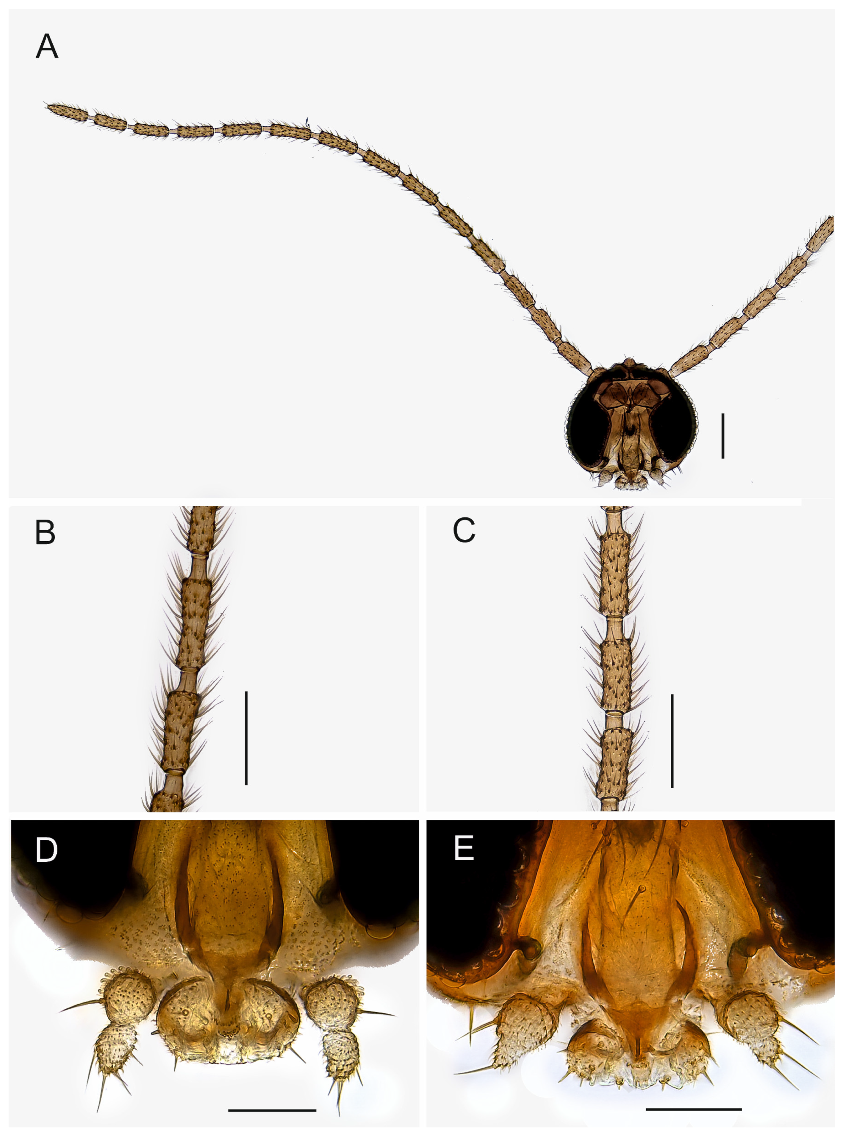
Description. Head (Figure 1). Normal, roundish. Eye bridge 1 to 2 facets wide. Anterior vertex non-setose. Face setose. Clypeus with 1–2 setae or non-setose. Antennal scape and pedicel normal. Antennal flagellum long, with 14 flagellomeres, bodies of flagellomeres subcylindrical, with smooth surface, body of 4th flagellomere 2.25–3.20× as long as subapically wide, necks longer than wide. Flagellomeral vestiture rather sparse, setae longer than width of flagellomeres. Mouth parts small. Maxillary palpus with 2 segments, 2nd segment strongly reduced, variably between species and even specimens; 1st segment with 1, rarely with 2 sharp setae; with long sparse sensilla dorsally.
Thorax (Figure 2). Brown; setae dark. Acrostichal setae few anteriorly, dorsocentral and lateral setae in indistinct rows of a few long and strong and short and fine setae. Scutellum with 2 long and strong setae and a few short and fine setae. Posterior pronotum non-setose. Anterior pronotum and prothoracic episternum with a few setae, other pleural sclerites non-setose. Mesothoracic katepisternum high with oblique anterior margin.
Wing (Figure 3A–C). Fumose. Length 1.6–2.2 mm. Anal lobe small. Veins distinct, except stM. Membrane non-setose, veins C, R and R1 setose, R5 with only dorsal setae, bM, r-m, stM, M and stCuA non-setose. c/w 0.70–0.90, R1/R 0.70–1.15, R1 joining C well before level of base of M-fork. Halter yellow, with long stalk.
Legs (Figure 3D,E). Yellow or pale brown, long, femora slender; setae dark; tibial spurs 1:2:2, fore tibial spur as long as tibial width or slightly longer, middle and hind left and right tibial spurs subequal in size, longer than tibial width. Fore tibia without spinose setae, middle tibia rarely with 1 spinose seta, hind tibia with dorsal row of strong spinose setae. The retrolateral apical setae of hind tibia fine. Fore tibial organ not impressed or proximally bordered, with small patch of a few setae. Tarsi unmodified, long, length of fore basitarsomere/length of fore tibia 0.47–0.63. Tarsal claws unmodified, without teeth.
Abdomen (Figure 4, Figure 5, Figure 6, Figure 7, Figure 8 and 
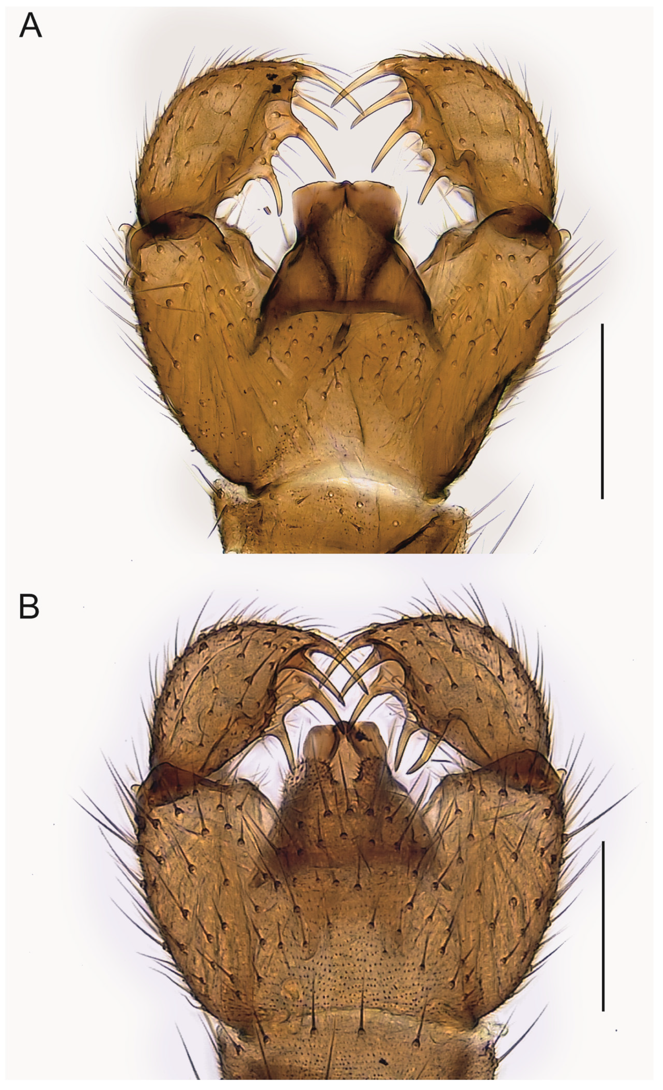
Figure 9). Pale brown or yellowish, normal, slender; setae dark, long and strong. Hypopygium brown or yellowish, as abdomen. Intergonocoxal area long or moderate, without lobe(s). Gonocoxa unmodified, longer than gonostylus, gonocoxae fused ventromedially; rather richly setose, setae at ventromedial margin short, medial membrane non-setose; with 1 elongated seta ventrally and dorsally at apicomedial corner. Gonostylus elongated, widest medially and distinctly narrowed towards apex or evenly wide and slightly narrowed towards apex, strongly excavated or at least slightly impressed medially, with lambda-shaped basomedial sclerotization; without apical tooth; with apical, subapical and medial megasetae, or only apical or subapical megasetae present; megasetae strong and with distinct basal bodies, in some species basalmost megaseta distinctly procurved; with some elongated setae medially and apically. Tegmen variable in shape, truncate or slightly curved apically, slightly curved basolaterally, constricted subapically, membranous or with sclerotized rim apically, with flat apicolateral parts; parameres sclerotized, approaching each other and joining to sclerotized rim or ending at apex separately. Apodemes of tegmen and aedeagal apodeme usually short, aedeagal teeth not detectable. Tergite 9 rather narrow, with long and strong setae. Tergite 10 (cercus) normal.
Female unknown.
Distribution. USA: California, Oregon; Canada: British Columbia.
Figure 4. Hypopygia, ventral view. (A). Holotype MZH GE.1928 of Spinopygina acerfalx sp. nov. (B). Holotype MZH GE.1932 of S. aurifera sp. nov. Scale bars = 0.1 mm.
Insects 14 00173 g004
Figure 5. Hypopygia, ventral view. (A). Holotype MZH GE.1935 of Spinopygina camura sp. nov. (B). Holotype MZH GE.1937 of S. edura sp. nov. Scale bars = 0.1 mm.
Insects 14 00173 g005
Figure 6. Hypopygia, ventral view. (A). Holotype MZH GE.1938 of Spinopygina peltata sp. nov. (B). Holotype MZH GE.1941 of S. plena sp. nov. Scale bars = 0.1 mm.
Insects 14 00173 g006
Figure 7. Hypopygium of Spinopygina quadracantha sp. nov. (A). Ventral view (holotype MZH GE.1944). (B). Dorsal view (paratype MZH GE.1956). Scale bars = 0.1 mm.
Insects 14 00173 g007
Figure 8. Hypopygium of Spinopygina uniceps (Hippa & Vilkamaa, 1994). (A). Ventral view (holotype CNC). (B). Dorsal view (specimen MZH GE.1962). Scale bars = 0.1 mm.
Insects 14 00173 g008
Figure 9. 
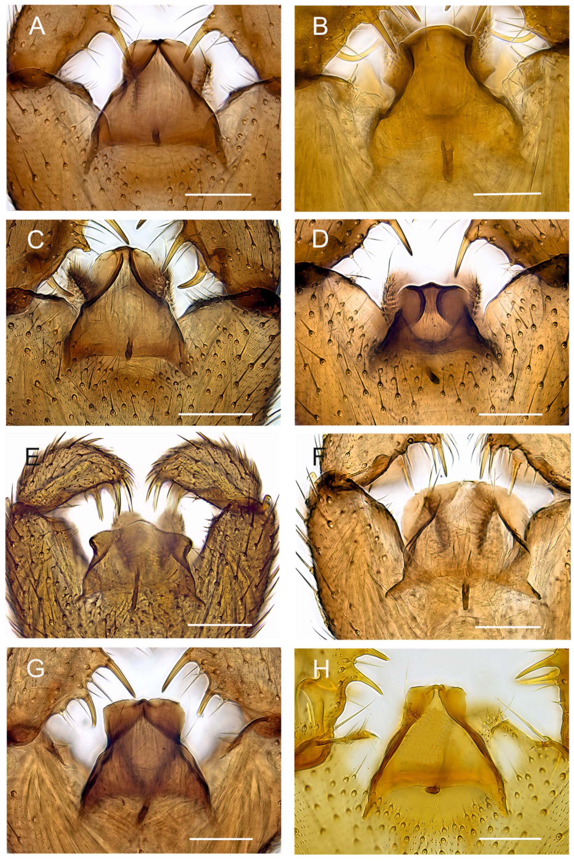
Tegmina, ventral view. (A). Holotype MZH GE.1928 of Spinopygina acerfalx sp. nov. (B). Holotype MZH GE.1932 of S. aurifera sp. nov. (C). Paratype MZH GE.1936 of S. camura sp. nov. (D). Holotype MZH GE.1937 of S. edura sp. nov. (E). Holotype MZH GE.1938 of S. peltata sp. nov. (F). Holotype MZH GE.1941 of S. plena sp. nov. (G). Holotype MZH GE.1944 of S. quadracantha sp. nov. (H). Holotype CNC of S. uniceps (Hippa & Vilkamaa, 1994) Scale bars = 0.05 mm.
Insects 14 00173 g009
Figure 10. Maximum likelihood hypothesis for relationships among selected taxa of Sciaridae (Diptera) based on DNA sequence data (18S, 28S, 16S, and COI), 4287 characters. Support numbers refer to ultrafast bootstrap values (ufboot) over 50.
Insects 14 00173 g010

3.2. Comparative Diagnosis of Spinopygina gen. nov.

Mohrig et al. [13] transferred Camptochaeta uniceps Hippa &Vilkamaa, 1994 into the Corynoptera spinifera group sensu [1]. Spinopygina gen. nov. resembles the C. spinifera group in having a rather similar lambda-shaped basomedial sclerotized structure of the impressed or excavated gonostylus, in lacking the apical tooth of gonostylus, in having the megasetae with strong basal bodies, in having an apical gonostylar megaseta in all and a strongly curved basal gonostylar megaseta in part of the species. Spinopygina differs from the C. spinifera group as well as from the Corynoptera parvula group sensu [1] in important characters: the long necks of antennal flagellomeres, the 2-segmented maxillary palpus without the sensory pit, the long stalk of the halter, the indistinct, not impressed fore tibial organ, the basoventrally united gonocoxae with distinctly shortened setae at their ventromedial margins and the tegmen with flat apicolateral parts and the apparent lack of the aedeagal teeth; see, e.g., Figure 4, Figure 5 and Figure 30A,B in Hippa & Vilkamaa (1994) [36]. Moreover, Spinopygina differs from Camptochaeta Hippa &Vilkamaa, 1994 in the sense of [1] in the lack of the apical tooth of the gonostylus in addition to the characters mentioned above.
Spinopygina resembles Claustropyga Hippa, Vilkamaa & Mohrig, 2003 in having the maxillary palpus 2-segmented (in part of Claustropyga), the sensilla scattered on the dorsal side of 1st segment of the maxillary palpus, in having a weakly modified fore tibial organ, in having the medial part of the gonostylus impressed or excavated, in having distinct basal bodies of the gonostylar megasetae and in having the megasetae strong at least in part of the species, in having the gonocoxae united basoventrally with the intergonocoxal area long or at least moderate. Spinopygina differs in having the basomedial part of gonostylus impressed with the lambda-shaped sclerotized structure (the impression lacks in Claustropyga), in having long flagellomeral necks and stalk of halter and in the lack of the apical tooth of gonostylus (lacks also in one known species of Claustropyga). Moreover, even if long, the intergonocoxal area of Spinopygina is not lobe-like produced as in most species of Claustropyga.
Spinopygina resembles Xylosciara Tuomikoski, 1960 in having a long intergonocoxal area, in having strong gonostylar megasetae including apical ones and in having a poorly differentiated patch of setae on the fore tibial organ. Spinopygina differs most strikingly in having 2-segmented maxillary palpus, longer necks and setae of antennal flagellomeres, in having strong and dark body vestiture and in having a modified tegmen and a strongly hollowed gonostylus.
Some species of Spinopygina resemble Keilbachia Mohrig, 1987 in having a strong procurved megaseta basally or subbasally on the gonostylus. Some species of Keilbachia lack the apical tooth of gonostylus and some species have a strongly modified tegmen reminiscent of that of Spinopygina, but Keilbachia has a 3-segmented maxillary palpus with a sensory pit on the 1st palpal segment, the fore tibial organ in depression at least in part of the species and it has ventromedially separated gonocoxae.
By the reduced maxillary palpus with sensilla scattered, long antennal flagellomeral bodies and their necks, long and slender legs, the long and oblique mesothoracic katepisternum, the strongly reduced anal lobe of wing and the poorly modified fore tibial organ, Spinopygina resembles Epidapus Haliday, 1851 and in lacking the apical tooth of gonostylus, especially Epidapus (Pseudoaptagogyna) absconditus group sensu [1]. Epidapus absconditus (Vimmer, 1926) has, like Spinopygina, strong apical-subapical gonostylar megasetae with distinct basal bodies. However, the megasetae of Epidapus absconditus are stout, straight and symmetrically narrowed at apex, not long and curved as in Spinopygina. The four megasetae of the related Epidapus quadrispinosus Mohrig & Mamaev, 1990 are all recurved. Moreover, Spinopygina differs from the E. absconditus group in having subequal left and right spurs on middle and hindtibia (unequal), in having the intergonocoxal area long with the gonocoxae united basoventrally (short and separate), with shortened setae at the ventromedial margin (equal), in having the tegmen strongly modified and with distinctly sclerotized parameres (simple, laterally and apically roundish and completely membranous tegmen in Pseudaptagogyna Vimmer, 1926) and in having the gonostylus impressed or excavated (in Pseudoaptagogyna convex medially) and in having (in part of the species) a curved basal megaseta in the gonostylus.
Spinopygina differs from all above-mentioned genera in having the tegmen with flat apicolateral parts and in lacking the aedeagal teeth (the teeth not detectable using a compound microscope.

3.3. Key to Species of Spinopygina gen. nov.

1.
Gonostylus with 5 megasetae …………………………….............. S. camura sp. nov.
Gonostylus with 3–4 megasetae …………………………………………………. 2
2
Gonostylus with 4 megasetae ………………………………………………….………. 3
Gonostylus with 3 megasetae …………………………………………………….. 7
3.
All megasetae at apical half of gonostylus, tegmen broad apically ….…………….. 4
One megaseta at basal half of gonostylus, tegmen narrow apically …………. 5
4.
Gonostylus much wider dorsally than ventrally, tegmen with large roundish apicolateral parts ………………………………………………......................S. plena sp. nov.
Gonostylus about as wide ventrally and dorsally, tegmen with small acuminate apicolateral parts …………………………………………..........S. peltata sp. nov.
5.
Tegmen distinctly constricted medially, roundish apically, parameres joining membraneous apex separately ………………………………................. S. aurifera sp. nov.
Tegmen slightly constricted subapically, truncate apically, parameres joining short sclerotized rim apically ………………………………………………………6
6.
Two medial megasetae at ventral margin of gonostylus, basalmost megaseta procurved and with long basal body, arising from medial excavation ………………………………..……………........... S. uniceps (Hippa& Vilkamaa, 1994)
One medial megaseta and basalmost megaseta at ventral margin of gonostylus, basalmost megaseta recurved and with moderate basal body …………… ………………….…………………………………………. S. quadracantha sp. nov.
7.
Tegmen small, its length less than half of length of gonostylus, parameres separately joining broad sclerotized rim apically, apex of tegmen largely sclerotized ………………………………………………….……................................ S. edura sp. nov.
Tegmen large, its length 2/3 of length of gonostylus, parameres united apically, joining narrow sclerotized rim apically, apex of tegmen largely membraneous ……………………………………………………….................... S. acerfalx sp. nov.

3.4. Descriptions of New Species

Spinopygina acerfalx sp. nov.Figure 2B, Figure 3A, Figure 4A and Figure 9A.
LSID urn:lsid:zoobank.org:act: 008D0F6D-919A-4915-99A8-8AB979A3DAD2.
Comparative diagnosis. By its tegmen, Spinopygina acerfalx sp. nov. is very similar to Spinopygina camura sp. nov., S. quadracantha sp. nov. and S. uniceps (Hippa & Vilkamaa). All have the tegmen narrowed subapically, with flat apicolateral parts and with the parameres joining a shortly sclerotized rim apically. Spinopygina acerfalx is similar to S. edura sp. nov. and differs from all above-mentioned species in having only three, not four or five, gonostylar megasetae. Moreover, in the form of the gonostylus and in the arrangement of the gonostylar megasetae Spinopygina acerfalx resembles S. edura, but the latter is distinct in having the tegmen much smaller and more sclerotized basally and in having the parameres wide apart, joining the broadly sclerotized rim separately at the apex.
Etymology. The name is a Latin noun in apposition, formed from acer, sharp, and falx, sickle, referring to the sharp and curved gonostylar megasetae.
Material examined. Holotype: USA, Oregon, ♂; Benton County; Corvallis; 1460 SW Allen St.; 44.550860° N, 121.270189° W; 4 March–20 April 2015; S. Fitzgerald leg.; Malaise trap; in Euparal; MZH GE.1928. Paratypes: USA, Oregon, 2 ♂♂; Same collection data as for holotype; in Euparal; MZH GE.1929, GE.1945. Oregon, 1 ♂; same collection data as for holotype but 5 April–3 June 2015; in Euparal; MZH GE.1976. Oregon, 2 ♂♂; same collection data as for holotype but 11 December 2014–14 January 2015; used for DNA extraction (samples No. SCI80, SCI81); in Euparal; MZH GE.1978, GE.1979. Oregon, 1 ♂; Benton County; 5 miles up Woods Creek Road from jct Highway 20; 44.544° N, 123.50° W; 25 April–15 May 2014; S. Fitzgerald leg.; Malaise trap; mixed forest; in Euparal; MZH GE.1930. Oregon, 1 ♂; Benton County; Corvallis; 6.4 miles up Woods Creek Road from jct Highway 20; 3 March–12 April 2015; Malaise trap; fir/alder/maple; in Euparal; MZH GE.1946. Oregon, 1 ♂; Coos County; Seven Devils’ Road; 43.3109° N, 124.3484° W; 7 February–15 May 2016; E. Boyd leg; Malaise trap; in Euparal; MZH GE.1931.
Description. Male. Head. Face and antenna uniformly brown, maxillary palpus pale brown. Eye bridge 1–2 facets wide. Body of 4th antennal flagellomere 2.60–3.20× as long as subapically wide, the neck longer than wide, the longest setae longer than the width of flagellomere. Face with 5–9 dark long and short setae. Clypeus non-setose or with 1 dark seta. Maxillary palpus with 2 segments; 2nd segment reduced; 1st segment with 1 sharp seta, with large indistinct dorsal patch of sensilla, 2nd segment with 1–5 setae.
Thorax (Figure 2B). Brown; setae dark. Anterior pronotum with 2 setae. Prothoracic episternum with 2–3 setae.
Wing (Figure 3A). Fumose. Length 1.8–2.0 mm. Anal lobe small. Width/length 0.40–0.45. R1/R 0.75–0.85. c/w 0.75–0.80. stM slightly longer than M-fork, bM shorter than or as long as r-m, stCuA shorter or as long as bM. bM and r-m non-setose. Halter yellow, with long stalk.
Legs. Yellow, long. Fore femur slender. Fore tibial organ not impressed or proximally bordered, with small patch of a few setae. Fore tibial spur longer than tibial width. Length of fore basitarsomere/length of fore tibia 0.63.
Abdomen. Pale brown; setae dark, long and strong. Hypopygium (Figure 4A) brown, as abdomen. Intergonocoxal area long, with short setosity. Gonocoxa longer than gonostylus; setae rather short, shorter towards medial margin. Gonostylus elongated, curved, the medial side strongly impressed; with a few elongated setae apically; without apical tooth, with 3 megasetae, 1 apical in ventral position, 1 subapical in dorsal position and 1 medial in ventral position, the megasetae long and strong, slightly recurved, with long basal bodies. Tegmen (Figure 9A) slightly shorter than wide, truncate apically, sharply narrowed subapically; with parameres joining short sclerotized rim apically; with flat apicolateral parts. Aedeagal apodeme short, aedeagal teeth not detectable.
Spinopygina aurifera sp. nov. Figure 1D, Figure 3B, Figure 4B and Figure 9B.
LSID urn:lsid:zoobank.org:act: B9089867-2FA5-4A03-92E4-5C99E60BD592.
Comparative diagnosis. In having the basal megaseta of the gonostylus strongly procurved, Spinopygina aurifera sp. nov. resembles S. camura sp. nov. and S. uniceps (Hippa and Vilkamaa) but by its form of the gonostylus and the tegmen, S. aurifera is not very similar to any other known species of the genus: the apicalmost megaseta is shifted to a subapical position and the two medial megasetae are in a lobe-like extension of the ventromedial margin of the gonostylus. The tegmen is characteristic in being strongly constricted at the middle, and in having the apex wide and roundish and in having the flat apicolateral parts small and triangular. Spinopygina aurifera differs from all other species of the genus in having a peculiar conical lobe apicodorsally in the gonocoxa.
Etymology. The name is a Latin noun in apposition, from the words auris, ear, and the suffix -fer, bearing, referring to the ear-like apicolateral parts of the tegmen.
Material examined. Holotype: USA, Oregon, ♂; Linn County; Hackleman Creek; 0.6 miles E of Tombstone Pass; 44.397501° N, 122.131401° E; 29 June–1 August 2016; S. Fitzgerald leg; Malaise trap; in Euparal; MZH GE.1932. Paratypes: USA, Oregon, 2 ♂♂; same collection data as for holotype; in Euparal; MZH GE.1933 and GE.1934. Oregon, 1 ♂; same collection data as for holotype; in Euparal; MZH GE.1970.
Description. Male. Head. Face and antenna uniformly brown, maxillary palpus pale brown. Eye bridge 1–2 facets wide. Body of 4th antennal flagellomere 2.25–2.55× as long as subapically wide, the neck longer than wide, the longest setae longer than the width of flagellomere. Face with 5–7 dark long and short setae. Clypeus non-setose or with 1 dark seta. Maxillary palpus (Figure 1D) with 2 segments; 2nd segment reduced; 1st segment with 1(2) sharp setae, with an indistinct dorsal patch of sensilla, 2nd segment with 3–5 setae.
Thorax. Brown; setae dark. Anterior pronotum with 2 setae. Prothoracic episternum with 2–4 setae.
Wing (Figure 3B). Fumose. Length 1.8–2.0 mm. Width/length 0.40–0.45. Anal lobe small. R1/R 0.75–0.90. c/w 0.70–0.75. stM slightly longer than M-fork, bM longer than r-m, stCuA as long as or shorter than bM. bM and r-m non-setose. Halter yellow, long.
Legs. Yellow, long. Fore femur slender. Fore tibial organ not impressed or proximally bordered, with small patch of a few setae. Fore tibial spur as long as tibial width. Length of fore basitarsomere/length of fore tibia 0.47.
Abdomen. Pale brown; setae dark, long and strong. Hypopygium (Figure 4B) brown, as abdomen. Intergonocoxal area long, with short setosity. Gonocoxa wide, slightly longer than gonostylus; setosity normal, at medial margin shorter. Gonostylus elongated, slightly curved, strongly excavated medially; with a few elongated setae apically and medially; without apical tooth, with 4 megasetae, 1 subapical and 2 medial at ventral margin and 1 basally arising from medial excavation, the megasetae long and strong, nearly straight except the basalmost one procurved, all with basal bodies. Tegmen (Figure 9B) longer than wide, curved apically, constricted laterally, with parameres ending wide apart at apex; with small triangular apicolateral parts. Aedeagal apodeme rather short, aedeagal teeth not detectable.
Spinopygina camura sp. nov. Figure 1C, Figure 5A and Figure 9C.
LSID urn:lsid:zoobank.org:act: 144A1DDD-712D-4B81-862A-735709641561.
Comparative diagnosis. Spinopygina camura sp. nov. is distinguished from all other species of the genus in having five instead of three or four gonostylar megasetae, the basalmost of which is strongly procurved (a paratype of S. quadracantha has five megasetae on one of its gonostyli) Spinopygina uniceps (Hippa and Vilkamaa) and S. aurifera sp. nov. also have a procurved basal megaseta on their gonostylus but they have only four megasetae. See also under S. acerfalx sp. nov. and S. quadracantha sp. nov.
Etymology. The name is a Latin adjective, camura, curved, referring to the curved basal megaseta of the gonostylus.
Material examined. Holotype: USA, Oregon, ♂; Benton County; 6.4 miles up Woods Creek Road from jct Highway 20; 6 March–12 April 2015; S. Fitzgerald leg.; Malaise trap; fir/alder/maple forest; in Euparal; MZH GE.1935. Paratype: USA, Oregon, 1 ♂; same collection data as for holotype; in Euparal; MZH GE.1936.
Description. Male. Head. Face and antenna uniformly brown, maxillary palpus pale brown. Eye bridge 2 facets wide. Body of 4th antennal flagellomere (Figure 1C) 2.75–2.90× as long as subapically wide, the neck longer than wide, the longest setae longer than the width of flagellomere. Face with 8–10 dark long and short setae. Clypeus with 1 dark seta. Maxillary palpus with 2 segments; 2nd segment reduced; 1st segment with 1 sharp seta, with an indistinct dorsal patch of sensilla, 2nd segment with 2–3 setae.
Thorax. Brown; setae dark. Anterior pronotum with 2 setae. Prothoracic episternum with 2–4 setae.
Wing. Fumose. Length 2.0 mm. Width/length 0.40–0.45. Anal lobe small. R1/R 0.80. c/w 0.80. stM slightly longer than M-fork, bM shorter than or as long as r-m, stCuA shorter than bM. bM and r-m non-setose. Halter yellow, long.
Legs. Yellow, long. Fore femur slender. Fore tibial organ not impressed or proximally bordered, with small patch of a few setae. Fore tibial spur longer than tibial width. Length of fore basitarsomere/length of fore tibia 0.60.
Abdomen. Pale brown; setae dark, long and strong. Hypopygium (Figure 5A) brown, as abdomen. Intergonocoxal area long, with short setosity. Gonocoxa longer than gonostylus; setae rather short, at medial margin shorter. Gonostylus elongated, curved laterally, strongly impressed medially; with a few elongated setae apically; without apical tooth, with 5 megasetae, 1 apical in ventral position, 1 subapical in dorsal position, 2 medially in ventral position and 1 basally arising from the medial excavation, megasetae long and strong, slightly recurved, the basalmost megaseta strongly procurved, all with long basal bodies. Tegmen (Figure 9C) slightly longer than wide, truncate apically, roundish laterally, with parameres joining to short sclerotized rim apically; with flat apicolateral parts. Aedeagal apodeme short, aedeagal teeth not detectable.
Spinopygina edura sp. nov. Figure 5B and Figure 9D.
LSID urn:lsid:zoobank.org:act: AE559992-0563-426C-B08C-7CEB91256997.
Etymology. The name is a Latin adjective, edura, hard, referring to the strongly sclerotized tegmen.
Comparative diagnosis. By its gonostylus with a sharp apex and three megasetae Spinopygina edura sp. nov. is very similar to S. acerfalx sp. nov. although the former has the medial megaseta slightly closer to apex of the gonostylus. Spinopygina edura differs from all the species of the genus in its small and more sclerotized tegmen with concave parameres joining a broadly sclerotized rim apically.
Material examined. Holotype: USA, Oregon, ♂; Benton County; Corvallis; 1460 SW Allen St.; 44.550860° N, 121.270189° W; ex larva, 19 October 2014, emerged 14 November 2014; S. Fitzgerald; in Euparal; MZH GE.1937.
Description. Male. Head. Face and antenna uniformly brown, maxillary palpus pale brown. Eye bridge 1–2 facets wide. Antennae in poor condition in the specimen studied, the neck longer than wide. Face with 10 dark long and short setae. Clypeus with 1 dark seta. Maxillary palpus with 2 segments; 2nd segment reduced; 1st segment with 1 sharp seta, with an indistinct dorsal patch of sensilla, 2nd segment with 3–4 setae.
Thorax. Brown; setae dark. Anterior pronotum with 1 seta. Prothoracic episternum with 3 setae.
Wing. Fumose. Length 2.2 mm. Width/length 0.40. Anal lobe small. R1/R 0.70. c/w 0.80. stM slightly longer than M-fork, bM shorter than r-m, stCuA shorter than bM. bM and r-m non-setose. Halter yellow, long.
Legs. Yellow, long. Fore femur slender. Fore tibial organ not impressed or proximally bordered, with small patch of a few setae. Fore tibial spur slightly longer than tibial width. Length of fore basitarsomere/length of fore tibia 0.60.
Abdomen. Pale brown; setae dark, long and strong. Hypopygium (Figure 5B) brown, as abdomen. Intergonocoxal area long, with short setosity. Gonocoxa longer than gonostylus; setae rather short, at medial margin shorter. Gonostylus elongated, curved, the medial side impressed; with a few elongated setae apically; without apical tooth, with 3 megasetae, 1 at very apex in dorsal position, 1 subapically in ventral position and 1 medially in ventral position; the megasetae long and strong, slightly recurved, with long basal bodies. Tegmen (Figure 9D) slightly shorter than wide, truncate apically, concave laterally, with strong basolateral sclerotizations, with concave parameres joining separately broad sclerotized rim apically; with large flat apicolateral parts. Aedeagal apodeme short, aedeagal teeth not detectable.
Spinopygina peltata sp. nov. Figure 1B, Figure 3D, Figure 6A and Figure 9E.
LSID urn:lsid:zoobank.org:act: E7434F57-4965-4501-B26C-3A0C49D11302.
Comparative diagnosis. In having four gonostylar megasetae all at the apical half of half of the gonostylus and in having a wide tegmen, Spinopygina peltata sp. nov. resembles S. plena sp. nov. but differs in having the tegmen constricted subapically and with acuminate flat apicolateral parts. Moreover, Spinopygina plena has the dorsal side of its gonostylus greatly enlarged.
Etymology. The name is formed from the Latin word pelta, shield, referring to the wide tegmen.
Material examined. Holotype: USA, Oregon, ♂; Benton County; Corvallis; 1460 SW Allen St.; 44.550860° N, 121.270189° W; 4 March–20 April 2015; S. Fitzgerald leg.; Malaise trap; in Euparal; MZH GE.1938. Paratypes: USA, Oregon, 6 ♂♂; same collection data as for holotype; MZH GE.1939, GE.1947–GE.1951. Oregon, 1 ♂; same collection data; in Euparal; MZH GE.1971. Oregon, 1 ♂; same collection data but 11 December 2014–15 January 2015; in Euparal; MZH GE.1980. Oregon, 2 ♂♂; same data as previous, used for DNA extraction (samples No. SCI82, SCI83), MZH GE.1973, GE.1974. Oregon, 1 ♂; Coos County; Seven Devils’ Road; 43.3109° N, 124.3484° W; 7 February–15 May 2016; E. Boyd leg; Malaise trap; in Euparal; MZH GE.1940. Oregon, 1 ♂; Coos County; Charleston; Seven Devils’ Road; 43.3132° N, 124.3485° W; 7 February–15 May 2016; S. Fitzgerald leg; Malaise trap; in Euparal; MZH GE.1972.
Description. Male. Head. Face brown, darker than antenna, maxillary palpus pale brown. Eye bridge 2 facets wide. Body of 4th antennal flagellomere 2.75–3.2× (Figure 1B) as long as subapically wide, the neck longer than wide, the longest setae longer than the width of flagellomere. Face with 5–9 dark long and short setae. Clypeus with 1 dark seta. Maxillary palpus with 2 segments; 2nd segment reduced; 1st segment with 1–2 sharp setae, with an indistinct dorsal patch of sensilla, 2nd segment with 2 setae.
Thorax. Brown; setae dark. Anterior pronotum with 1–2 setae. Prothoracic episternum with 2–5 setae.
Wing. Fumose. Length 1.7–2.1 mm. Width/length 0.40–0.45. Anal lobe small. R1/R 0.80–1.15. c/w 0.75–0.90. stM slightly longer than M-fork, bM longer than or as long as r-m, stCuA shorter than r-m. bM and r-m non-setose. Halter yellow, long.
Legs (Figure 3D). Yellow, long. Fore femur slender. Fore tibial organ not impressed or proximally bordered, with small patch of a few setae. Fore tibial spur longer than tibial width. Length of fore basitarsomere/length of fore tibia 0.58.
Abdomen. Pale brown; setae dark, long and strong. Hypopygium (Figure 6A) brown, as abdomen. Intergonocoxal area long, with short setosity. Gonocoxa rather narrow, much longer than gonostylus; setae rather short, at medial margin shorter. Gonostylus elongated, rather straight, the medial side impressed; with a few elongated setae apically; without apical tooth, with 4 megasetae, 1 at apex in dorsal position, 1 subapically in ventral position and 2 slightly more mediad in dorsal and ventral positions, the megasetae long and strong, nearly straight, with short basal bodies. Tegmen (Figure 9E) shorter than wide, hyaline and slightly produced apically, roundish basolaterally and slightly constricted subapically, with parameres ending separately wide apart at apex; with flat apicolateral parts. Aedeagal apodeme long, aedeagal teeth not detectable.
Spinopygina plena sp. nov.Figure 1A,E, Figure 3E, Figure 6B and Figure 9F.
LSID urn:lsid:zoobank.org:act: 8F5CAF1D-108C-4008-A048-35CA415C8E19.
Comparative diagnosis. Spinopygina plena sp. nov. resembles S. peltata sp. nov., see under the latter.
Etymology. The name is a Latin adjective, plena, plump, referring to the tumid gonostylus.
Material examined. Holotype: USA, California, ♂; Los Angeles County; 9 km N of La Canada; 34°25′ N, 118°19′ W; 900 m; 14 December 1994; B.V. Brown leg.; Malaise trap; ravine with oak forest; in Euparal; MZH GE.1941. Paratypes: USA, California, 2 ♂♂; same collection data as for holotype; in Euparal; MZH GE.1942, GE.1943. California, 1 ♂; same collection data as for holotype but 20 January–2 February 1995; B.V. Brown leg.; in Euparal; MZH GE.1981.
Description. Male. Head (Figure 1A). Face brown, darker than antenna, maxillary palpus pale brown. Eye bridge 2 facets wide. Body of 4th antennal flagellomere 2.3–2.9× as long as subapically wide, the neck longer than wide, the longest setae longer than the width of flagellomere. Face with 6–10 dark long and short setae. Clypeus non-setose or with 1 dark seta. Maxillary palpus (Figure 1E) with 2 segments; 2nd segment reduced; 1st segment with 1–2 sharp setae, with an indistinct dorsal patch of sensilla, 2nd segment with 1–2 setae.
Thorax. Brown; setae dark. Anterior pronotum with 2–4 setae. Prothoracic episternum with 3–5 setae.
Wing. Fumose. Length 1.6–1.7 mm. Width/length 0.40. Anal lobe small. R1/R 0.80–0.95. c/w 0.75–0.80. stM slightly longer than M-fork, bM longer than r-m, stCuA shorter than r-m. bM and r-m non-setose. Halter yellow, long.
Legs. Yellow, long. Fore femur slender. Fore tibial organ (Figure 3E) not impressed or proximally bordered, with indistinct patch of a few setae. Fore tibial spur longer than tibial width. Length of fore basitarsomere/length of fore tibia 0.50.
Abdomen. Pale brown; setae dark, long and strong. Hypopygium (Figure 6B) brown, as abdomen. Intergonocoxal area long, with short setosity. Gonocoxa longer than gonostylus; setosity rather sparse, at medial margin shorter. Gonostylus wide, curved laterally, strongly impressed medially, distinctly lobe-like produced dorsomedially, with a few elongated setae apically without apical tooth, with 4 megasetae, 1 at the very apex, 2 subapically, 1 medially, the megasetae rather long and strong, nearly straight, with distinct basal bodies. Tegmen (Figure 9F) slightly shorter than wide, truncate apically, roundish laterally, with lateral sclerotizations (parameres) joining wide apart at apex; with large flat apicolateral parts. Aedeagal apodeme short, aedeagal teeth not detectable.
Spinopygina quadracantha sp. nov.Figure 7 and Figure 9G.
LSID urn:lsid:zoobank.org:act: E8ADE6BD-FF9F-4F09-931A-3DAD514FDCEA.
Comparative diagnosis. In having all gonostylar megasetae long and slightly recurved, Spinopygina quadracantha sp. nov. resembles S. acerfalx sp. nov. and S. edura sp. nov. but differs in having four instead of three megasetae. However, two of the studied specimens have an additional procurved megaseta at the basal part in one of their gonostyli (Figure 7B). These gonostyli with 5 megasetae are difficult to distinguish from those of Spinopygina camura sp. nov. but the tegmen of S. quadracantha is bigger with larger apicolateral flat parts. A large tegmen with convex parameres joining into a short sclerotized rim apically is fairly similar to that of S. acerfalx and differs distinctly from that of S. edura sp. nov. See also under the latter.
Etymology. The name is a Latin noun in apposition, from the words quadrus, four, and acantha, spine, referring to the four gonostylar megasetae.
Material examined. Holotype: Canada, British Columbia, ♂; Vancouver Island; Rocky Point; 19 March 1995; N. Winchester leg.; Malaise trap; in Euparal; MZH GE.1944.
Paratypes: Canada, British Columbia, 7 ♂♂; same collection data as for holotype but 29 October 1995; MZH GE.1952 to GE.1958. British Columbia, 2 ♂♂; same collection data as previous; in Euparal, MZH GE.1974, GE.1975. British Columbia, 2 ♂♂; same collection data as previous but 15 January 1995; in Euparal; MZH GE.1959, GE.1960.
Description. Male. Head. Face and antenna uniformly brown, maxillary palpus pale brown. Eye bridge 2 facets wide. Body of 4th antennal flagellomere 2.40–3× as long as subapically wide, the neck longer than wide, the longest setae longer than the width of flagellomere. Face with 4–10 dark long and short setae. Clypeus with 1 dark seta. Maxillary palpus with 2 segments; 2nd segment reduced; 1st segment with 1–2 sharp setae, with an indistinct dorsal patch of sensilla, 2nd segment with 1–3 setae.
Thorax. Brown; setae dark. Anterior pronotum with 2 setae. Prothoracic episternum with 3 setae.
Wing. Fumose. Length 1.8–2.1 mm. Width/length 0.40–0.45. Anal lobe small. R1/R 0.85–1.0. c/w 0.70–0.90. stM slightly longer, as long as or shorter than M-fork, bM longer or shorter than r-m, stCuA shortest. bM and r-m non-setose. Halter yellow, long.
Legs. Yellow, long. Fore femur slender. Fore tibial organ with weak vestiture, forming an indistinct patch of few setae. Fore tibial spur longer than tibial width. Length of fore basitarsomere/length of fore tibia 0.60.
Abdomen. Pale brown; setae dark, long and strong. Hypopygium (Figure 7) brown, as abdomen. Intergonocoxal area long, with short setosity. Gonocoxa longer than gonostylus; setae rather short, at medial margin shorter. Gonostylus elongated, curved, strongly impressed medially; with a few elongated setae apically and medially; without apical tooth, with 4 (rarely 5) megasetae, 1 at the very apex, 1 subapically and 2 medially wide apart, the megasetae long and strong, slightly recurved, with long basal bodies. Tegmen (Figure 9D) longer than wide, truncate apically, constricted laterally, with parameres joining to short sclerotized rim apically; with large flat apicolateral parts. Aedeagal apodeme short, aedeagal teeth not detectable.
Spinopygina uniceps (Hippa & Vilkamaa, 1994) comb. nov. Figure 2A, Figure 8 and Figure 9H. Camptochaeta uniceps Hippa & Vilkamaa, 1994: 56, Figure 30A,B.
Corynoptera uniceps Mohrig et al. (2013): 195.
Short redescription. Male. Wing length 1.6–2.2 mm (Figure 2A). 1st segment of maxillary palpus with 1 seta, 2nd segment of with 2–5 setae. Gonostylus (Figure 8) widest medially, strongly narrowed towards apex, strongly excavated medially, apex with rather short apical megaseta; with two medial megasetae at dorsomedial margin, basal megaseta with long basal body arising from medial excavation. Tegmen (Figure 9H) with flat apicolateral parts, parameres joining short sclerotized rim apically.
Comparative diagnosis. By its form of gonostylus with four megasetae and by the tegmen with flat apicolateral parts and the parameres joining a short apical rim, Spinopygina uniceps (Hippa & Vilkamaa, 1994) most resembles S. quadracantha sp. nov. but differs in having a shorter apical megaseta of the gonostylus, in having the basal megaseta distinctly procurved and with a long basal body and placed in the medial excavation instead at the ventromedial margin of the gonostylus. Spinopygina uniceps has the apex of tegmen narrower and with smaller flat apicolateral parts (Figure 7 and Figure 8). See also under Spinopygina acerfalx.
Material examined. Holotype: Canada, British Columbia, ♂; Vancouver Island; Upper Carmanah Valley; 30 July 1991; N. Winchester leg.; Malaise trap; forest floor; in Euparal; CNC. Paratypes. Canada, British Columbia, 3 ♂♂; same collection data as for holotype; in Euparal; MZH GE.1116 to GE.1118. British Columbia, 1 ♂; same collection data as for holotype; in Euparal; RBCM.
New material. Canada, British Columbia, 3 ♂♂; Vancouver Island; Upper Carmanah Valley; 10–29 September 1991; N. Winchester leg; Malaise trap, forest floor; in Euparal; MZH GE.1961 to GE.1963. British Columbia, 5 ♂♂; same data as previous but 17–26 October 1991; in Euparal; MZH GE.1964 to GE.1968. British Columbia, 1 ♂; same collection data as previous; in ethanol; MZH GE.1969. British Columbia, 1 ♂; same collection data as previous; in Euparal; GE.1977.

3.5. Phylogeny

In general, the present hypothesis (Figure 10), based on the same gene markers, is similar to the earlier published phylogenies [9,10,12]. The proposed subfamilies Sciarinae, Chaetosciarinae, Cratyninae and Megalosphyinae appear with good supports, as well as the polyphyletic Pseudolycoriella group of genera.
In this study, Spinopygina nested with Claustropyga as its sister group. As in the analysis of [12], Claustropyga in its original sense [3] was not monophyletic, C. refrigerata (Lengersdorf, 1930), in the earlier morphological cladistic analysis [3] appearing as the sister group of all other species of Claustropyga, now appeared in the Megalosphyinae clade. Moreover, Pseudolycoriella porotaka nested in the Cratyninae clade, although with low support. The placement of this genus was different also in the previous molecular analyses [9,12]. Morphologically, Pseudolycoriella is not very similar to any other genus of Sciaridae.
The former placement [13] of S. uniceps into the Corynoptera spinifera group proposed by Menzel and Mohrig (2000) [1] is not supported, and that group appears as polyphyletic. Interestingly, the two analyzed species of the group, C. spinifera Tuomikoski, 1960 and C. verrucifera (Lengersdorf, 1952), included in Camptochaeta [36] and subsequently transferred into the C. spinifera group [1], in the present hypothesis appear in the Camptochaeta clade with strong support. Moreover, the present hypothesis supports the close relationship between Keilbachia and Camptochaeta. The consequent changes in the classification as well as the description of the new species of Camptochaeta with an exceptional tegmen will be done in another publication (Vilkamaa et al. in prep.) Morphological similarities and differences in Spinopygina to Claustropyga, Corynoptera spinifera group and other relevant taxa are discussed under Comparative diagnosis of Spinopygina.

4. Discussion

Spinopygina gen. nov. has the structure of its tegmen as a unique character. By the combination of this and other characters, although the latter shared with many other genera, the genus is easy to identify.
Spinopygina shares quite a few morphological characters with many other genera, and a hypothesis of its phylogenetic position and of the sister group relationship between Spinopygina and Claustropyga were only possible by analyzing molecular characters. Although the molecular data were available from only two of the eight species, these species represent two different morphological types of Spinopygina, and the monophyly of the eight known species is well founded.
Judging from the few collection localities of the material for the present study, it is highly probable that more species of the new genus will be found in the western Nearctic region.
Regarding the whole sciarid fauna, the Nearctic region is far less known than the Palaearctic fauna [1,13] and it is possible that more new genera remain to be discovered in the region, especially in the less studied western parts.

Author Contributions

Conceptualization, P.V. and J.Š.; methodology, P.V., J.Š. and N.B.; software, N.B.; validation, P.V. and J.Š.; formal analysis, N.B.; investigation, P.V. and J.Š.; resources, P.V. and J.Š.; data curation, P.V., J.Š. and N.B.; writing—original draft preparation, P.V., J.Š. and N.B.; writing—review and editing, P.V., J.Š. and N.B.; visualization, P.V., J.Š. and N.B.; supervision, P.V. and J.Š.; funding acquisition, P.V. and J.Š. All authors have read and agreed to the published version of the manuscript.

Funding

The research by J. Ševčík was supported by the Ministry of Culture of the Czech Republic by institutional financing of long-term conceptual development of research institution (the Silesian Museum, MK000100595).

Data Availability Statement

The data presented in this study are available on request from the corresponding author. The data are not publicly available due to ongoing research.

Acknowledgments

Brian Brown (Los Angeles), Claudia Copley and Neville Winchester (Victoria), Scott Fitzgerald (Corvallis) and Jukka Salmela (Rovaniemi) are thanked for material. Max Söderholm (Helsinki) is thanked for Figure 2A and Pentti Halenius (Helsinki) for processing other photographs.

Conflicts of Interest

The authors declare no conflict of interest. The funders had no role in the design of the study; in the collection, analyses, or interpretation of data; in the writing of the manuscript; or in the decision to publish the results.

References

  1. Menzel, F.; Mohrig, W. Revision der paläarktischen Trauermücken (Diptera: Sciaridae). Stud. Dipterol. 2000, 1999 (Suppl. 6), 1–761. [Google Scholar]
  2. Hippa, H.; Vilkamaa, P.; Heinakroon, A. The genus Pseudozygoneura Steffan (Diptera, Sciaridae). Acta Zool. Fenn. 1998, 210, 1–86. [Google Scholar]
  3. Hippa, H.; Vilkamaa, P.; Mohrig, W. Phylogeny of Corynoptera Winnertz and related genera, with the description of Claustropyga gen. nov. (Diptera, Sciaridae). Stud. Dipterol. 2003, 9, 469–511. [Google Scholar]
  4. Vilkamaa, P. Phylogeny of Prosciara Frey and related genera (Diptera, Sciaridae). Syst. Entomol. 2000, 25, 47–72. [Google Scholar] [CrossRef]
  5. Hippa, H.; Vilkamaa, P. The genus Xylosciara Tuomikoski (Diptera, Sciaridae): Phylogeny and review of species. Acta Zool. Fenn. 2004, 214, 1–38. [Google Scholar]
  6. Vilkamaa, P.; Hippa, H. The genus Pnyxiopalpus gen. n. (Diptera: Sciaridae). Syst. Entomol. 1999, 24, 209–241. [Google Scholar] [CrossRef]
  7. Vilkamaa, P.; Hippa, H. The Genus Xenosciara gen. n. and the phylogeny of the Sciaridae (Diptera). Zootaxa 2004, 699, 1–24. [Google Scholar] [CrossRef]
  8. Vilkamaa, P.; Hippa, H. Phylogeny of Peyerimhoffia Kieffer, with the revision of the species (Diptera: Sciaridae). Insect Syst. Evol. 2005, 35, 457–480. [Google Scholar]
  9. Shin, S.; Jung, S.; Menzel, F.; Heller, K.; Lee, H.; Lee, S. Molecular phylogeny of black fungus gnats (Diptera: Sciaroidea: Sciaridae) and the evolution of larval habitats. Mol. Phylogenet. Evol. 2013, 66, 833–846. [Google Scholar] [CrossRef]
  10. Shin, S.; Lee, H.; Lee, S. Proposal of a new subfamily of Sciaridae (Diptera: Sciaridae), with description of one new species from South Korea. Zootaxa 2019, 4543, 127–136. [Google Scholar] [CrossRef]
  11. Miao, X.; Huang, J.; Menzel, F.; Wang, Q.; Wie, Q.; Liu, X.-L.; Wu, H. Five mitochondrial genomes of black fungus gnats (Sciaridae) and their phylognetic implications. Int. J. Biol. Macromol. 2020, 150, 200–205. [Google Scholar] [CrossRef] [PubMed]
  12. Arthofer, W.; Gollner, A.; Heller, K.; Steiner, F.M.; Schlick-Steiner, B.C.; Seeber, J. Contribution to the molecular phylogeny of Sciaridae (Diptera) with special attention to the genera Bradysia and Corynoptera. Zootaxa 2021, 4951, 391–400. [Google Scholar] [CrossRef] [PubMed]
  13. Mohrig, W.; Heller, K.; Hippa, H.; Vilkamaa, P.; Menzel, F. Revision of black fungus gnats (Diptera: Sciaridae) of North America. Stud. Dipterol. 2013, 19, 1–286. [Google Scholar]
  14. Deady, R.; Heller, K.; Work, T.; Venier, L. Peyerimhoffia jaschhoforum (Diptera, Sciaridae), a new deadwood inhabiting species from Canada. Biodivers. Data J. 2014, e4200. [Google Scholar] [CrossRef]
  15. Eiseman, C.S.; Heller, K.; Rulik, B. A new leaf-mining Dark-winged fungus gnat (Diptera: Sciaridae), with notes on other insects associates of Marsh Marigold (Ranunculaceae: Caltha palustris L.). Proc. Entomol. Soc. Wash. 2016, 118, 519–532. [Google Scholar] [CrossRef]
  16. Heller, K.; Hippa, H.; Vilkamaa, P. Taxonomy of Bradysia Winnertz (Diptera, Sciaridae) in the Northern Holarctic, with the description of four new species. Eur. J. Taxon. 2015, 122, 1–15. [Google Scholar] [CrossRef] [Green Version]
  17. Hippa, H.; Vilkamaa, P. New species of Claustropyga Hippa, Vilkamaa & Mohrig (Diptera, Sciaridae) from the Holarctic region. Zootaxa 2016, 4088, 594–600. [Google Scholar]
  18. Mohrig, W.; Kauschke, E. New Black Fungus Gnats (Diptera, Sciaridae) of North America Part I. Genus Scatopsciara Edwards, 1927. Zootaxa 2016, 4150, 401–435. [Google Scholar] [CrossRef]
  19. Mohrig, W.; Kauschke, E. New Black Fungus Gnats (Diptera, Sciaridae) of North America Part II. Genus Bradysiopsis Tuomikoski, 1960. Zootaxa 2016, 4154, 293–302. [Google Scholar] [CrossRef]
  20. Mohrig, W.; Kauschke, E. New Black Fungus Gnats (Diptera, Sciaridae) of North America Part III. Genera Camptochaeta Hippa & Vilkamaa, Claustropyga Hippa, Vilkamaa & Mohrig and Dichopygina Vilkamaa, Hippa & Komarova. Zootaxa 2017, 4258, 301–326. [Google Scholar]
  21. Mohrig, W.; Kauschke, E. New Black Fungus Gnats (Diptera, Sciaridae) of North America Part IV. Genera Eugnoriste Coquillett and Keilbachia Mohrig. Zootaxa 2017, 4319, 53–76. [Google Scholar] [CrossRef]
  22. Mohrig, W.; Kauschke, E. New Black Fungus Gnats (Diptera, Sciaridae) of North America. Part V. Genera Pseudolycoriella Menzel Mohrig and Phytosciara Frey. Zootaxa 2019, 4543, 261–283. [Google Scholar] [CrossRef] [PubMed]
  23. Vilkamaa, P.; Hippa, H.; Taylor, S. The genus Camptochaeta in Nearctic caves, with the description of C. prolixa sp. n. (Diptera, Sciaridae). ZooKeys 2011, 135, 69–75. [Google Scholar] [CrossRef] [PubMed]
  24. Vilkamaa, P.; Hippa, H.; Heller, K. Notes on the taxonomy of the Holarctic Corynoptera Winnertz sensu lato (Diptera, Sciaridae), with description of six new species. Zootaxa 2013, 3710, 322–332. [Google Scholar] [CrossRef]
  25. Vilkamaa, P.; Menzel, F. Descriptions of new species of the genera Camptochaeta Hippa & Vilkamaa and Corynoptera Winnertz (Diptera, Sciaridae) from the Holarctic. Zootaxa 2017, 4353, 347–359. [Google Scholar]
  26. Vilkamaa, P.; Menzel, F. Re-classification of Lycoriella Frey sensu lato (Diptera, Sciaridae), with description of Trichocoelina gen. n. and twenty new species. Zootaxa 2019, 4665, 1–67. [Google Scholar] [CrossRef]
  27. Hebert, P.D.N.; Ratnasingham, S.; Zakharov, E.V.; Telfer, A.C.; Levesque-Beaudin, V.; Milton, M.A.; Pedersen, S.; Jannetta, P.; DeWaard, J.R. Counting animal species with DNA barcodes: Canadian insects. Philos. Trans. R. Soc. Lond. B Biol. Sci. 2016, 371, 20150333. [Google Scholar] [CrossRef] [PubMed]
  28. Hippa, H.; Vilkamaa, P. The genus Prosciara Frey (Diptera, Sciaridae). Entomol. Fenn. 1991, 2, 113–155. [Google Scholar] [CrossRef]
  29. Vilkamaa, P.; Hippa, H. Review of the genus Prosciara Frey (Diptera, Sciaridae) in the Indomalayan region. Acta Zool. Fenn. 1996, 203, 1–57. [Google Scholar]
  30. Mohrig, W. Die Trauermücken (Diptera: Sciaridae) von Papua-Neuguinea. Teil I. Gattungen Sciara, Schwenckfeldina, Aerumnosa gen. nov., Cratyna, Phytosciara und Chaetosciara. Stud. Dipterol. 1999, 6, 153–203. [Google Scholar]
  31. Mohrig, W. Black fungus gnats of Central America Part I. (Diptera, Sciaridae). Beitr. Entomol. 2003, 53, 1–69. [Google Scholar] [CrossRef]
  32. Mohrig, W. Die Trauermücken (Diptera: Sciaridae) von Papua-Neuguinea. Teil II. Gattungen Scythropochroa, Cratyna, Pseudozygomma, Epidapus, Hyperlasion, Corynoptera, Keilbachia, Scatopsciara, Pellicipalanta gen. nov. und Pseudozygomma gen. nov. Stud. Dipterol. 2004, 11, 129–174. [Google Scholar]
  33. Mohrig, W. Die Trauermücken (Diptera: Sciaridae) von Papua-Neuguinea. Teil III. Gattungen Ctenosciara und Pseudolycoriella. Stud. Dipterol. 2013, 20, 123–168. [Google Scholar]
  34. Vilkamaa, P.; Rudzinski, H.-G.; Ševčík, J.; Burdiková, N. Phylogenetic position of Aerumnosa Mohrig (Diptera, Sciaridae) as revealed by multigene analysis, with the description of four new Oriental species. Zootaxa 2018, 4399, 248–260. [Google Scholar] [CrossRef] [PubMed]
  35. Vilkamaa, P.; Halenius, P.; Ševčík, J. Review of Pseudoaerumnosa Rudzinski (Diptera, Sciaridae), with the description of twenty-four new species. Zootaxa 2019, 4656, 1–42. [Google Scholar] [CrossRef]
  36. Hippa, H.; Vilkamaa, P. The genus Camptochaeta gen. n. (Diptera, Sciaridae). Acta Zool. Fenn. 1994, 194, 1–85. [Google Scholar]
  37. Hippa, H.; Vilkamaa, P.; Heller, K. Review of the Holarctic Corynoptera Winnertz, 1867, s. str. (Diptera, Sciaridae). Zootaxa 2010, 2695, 1–197. [Google Scholar] [CrossRef]
  38. Stucky, B.J. SeqTrace: A Graphical Tool for Rapidly Processing DNA Sequencing Chromatograms. J. Biomol. Tech. 2012, 23, 90–93. [Google Scholar] [CrossRef]
  39. Katoh, K.; Standley, D.M. MAFFT multiple sequence alignment software version 7: Improvements in performance and usability. Mol. Biol. Evol. 2013, 30, 772–780. [Google Scholar] [CrossRef]
  40. Castresana, J. Selection of conserved blocs from multiple alignments for their use in phylogenetic analysis. Mol. Biol. Evol. 2000, 17, 540–552. [Google Scholar] [CrossRef] [PubMed]
  41. Nguyen, L.T.; Schmidt, H.A.; von Haeseler, A.; Minh, B.Q. IQ-TREE: A fast and effective stochastic algorithm for estimating maximum likelihood phylogenies. Mol. Biol. Evol. 2015, 32, 268–274. [Google Scholar] [CrossRef] [PubMed]
  42. Hoang, D.T.; Chernomor, O.; von Haeseler, A.; Minh, B.Q.; Vinh, L.S. UFBoot2: Improving the ultrafast bootstrap approximation. Mol. Biol. Evol. 2017, 35, 512–522. [Google Scholar] [CrossRef] [PubMed]
  43. Letunic, I.; Bork, P. Interactive Tree of Life (iTOL) v5: An online tool for phylogenetic tree display and annotation. Nucleic Acids Res. 2021, 49, 293–296. [Google Scholar] [CrossRef] [PubMed]
Figure 1. (A). Head, frontal view (holotype MZH GE.1941 of Spinopygina plena sp. nov.). (B). Antennal flagellomeres 3–4, frontal view (paratype MZH GE.1940 of S. peltata sp. nov.). (C). Antennal flagellomeres 2–4, frontal view (paratype MZH GE.1936 of S. camura sp. nov.). (D). Mouth parts, frontal view (holotype MZH GE.1932 of S. aurifera sp. nov.). (E). Mouth parts, frontal view, (holotype MZH GE.1941 of S. plena sp. nov.). Scale bars for (AC) = 0.1 mm, for (D,E) = 0.05 mm.
Insects 14 00173 g001
Figure 2. (A). Habitus, ventral view, specimen MZH GE.1969 of Spinopygina uniceps (Hippa & Vilkamaa; 1994). (B). Thorax, lateral view (paratype MZH GE.1931 of S. acerfalx sp. nov.). Scale bars for (A) = 1.0 mm, for (B) = 0.50 mm.
Insects 14 00173 g002
Figure 3. (AC). Wings, ventral view. (A). Paratype MZH GE.1929 of Spinopygina acerfalx sp. nov. (B). Paratype MZH GE.1933 of S. aurifera sp. nov. (C). Paratype MZH GE.1981 of S. plena sp. nov. (D). Foreleg, prolateral view (holotype MZH GE.1938 of S. peltata sp. nov.) (E). Apical part of fore tibia, prolateral view (paratype MZH GE.1942 of S. plena sp. nov. Scale bars for (AC) = 1.0 mm, for (D) = 0.5 mm, for (E) = 0.05 mm.
Insects 14 00173 g003
Table 1. List of specimens used for the phylogenetic analysis, with GenBank accession numbers. The voucher codes are stated only in the newly sequenced species.
SpeciesVoucher16S28S18SCOI
Bradysia hilaris JQ613922JQ613726JQ613630JQ613822
Camptochaeta camptochaeta JQ613970JQ613773JQ613675JQ613870
Camptochaeta exquisitaSCI84OQ024861OQ024849OQ024870OQ024762
Camptochaeta mixtaSCI89OQ024864OQ024852OQ024873OQ024765
Camptochaeta pellaxSCI85OQ024862OQ024850OQ024871OQ024763
Camptochaeta sp.SCI87OQ024863OQ024851OQ024872OQ024764
Catotricha subobsoleta MG554124MG554155KP288784KT316873
Chaetosciara umbalis JQ613905JQ613709JQ613613JQ613805
Claustropyga abblanda MG554121MG554146MG554134MG554165
Claustropyga brevichaetaSCI113OQ024865OQ024853OQ024874OQ024766
Claustropyga corticisSCI122---OQ024769
SCI123OQ024867OQ024856OQ024877-
Claustropyga refrigerata N/AKU949253N/AKU923133
Corynoptera blanda JQ613965JQ613768JQ613670JQ613865
Corynoptera boletiphaga JQ613974JQ613777JQ613679JQ613874
Corynoptera deserta KU949092KU949259N/AKU923138
Corynoptera obscuripila ---MZ625500
Corynoptera spiniferaBOLDN/AN/AN/ASCINO533-15
Corynoptera subdentataSCI118OQ024866OQ024855OQ024876OQ024768
Corynoptera subtilis JQ613978JQ613781JQ613682JQ613878
Corynoptera verruciferaSCI114N/AOQ024854OQ024875OQ024767
Cratyna vagabunda JQ613968JQ613771JQ613673JQ613968
Cratyna ambigua JQ613929JQ613733JQ613637JQ613829
Diadocidia ferruginosa MG554126MG554157KP288786KC435634
Ditomyia fasciata MG554125MG554156MG554141MG554168
Epidapus absconditus N/AKU949293N/AKU923211
Epidapus atomarius JQ613971JQ613774JQ613676JQ613871
Exechia fusca MG554126MG554158MG684611MG684785
Hemineurina flavicornis JQ613885JQ613689JQ613593JQ613786
Heterotricha takkae MG554128MG554159MG684612MG684786
Keilbachia subacumina JQ613951JQ613755N/AJQ613851
Keroplatus testaceus MG554129MG554160KP288746KT316834
Leptosciarella trochanterata JQ613941JQ613745JQ613648JQ613841
Lycoriella ingenua JQ613901JQ613705JQ613609JQ613802
Pseudolycoriella porotaka MK906478MK906568N/AMK906375
Scatopsciara atomaria JQ613973JQ613776JQ613678JQ613873
Scatopsciara vagula JQ613967JQ613770JQ613672JQ613867
Schwenckfeldina carbonaria MG554120MG554145MG554133MG554164
Sciara helvola JQ613911JQ613715JQ613619JQ613811
Spinopygina acerfalxSCI80OQ024859OQ024847OQ024868OQ024760
Spinopygina peltataSCI82OQ024860OQ024848OQ024869OQ024761
Trichosia splendens JQ613969JQ613772JQ613674JQ613869
Xylosciara betulae JQ613963JQ613766JQ613668JQ613863
Zygoneura sciarina JQ613909JQ613713JQ613617JQ613809
Disclaimer/Publisher’s Note: The statements, opinions and data contained in all publications are solely those of the individual author(s) and contributor(s) and not of MDPI and/or the editor(s). MDPI and/or the editor(s) disclaim responsibility for any injury to people or property resulting from any ideas, methods, instructions or products referred to in the content.

