Article

Melinacidin-Producing Acrostalagmus luteoalbus, a Major Constituent of Mixed Mycobiota Contaminating Insulation Material in an Outdoor Wall

1
Department of Civil Engineering, Aalto University, P.O. Box 12100, FI-00076 Aalto, Finland
2
Department of Microbiology, Faculty of Science and Informatics, University of Szeged, Közép fasor 52, H-6726 Szeged, Hungary
3
Department of Civil Engineering and Architecture, Tallinn University of Technology, Ehitajate tee 5, 19086 Tallinn, Estonia
4
International Laboratory for Air Quality and Health, Queensland University of Technology, 2 George Street, Brisbane, QLD 4001, Australia
*
Author to whom correspondence should be addressed.
Pathogens 2021, 10(7), 843; https://doi.org/10.3390/pathogens10070843
Submission received: 21 May 2021 / Revised: 28 June 2021 / Accepted: 29 June 2021 / Published: 4 July 2021
(This article belongs to the Special Issue Detection of Indoor Fungi)

Abstract

:
Occupants may complain about indoor air quality in closed spaces where the officially approved standard methods for indoor air quality risk assessment fail to reveal the cause of the problem. This study describes a rare genus not previously detected in Finnish buildings, Acrostalagmus, and its species A. luteoalbus as the major constituents of the mixed microbiota in the wet cork liner from an outdoor wall. Representatives of the genus were also present in the settled dust in offices where occupants suffered from symptoms related to the indoor air. One strain, POB8, was identified as A. luteoalbus by ITS sequencing. The strain produced the immunosuppressive and cytotoxic melinacidins II, III, and IV, as evidenced by mass spectrometry analysis. In addition, the classical toxigenic species indicating water damage, mycoparasitic Trichoderma, Aspergillus section Versicolores, Aspergillus section Circumdati, Aspergillus section Nigri, and Chaetomium spp., were detected in the wet outdoor wall and settled dust from the problematic rooms. The offices exhibited no visible signs of microbial growth, and the airborne load of microbial conidia was too low to explain the reported symptoms. In conclusion, we suggest the possible migration of microbial bioactive metabolites from the wet outdoor wall into indoor spaces as a plausible explanation for the reported complaints.

1. Introduction

The term “microbiota” refers to the microbial community in a defined environment. The term “microbiome” refers to the total genome of such microbiota. In buildings, the colonizing microbiota and the microbiome are global and uniform compared to those outdoors, which are local and diverse [1,2]. In urban environments in high-income countries, the major microbial exposure by humans over their lifetime is to uniform building microbiota [3,4,5]. An indoor lifestyle leaves occupants at the mercy of uniform building microbiota, where microbial exposomes trapped indoors can reach higher concentrations and persist longer than those outdoors [1,3,6,7,8]. Microbes including Aspergillus, Penicillium, Trichoderma, Fusarium, Chaetomium, Streptomyces, Bacillus, and Nocardiopsis species, which produce bioreactive metabolites such as mycotoxins [9,10,11,12,13,14,15], immunoreactive substances [16,17,18], mitochondrial and ionophoric toxins, and fungicides and antibiotics, contribute to the building exposome in wet buildings worldwide [19,20,21,22,23,24,25,26,27,28,29,30,31,32,33].
The loss of biological diversity promotes dysbiosis, imbalances in the microbial ecosystem on or within the body, and loss of tolerance to environmental microbes [34,35,36,37,38,39,40,41,42,43]. Dysbiosis decreases resilience against changes in environmental exposure and promotes systemic subclinical inflammation, allergy, and asthma [44,45,46,47], symptoms also associated with wet buildings [48,49,50,51,52,53,54,55,56,57,58]. The pathophysiological mechanism behind the morbidity associated with wet buildings is still an open question [59,60,61,62,63], but enhancement of dysbiosis, loss of tolerance to environmental microbes, and activation of inflammasomes by exposure to immunoreactive antimicrobial substances from wet, “moldy” buildings cannot be excluded [13,35,44,51,54,56,57,58,64].
Recently, new indoor toxigenic fungi and their toxins have been identified using toxicity screening of culturable indoor fungal isolates [22,65,66]. Most metabolically active fungi found in indoor environments are detectable by cultivation [1,67]. Because fungi digest before they ingest, their metabolites are secreted into the environment. The secreted metabolites, including enzymes, signaling molecules, surfactants, etc., consist of much more than conidia and hyphal fragments [68,69]. New mechanisms of secretion and emissions of bioreactive substances into indoor air have been proposed, stressing the relevance of the viability and metabolic state of indoor microbes [6,70,71,72].
Occupants may complain about the indoor air quality in closed spaces where there are no visible signs of microbial growth and the airborne load of microbial particles is too low to explain the reported symptoms [1,73]. This study is a continuation of earlier studies in 2014–2020 concerning problems related to indoor air in a public building in Finland [65,66,71,73]. The aim of this study was to develop methods for elucidating the possible migration of secreted microbial metabolites from outer walls to offices with regard to indoor-related health complaints.

2. Results

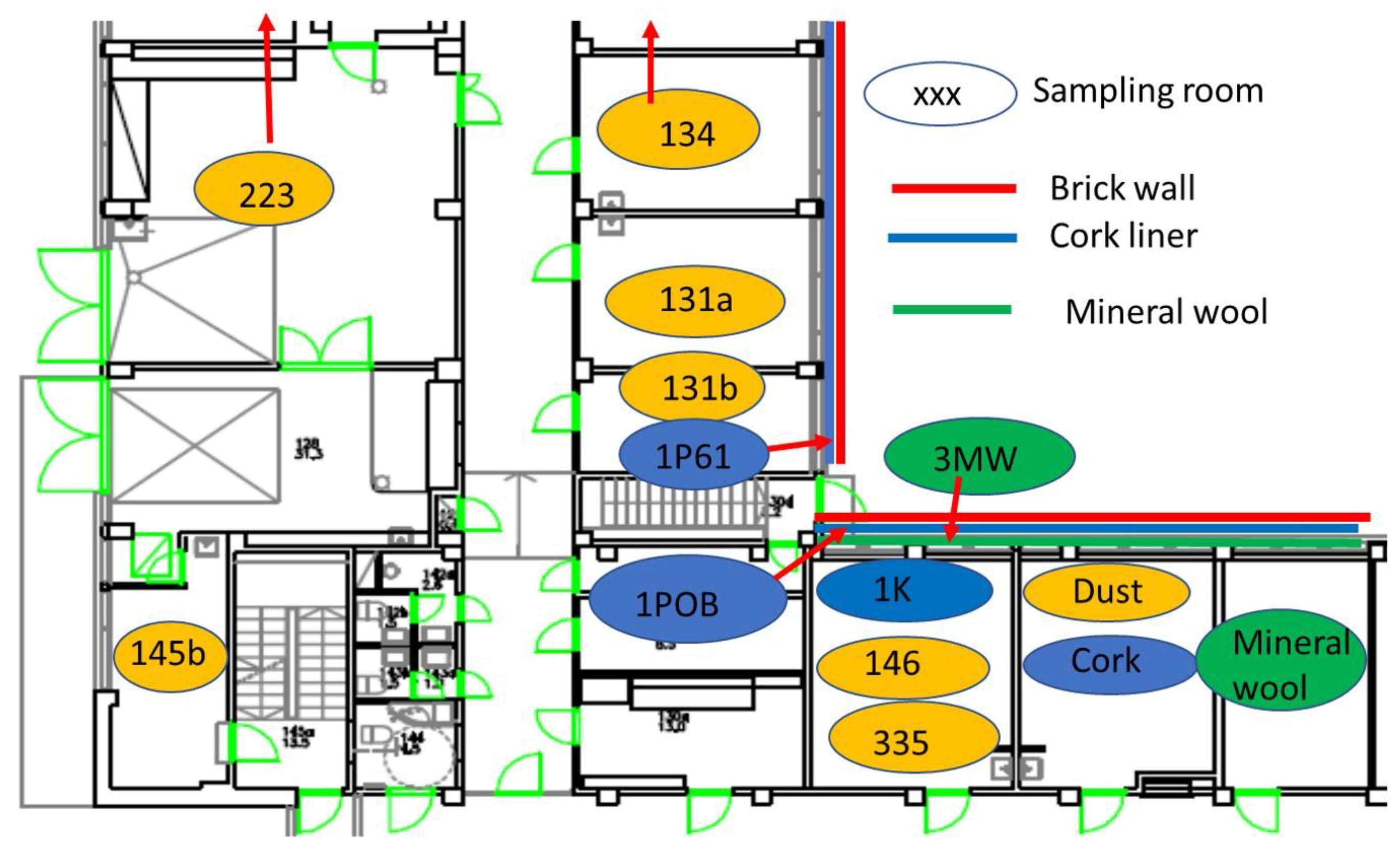
2.1. Building Inspection Connected to Five Problematic Rooms

Four office rooms in a public building, 131a, 131b, 335, and 145b, [73], were associated with indoor-air-related health symptoms and abandoned by their occupants. One office room, 146, was provided with an air cleaner. The building and a floor plan showing the investigated rooms are described in Section 4.1. A building inspection revealed that the outdoor wall outside the problematic rooms was damaged and that rainwater had penetrated into the wall structure. A cork liner used as isolation inside the plinth in the outer wall was moist and degraded by microbes. Stereomicroscopic inspection of the liner indicated the growth of molds including Aspergillus, Chaetomium, and Trichoderma and an unrecognized fungus, as shown in Figure 1. Mineral wool insulation inside the outer wall was also moist and contaminated with mold. The inner surfaces of the five rooms and the collected samples of hard boards and gypsum liners exhibited no visible water damage or mold growth.

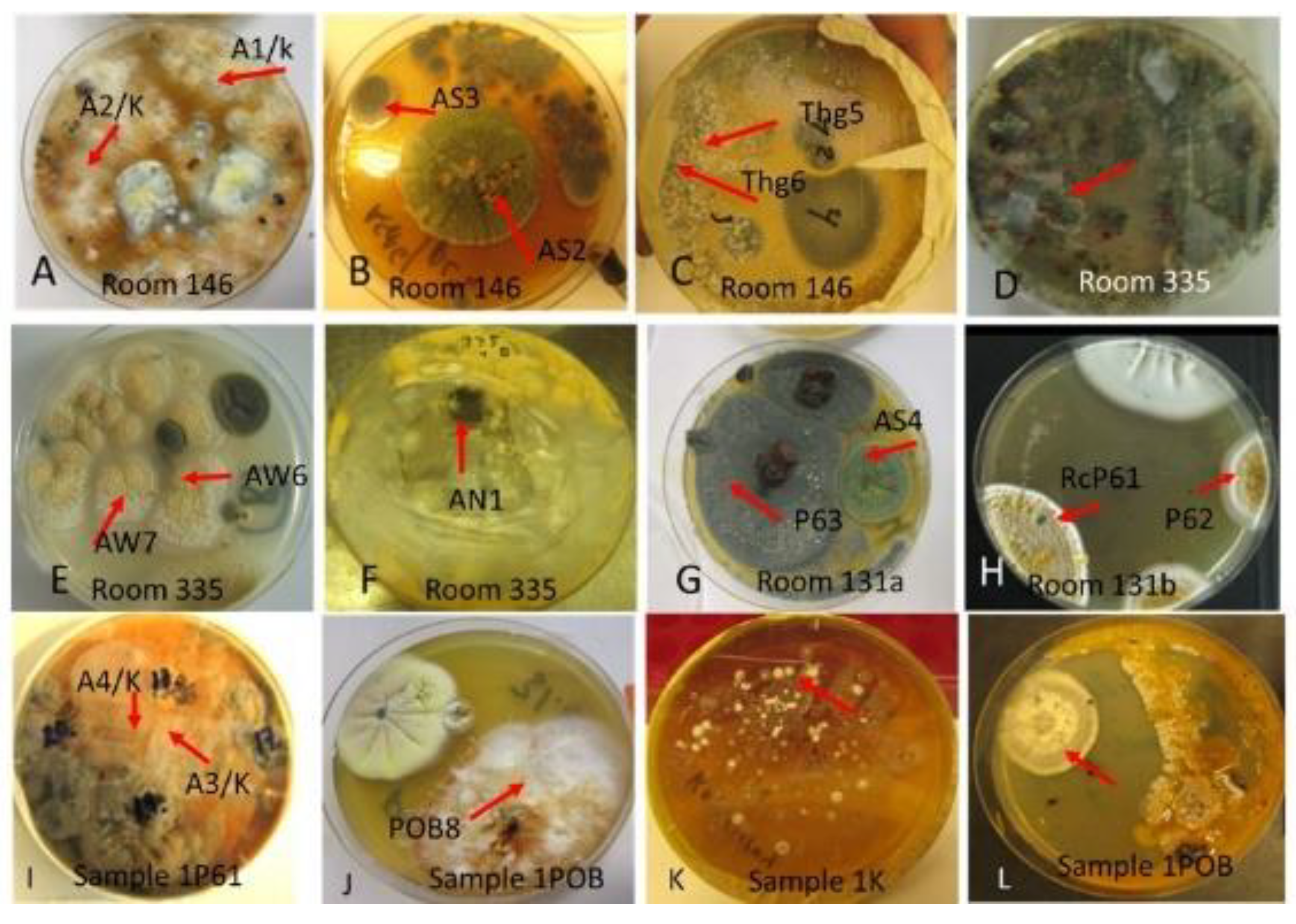
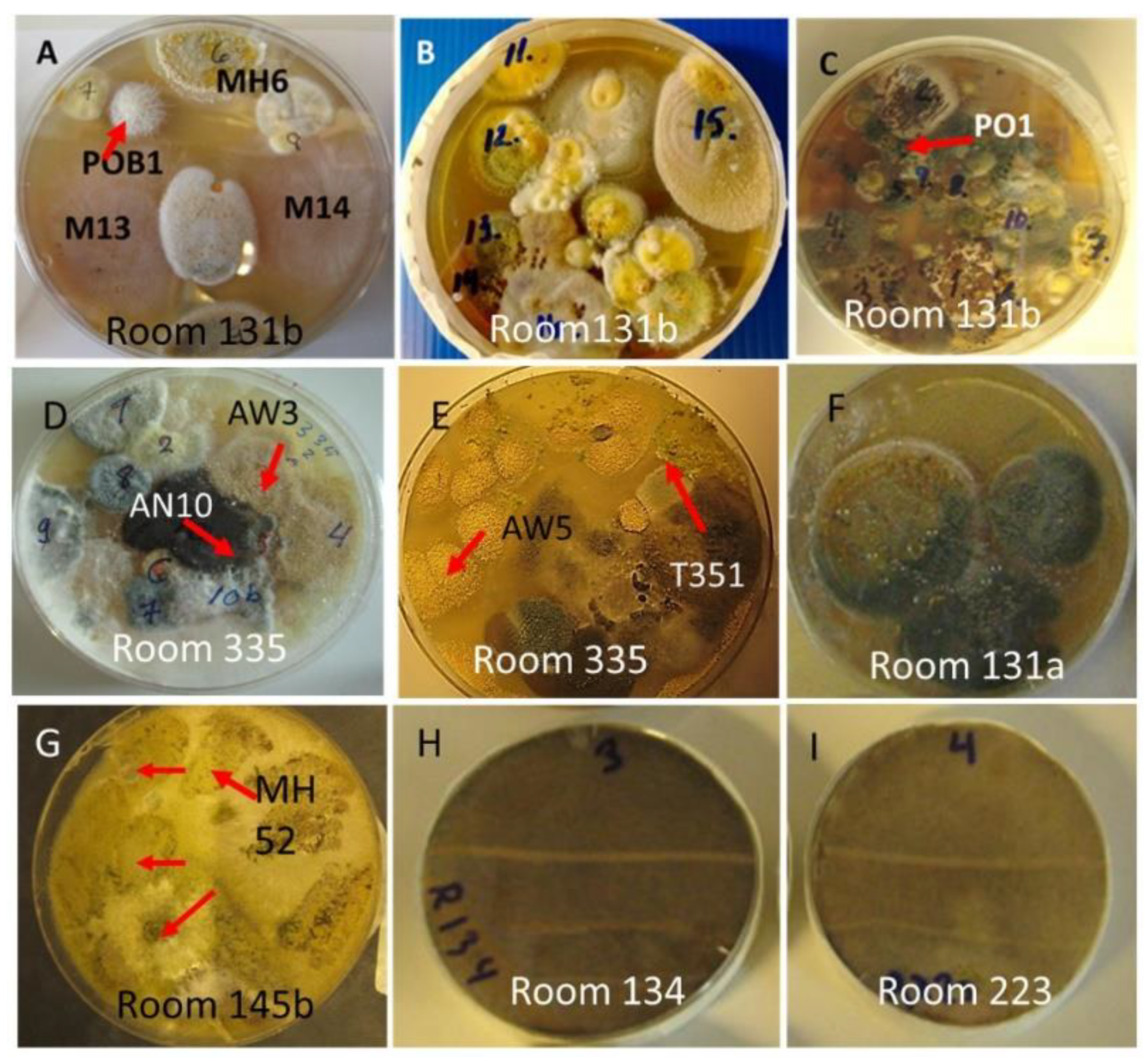
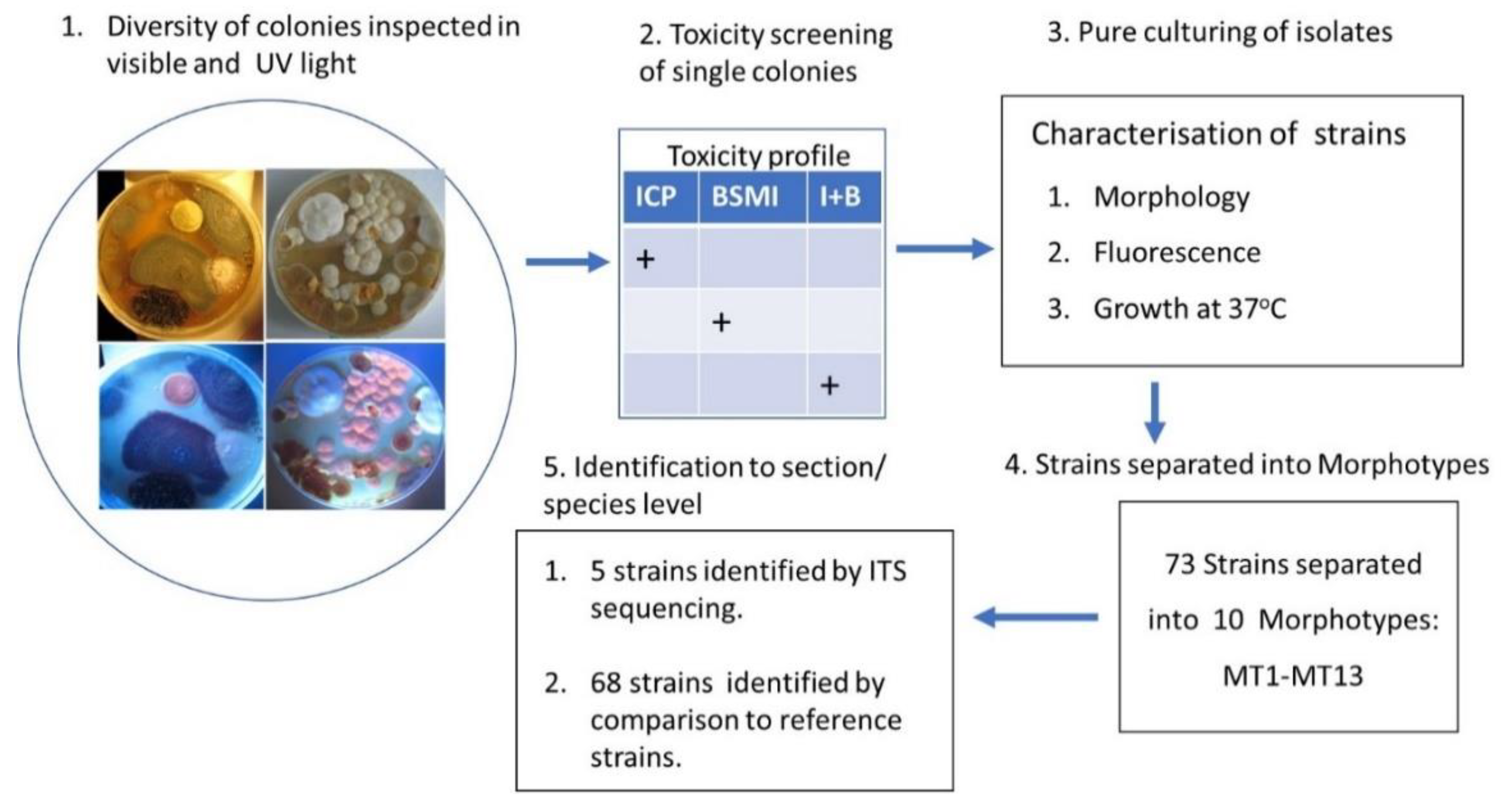
2.2. Diversity Tracking of Molds Cultivated from Wall Structures

The massive microbial growths cultivated on pieces of the moist cork liner (samples 1P61, 1K, 1POB) and mineral wool (sample 3MW) collected from outside the problematic rooms are shown in Figure 2. The major fungal colony types obtained on malt extract agar were light brown-white colonies (Figure 2A,I,J), green colonies (Figure 2B,G), green mycoparasitic colonies (Figure 2C,D), yellow and black colonies (Figure 2E,F), and gray yellow-green colonies (Figure 2G,H). An odd antifungal colony that presented only on single plates is shown in Figure 2K. Figure 2L shows the major bacterial colonizer of the cork liner cultivated on tryptic soy agar (spore-forming actinobacterium).
The diversity of the major fungal isolates was characterized as follows: biomass lysates of five colonies from each plate were tested for toxic responses by two rapid screening bioassays, boar sperm motility inhibition (BSMI) assay and inhibition of cell proliferation (ICP), and fluorescence emission. These bioassays showed that more than 70% of the tested colonies were toxic. Conidiophores and conidia/spores were inspected using a phase contrast microscope. The toxigenic colonies from plates A to K (Figure 2) were grouped into 10 morphotypes as shown in Table 1: brown-white colonies with Acrostalagmus-like conidiophores (MT1), tree Aspergillus morphotypes differing in fluorescence emission and toxic response (MT2, MT3, MT4), two Trichoderma morphotypes differing in conidia size (MT5, MT6), and one toxigenic Penicillium morphotype (MT7). The final morphotype was obtained by microscopic inspection of the cork liner, representing a potentially toxic ascomata-producing Chaetomium-like morphotype (MT11). The major bacterial colonizers were spore-forming actinobacteria that were toxic in either BSMI or ICP or both (MT8-MT10).
Representatives of morphotypes MT1 to MT7 were identified by ITS sequencing as Acrostalagmus luteoalbus (MT1), Trichoderma atroviride (MT5), Trichoderma trixiae (MT6), and Penicillium expansum (MT7). The isolates assigned to morphotypes MT2, MT3, and MT4 were identified as belonging to Aspergillus section Versicolores, section Circumdati, and section Nigri, respectively, based on similarity to the reference strains SL/3, PP2, and HAMBI-1271. The spore-forming actinobacteria were morphologically identical to each other but were separated into three morphotypes, MT8-MT10, based on their toxic response in the bioassays.

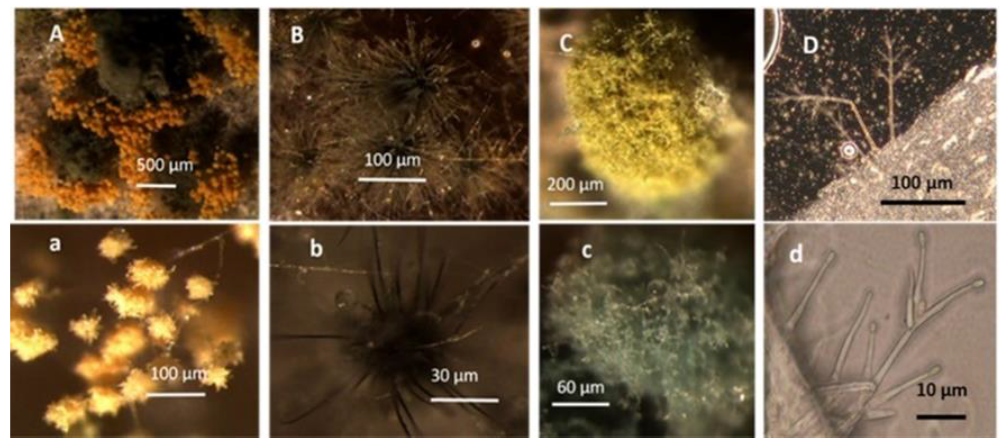
2.3. Cultured Settled Dust from Problematic Rooms Revealed Five Major Morphotypes

We looked for representatives of the genera colonizing the cork liner and mineral wool, Acrostalagmus, Aspergillus, Trichoderma, Penicillium, and Chaetomium, in settled dust collected from problematic rooms 131a, 131b, 335, and 145b, from nonproblematic rooms 134 and 223, and from room 146, where the occupant did not complain but had an air cleaner installed. The plates containing cultured settled dust are shown in Figure 3.
A majority (>70%) of the tested colonies in dust from the problematic rooms and room 146 showed a toxic response in the bioassays. The results in Table 2 show the toxigenic morphotypes in settled indoor dust: isolates similar to Acrostalagmus (MT1); Aspergillus section Versicolores (MT2), Circumdati (MT3), and Nigri (MT4); mycoparasitic isolates similar to T. trixiae (MT5) and T. atroviride (MT6); and P. expansum (MT7) and Chaetomium isolates (MT11). One isolate, MH52, was identified by ITS sequencing as Chaetomium globosum. The results in Table 1 and Table 2 show that the toxigenic fungal morphotypes mainly found in the cork liner were also detected in the settled dust. Settled dust from room 146 looked similar to the dust shown in Figure 3G, containing mainly toxigenic Chaetomium-like colonies. Settled dust from rooms 223 and 134, located farther from the water-damaged site of the wall, contained mainly nontoxic Rhizopus; none of the 40 tested biomass dispersals were toxic in the bioassays, and the occurrence of toxic colonies was <5%.

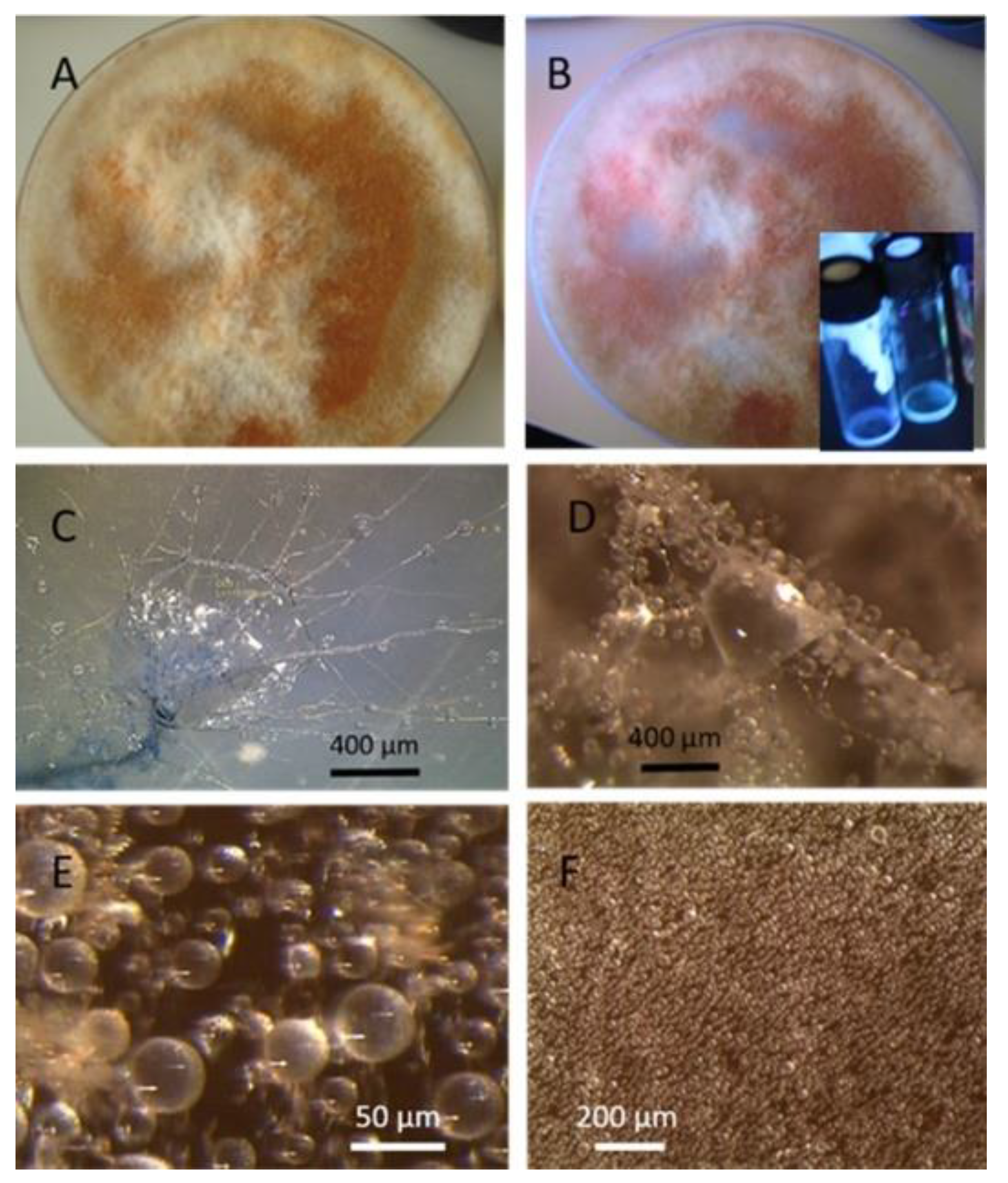
2.4. Characterization of Acrostalagmus luteoalbus Strain POB8

The strains representing morphotype MT1, similar to A. luteoalbus strain POB8 shown in Figure 4, were isolated as a major colonizer from cork liner 1P61 and 1POB and from settled dust from room 131b.
Ethanol extracts prepared from plate-grown biomass of strains POB8, A1/K, A2/K, A3/K, A4/K, and POB1 exhibited similar blue fluorescence to the biomass dispersal shown in Table 1. The bioreactivity of extracts of biomass from the plate of strain POB8 shown in Figure 4 and the five strains representing the same morphotype was tested.
To reveal the diversity of the five Acrostalagmus sp. strains compared with Acrostalagmus luteoalbus POB8 isolated from the cork liners and indoor dust, they were tested with four complementary bioassays. The tests are described and referenced in Section 4.4.1 and Section 4.4.2. Briefly, the two BSMI assays measured sublethal toxicity as inhibition of motility in exposed sperm cells. The spermatozoa membrane integrity disruption (SMID) assay measured lethal toxicity as a loss of plasma membrane integrity. The ICP assay measured cytostatic toxicity as a loss of the proliferating ability of a somatic cell line, PK-15. The results in Table 3 enable a comparison of the EC50 concentrations for the toxicity endpoints from the assays measuring different biological activities. A comparison of the EC50 concentrations obtained in the four bioassays revealed a characteristic and uniform toxicity profile for the five Acrostalagmus sp. isolates and A. luteoalbus strain POB8. For the six Acrostalagmus strains, the toxic endpoints in terms of EC50 concentrations were similar in the three assays, around 10 µg mL−1, indicating that the Acrostalagmus extracts, in contrast to the extracts of reference strains, inhibited sperm motility and cell proliferation at the same concentrations after 1 and 3 d, respectively. The low toxic endpoints in the SMID assay also indicated a rapid lethal effect in sperm cells exposed at 37 °C. Briefly, the toxicity profile revealed by the bioassays indicated that the Acrostalagmus extracts exhibited lethal and cytostatic toxicity when exposed to cells at 37 °C and a motility-inhibiting effect when exposed to cells at 22 °C for 1 d. None of the Acrostalagmus strains exhibited a rapid motility-inhibiting effect after 20 min of exposure at 22 °C. This uniform toxicity profile of the six Acrostalagmus sp. isolates (A1/K, A2/K, A3/K, A4/K, and POB1) and A. luteoalbus POB8 separated them from the reference strains. This indicates that the strains may have produced the same bioactive metabolites, strengthening the hypothetical identity of the five Acrostalagmus sp. strains as A. luteoalbus.
Stereomicrographs of A. luteoalbus strain POB8 (Figure 4) show that the metabolically active biomass secreted exudates and vesicles. The toxic endpoints of the liquid exudate collected from strain POB8 exhibited a toxicity profile and blue fluorescence similar to those of the ethanol extracts. This indicates that the blue fluorescing exudates possibly contained the same substances as the blue fluorescing ethanol extracts. The liquid exudates of the reference strains of genus Aspergillus exhibited no toxicity in the bioassays, whereas exudates of the Penicillium, Stachybotrys, and Chaetomium strains secreted toxins in their guttation droplets and/or exudates.

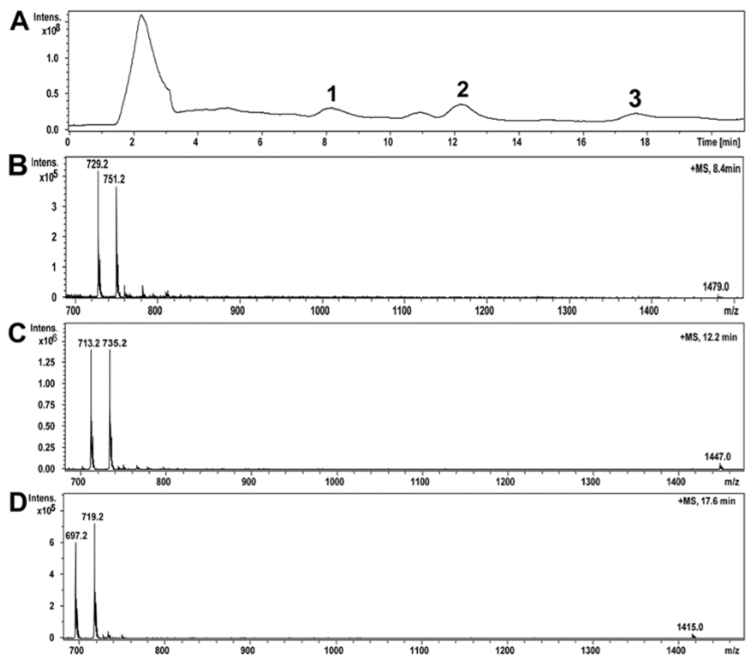
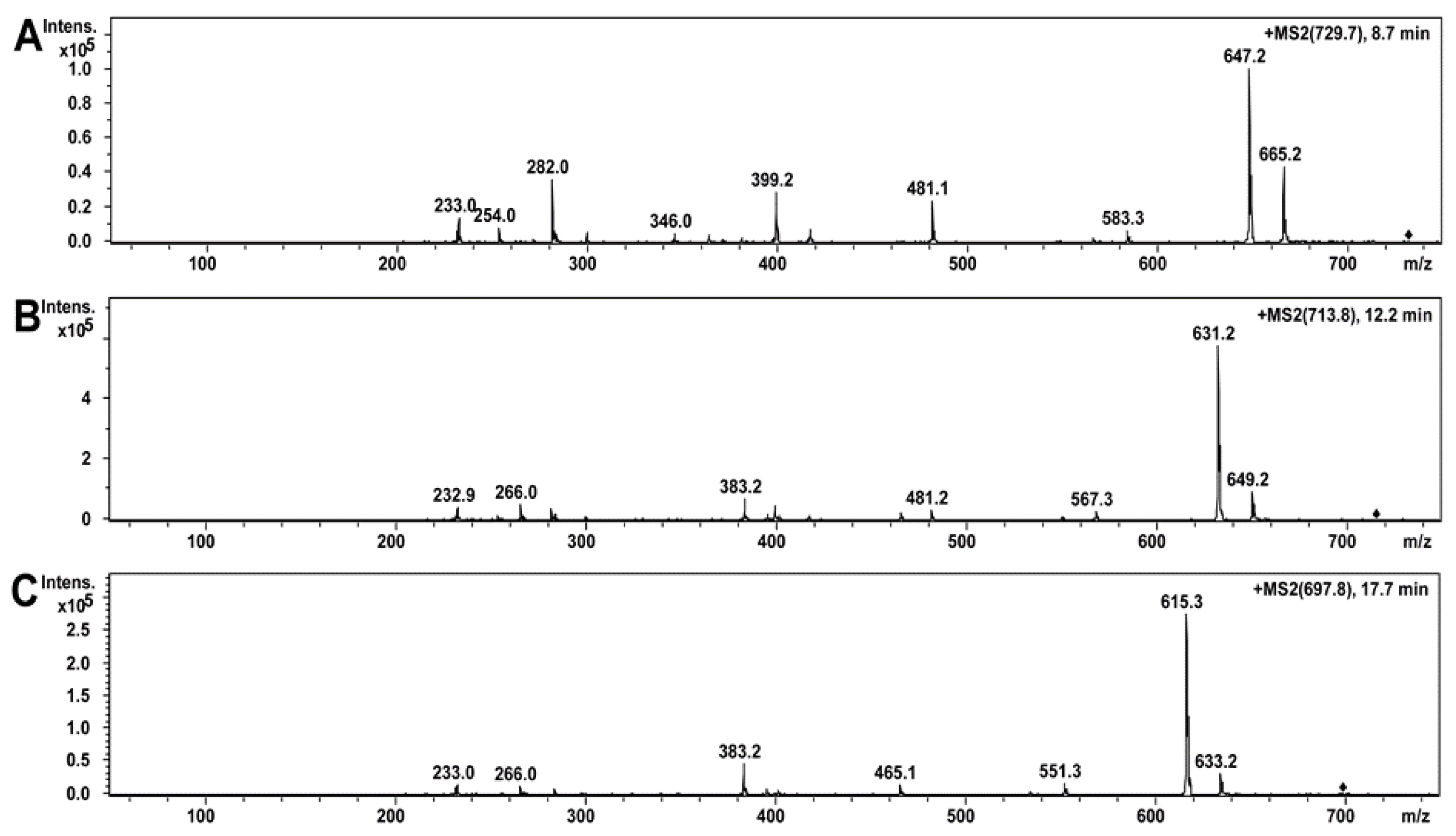
2.5. Compounds of Toxic A. luteoalbus POB8 Ethanol Extract Were Identified as Melinacidins II, III, and IV

The ethanol extract of A. luteoalbus POB8 was analyzed using high-performance liquid chromatography–electrospray ionization mass spectrometry (HPLC-ESI-MS) (Figure 5A–D). On the HPLC-MS total ion chromatograms of the ethanol extract of A. luteoalbus, three compounds were identified, as shown in Figure 5A. Compound 1, with a retention time of 8.5 min, had a protonated mass ion [M + H]+ at m/z 729.2, a sodiated mass ion [M + Na]+ at m/z 751.2, and a sodiated mass ion of dimer [2M + Na]+ at m/z 1479.0 (Figure 5B). Compound 2, with a retention time of 12.2 min, had a protonated mass ion [M + H]+ at m/z 713.2, a sodiated mass ion [M + Na]+ at m/z 735.2, and a sodiated mass ion of dimer [2M + Na]+ at m/z 1447.0 (Figure 5C). Compound 3, with a retention time of 17.7 min, had a protonated mass ion [M + H]+ at m/z 697.2, a sodiated mass ion [M + Na]+ at m/z 719.2, and a sodiated mass ion of dimer [2M + Na]+ at m/z 1415.0, with retention time of 17.7 min (Figure 5D). The obtained mass spectrometry data of compounds 13 matched the dimeric epipolythiodioxopiperazines melinacidin IV (728 g/mol), III (712 g/mol), and II (696 g/mol), respectively, reported earlier [74,75].
The obtained MS/MS data of compounds 13 were similar to those reported for the dimeric epipolythiodioxopiperazines [76,77,78]. The MS/MS spectrum fragmentation patterns (Figure 6A) of compound 1 were identical to the reported MS/MS spectrum of melinacidin IV [76]. Furthermore, the mass spectra and fragmentation patterns of melinacidins IV, III, and II highly resemble each other (Figure 6A–C). According to the obtained and reported MS and MS/MS mass spectrometry data, compounds 1, 2, and 3 of A. luteoalbus were identified as melinacidin IV, III, and II, respectively. The amounts of melinacidin IV (230 µg mL−1), III (290 µg mL−1), and II (120 µg mL−1) in the ethanol extract of A. luteoalbus were calculated from the total absorbance (220 nm) of the HPLC-UV chromatograms.

3. Discussion

This article describes the detection of cultivable toxigenic fungi in wet outdoor walls and indoor dust in a building with indoor air quality problems. One of the species found, melinacidin-producing Acrostalagmus luteoalbus, was detected as building-associated mold for the first time. This study illustrated that the mixed mycobiota cultivated from the wet wall and from settled indoor dust in problematic rooms seemed to contain the same major fungal species and genera. This indicates a possible connection between outdoor structures and indoor spaces (Figure 1, Figure 2 and Figure 3, Table 1 and Table 2).
Interestingly, a rare mold genus identified as Acrostalagmus, which was the major constituent of the mixed mycobiota in the wet cork liner from the water-damaged outdoor wall, was also present in indoor dust. One representative strain, POB8, was identified at the species level as A. luteoalbus by ITS sequencing. This strain has been used as a reference strain [65,66,79,80], but otherwise there are no reports of Acrostalagmus sp. strains growing on building materials, globally or in Finland. Based on its distinct morphology and phylogenetic distance, A. luteoalbus (basionym Sporotrichum luteoalbum) was introduced as a generic distinction between the former sections Verticillium and Nigrescentia [81,82,83,84]. The occurrence of Verticillium in building materials and cork has been described, but no isolates have been identified as Acrostalagmus species [10,85]. The production of immunosuppressive and cytotoxic melinacidin by an A. luteoalbus isolate has been shown [74,86]. The detected bioreactivity in biomass and guttation droplets indicate the possibility of a bioreactive microbial secretome including melinacidins migrating from a wet outdoor wall into indoor spaces.
This study presents the mass spectrometry data characteristics of melinacidins and shows that A. luteoalbus strain POB8 produces melinacidins II, III, and IV. Melinacidin derivatives were previously reported from a variety of fungi including Acrostalagmus luteoalbus (syn. A. cinnabarinus) [74,86]. Melinacidins belonging to the epipolythiodioxopiperazines (ETPs) are related but not identical to verticillin, chaetocin, and gliotoxin, the best-known ETPs produced by Aspergillus fumigatus. Epipolythiodioxopiperazines have antiproliferative, cytotoxic, immunomodulatory, antiviral, and antimicrobial activity in vitro [77,87] and have been shown to be toxic to mammals; the LD in mice (i.p.) is 2–4 mg/kg [75,77,88,89,90]. The toxicity of ETPs depends on a disulfide bridge, with inactivating enzymes as methyl transferases via reactions with thiol groups. They also generate reactive oxygen species by redox cycling, inducing oxidative stress and mitochondrial damage [88,91]. This may explain the newly observed toxicity to sperm cells. However, this has yet to be confirmed with pure melinacidins.
Assuming that melinacidins were the only toxic substances in the ethanol extracts, the EC50 concentration in the BMSI and ICP assays for the tested melinacidin mixture was calculated as 0.3 to 0.6 µg mL−1. Based on a similar toxicity profile and blue fluorescence, it is possible, but not proven, that the liquid exudates secreted from the growing biomass of POB8 (Figure 4B) also contained melinacidins. Melinacidin concentration in the exudate, when calculated based on the toxic response, would be around 40 to 80 µg melinacidins per mL exudate. The hypothetically calculated melinacidin content in the biomass (wet wt) and exudates would be around 0.8 to 2 µg and 0.4 to 0.8 µg melinacidins mg−1, respectively. This means that similar amounts of melinacidins could migrate with the exudate as with fungal particles.
Four other genera were found in both the wet outdoor wall and the indoor dust from the three problematic rooms in the old wing of the building (and room 146 with the air cleaner) close to the wet wall, but not from dust from the two more remote nonproblematic rooms. The fungi representing the genera Aspergillus, Trichoderma, Penicillium, and Chaetomium are common constituents of mycotoxin-producing indoor mycobiota and common colonizers of indoor building materials [1,65,66,72,79,92,93]. These fungi are also listed as indicator species for water damage [1,94,95].
Occupants in the rooms complained about indoor air quality even though no signs of microbial growth or water damage were detected, and the airborne load of microbial particles (<4 CFU m−3) was too low to explain the reported symptoms. However, the negative pressure of 3–4 Pa and a sealing repair that was performed led to the suspicion of air leakage through the building structure [1,73]. Although microbial contamination inside buildings does not necessarily have direct contact with the indoor air, microbial growth within the exterior walls can affect indoor air quality. This can happen if, as a consequence of fluctuations in wind and indoor air pressure, the infiltration airflow drifts through a contaminated wall structure [96,97,98,99,100].
The occurrence of the same molds in the wet wall and indoor dust indicates possible transmission of conidia and spores into the problematic rooms and enrichment in settled dust. Tiny, potentially antibiotic-producing spores of Streptomyces migrated from the construction material into the indoor air [96]. In addition to conidia and fragments of microorganisms, the substances of microbial metabolism [6,72,79], secreted in liquids in guttation droplets and vesicles, may be trapped within building insulation and structural elements. The fungal secretome and fungal metabolites (including proteins, peptides, surface active substances, mycotoxins, VOCs, etc.) may be transported to interior spaces via liquid and vapor fluxes within materials and via airflow (negative pressure) within ventilation systems and rooms [1,96,97,98,99,100,101,102,103,104,105]. The microbial liquid metabolites, secretome, and VOC emissions [106,107] may enhance and exceed the indoor concentrations of bioreactive agents provided by airborne conidia and fragments [9,13,65,68,79]. Fungal protein homologues of human proteins that initiate or signal tissue damage, mycotoxins, and microbial mitochondrial toxins have all been reported to enhance and provoke inflammatory responses [103,104,105]. Indoor microbes may produce and emit airborne surface-active agents, enhancing the immunoreactivity of inhaled allergens [6,106,107]. The occupants of the problematic rooms may have been exposed to indoor air polluted with immunoreactive metabolites and surface-active agents from microbial species actively growing on the cork liner and mineral wool in the wet outdoor wall. This exposure combined with a low tolerance to environmental microbes may have decreased their resilience and would explain the symptoms they experienced. The noncomplaining occupant in room 146 may have been more resilient to the exposure, or the air in the room may have been efficiently cleaned by the air cleaner.
In conclusion, we suggest that the outdoor wall was the potential emission source for the indoor mold contamination detected in the problematic rooms. In addition to viable conidia, the mixed microbiota colonizing the wet outer wall possibly also emitted immunoreactive microbial metabolites among the exudates, vesicles, and fungal fragments into indoor air exposing the occupants of the problematic rooms. We also suggest that exposure to the immunoreactive metabolites may have attenuated the immunological tolerance to commensal and environmental microbes. There are no methods yet for measuring the total load of airborne immunoreactive fungal metabolites in indoor air, or for measuring decreased resilience to potentially hazardous exposure. Our proposed connection in this study between microbial emissions from the wet outer wall and the reported complaints concerning the indoor air quality is speculative but cannot be excluded.

4. Materials and Methods

4.1. A Public Building from the 1960s Investigated for Mold Contamination

A public building in southern Finland that was involved in indoor-air-related complaints was investigated for mold contamination during 2013–2014, as described in [73]. The building consisted of two parts: an old wing built in 1957 (rooms 145b, 146, and 335) and a new wing erected in 1963 (rooms 131a, 131b, 223, and 134); it was concrete-framed and brick-lined and had mineral wool as isolation material. The building still contained the original construction materials, such as a cork liner in the building network (Figure 7 and Figure 8), and mechanical ventilation was installed in the 1970s. Ventilation was turned off from 8:00 pm to 6:00 am on workdays and during weekends. The floor plan of the building, the sites of the investigated rooms, and the structure of the outer wall are shown in Figure 1. The building underwent an indoor air survey in 2010 and problematic rooms were abandoned. Reported symptoms included respiratory distress, increased oxygen demand, the need for a portable oxygen cylinder, and skin symptoms (personal communication from the occupants).
A new survey of rooms 131a and 131b early in 2013 revealed a negative pressure of 3–4 Pa in relation to outdoor pressure, so sealing repair was performed. No elevated concentrations of airborne microbes (<4 cfu m3) were detected, but the indoor air quality complaints continued. Samples had been taken from the rooms by methods approved for official use in Finland for health risk assessment of indoor air, i.e., VOC and airborne microbes with the Andersen impactor [108,109]. The results obtained by analyzing these samples did not indicate the causative agents for the symptoms experienced by the occupants or explain their complaints.
The rooms classified as problematic remained abandoned. After renovation and removal of the insulation material in the outer wall in 2016–2018, rooms 131a and 145b were returned to ordinary use, whereas rooms 131b and 335 were used for equipment storage.

4.2. Collection of Samples and Diversity Tracking of Microbial Constituents in Building Materials and Settled Dust

The sampling sites and the degraded cork liner colonized by microbes are shown in Figure 7 and 8. The samples of the building network were taken by drilling with a shock drill through the wall (Figure 8A). When the drill penetrated the concrete, the concrete dust was removed by vacuuming. Then, a tube was fixed to the drill (Figure 8B) and pressed into the cork liner, filling the tube with cork. The cork trapped in the tube was aseptically removed into sterile plastic bags, as shown in Figure 8C.
Mineral wool was collected from inside the wall on the third floor (Figure 7), and pieces of hard board, gypsum liners, and settled dust from inner surfaces 1–2 m above floor level in the problematic rooms were collected into sterile plastic bags. The material samples were inspected with a stereomicroscope and fluorescence microscope, using 400× magnification (Nikon Eclipse E600, Nikon Corporation, Tokyo, Japan) with BP330-380 nm/LP400 nm filters, and stored at −20 °C before cultivation. Staining with fluorescent Hoechst and propidium iodide stains (Figure 8A–C) was described in Andersson et al. [6].

4.3. Experimental Design for Cultivation and Identification of Microbial Isolates

The methods used for cultivating the mold colonies were described in [6]. Pieces of the material samples, about 2 mm × 2 mm × 2 mm, were rubbed on the surface of malt extract agar plates (15 g malt extract from Sharlab, Barcelona, Spain, and 12 g of agar from Amresco, Solon, OH, USA, in 500 mL of H2O) and on tryptic soy agar plates (Sharlab, Barcelona, Spain). Settled dust collected on cotton swabs was streaked on malt extract agar. Plates were incubated for 4 weeks at 22–24 °C.
A scheme illustrating the experimental design for tracking the diversity of major microbial constituents in building material and dust is shown in Figure 9.
Separation of the isolates into morphotypes (MTs) was based on the toxicity profile and morphology of conidiophores, conidia, ascomata, and ascospores obtained with a phase-contrast microscope (400× magnification; Olympus CKX41, Tokyo, Japan) and image recording software (cellSens® standard v. 11.0.06, 2012, Olympus Soft Imaging Solutions GmbH, Münster, Germany) and compared to reference strains according to Samson [110,111] as described in [65,66].
Selected representatives of the morphotypes were identified in previous studies by ITS or tef1α sequencing with the primer pairs ITS1 (5′-TCCGTAGGTGAACCTGCGG-3′)/ITS4 (5′-TCCTCCGCTTATTGATATGC-3′) and EF595F (5′-CGTGACTTCATCAAGAAGATG-3′)/EF1160R (5′-CCGATCTTGTAGACGTCCTG-3′), respectively [79].

4.4. Toxicity Assays

4.4.1. Rapid Screening Test of Single Colonies

Rapid screening tests applied directly to the primary sampling plates to measure (a) the toxins affecting the cellular energy metabolism, mitochondria, and ion homeostasis based on inhibition of boar spermatozoa motility (BSMI), and (b) the toxins affecting macromolecular synthesis and cytostatic activity based on inhibition of the proliferation of somatic cell line PK-15 (ICP) were previously described in detail [65,79]. In this study, the colony biomass was suspended in 200 μL of ethanol and heated in a water bath to 55–60 °C for 10 min. A colony was considered very toxic in the BSMI assay when <2.5 vol.% of its biomass suspension inhibited boar sperm motility after 30 min to 1 day of exposure, and slightly toxic if motility inhibition occurred after 3 days of exposure. A colony was considered toxic in the in vitro ICP assay when <5 vol.% resulted in inhibition of cell proliferation of porcine kidney (PK-15) cells after 2 days of exposure in ICP assays.

4.4.2. Toxicity Assays for Ethanol-Extracted Pure Fungal Cultures

Toxicity assays involving the ethanol extraction of lipophilic bioactive peptides and mycotoxins [22,31,65,66,80,93] obtained from pure fungal cultures were performed using porcine cells (sperm and somatic cell line PK-15) as indicators according to previously described methods [79,80]. The toxic response in the bioassays was measured for the ethanol extracts as toxic endpoints defined as EC50 concentrations, i.e., the lowest concentration of ethanol-soluble dry substances per mL of target cell suspension causing an adverse (toxic) effect in 50% of exposed cells. The test procedures and calculation of EC50 for the ethanol-dry substances and pure mycotoxins in the BSMI, SMID, and ICP assays were described previously [79,80,112]. The BSMI assay measured sublethal toxicity as disturbance in the mitochondrial activity, ion homeostasis, and energy supply of the exposed sperm cells. The BSMIM and BSMIR assays measured motility after exposure of motile and resting spermatozoa, respectively. The SMID assay measured lethal toxicity as loss of plasma membrane integrity. The ICP assay measured cytostatic toxicity and cell death as the loss of the proliferating ability of growing somatic cells, which may be caused by inhibition of macromolecular synthesis and/or induction of necrotic or apoptotic cell death [65,66,73,80].

4.5. Chemical Analysis

The biomass of A. luteoalbus strain POB8 was harvested from a malt extract agar (MEA) plate incubated at room temperature for 10 days. The collected biomass, about 200–400 mg wet weight of A. luteoalbus, was extracted with ethanol, and the toxic ethanolic extract was analyzed by high-performance liquid chromatography–ion trap mass spectrometry (HPLC-IT-MS) as described by Salo et al. [65]. HPLC–electrospray ionization ion trap mass spectrometry analysis (ESI-IT-MS) was performed using an MSD-Trap-XCT plus ion trap mass spectrometer equipped with an Agilent ESI source and Agilent 1100 series LC (Agilent Technologies, Wilmington, DE, USA) in positive mode with a mass range of m/z 50–2000. ESI source parameters used for analysis were: nebulizer gas pressure, 35 psi; drying gas flow rate, 8 L min−1; drying gas temperature, 350 °C; and capillary voltage, 5000 V. The column used was a SunFire C18, 2.1 × 50 mm, 2.5 μm (Waters, Milford, MA, USA). Separation of compounds from the ethanol extract of biomass of A. luteoalbus strain was done using an isocratic method of solution A, H2O with 0.1% (v/v) formic acid, and B, methanol in a ratio of 40/60 (v/v), for 15 min and a gradient of 100% B for 50 min at a flow rate of 0.2 mL min−1. Identification of compounds of the toxic ethanolic extract was done by HPLC-IT-MS and MS/MS analysis. The amounts of identified compounds were calculated from total absorbance (220 nm) of the HPLC-UV chromatogram of the A. luteoalbus POB8 ethanol-extracted biomass.

Author Contributions

Conceptualization, J.S. and M.A.A.; data curation, J.S., T.M., L.K., R.M., and M.A.A.; formal analysis, J.S., T.M., R.M. and M.A.A.; funding acquisition, J.K. and H.S.; investigation, J.S., T.M. and M.A.A.; methodology, J.S. and M.A.A.; project administration, J.K. and H.S.; resources, M.A.; supervision, J.K., L.K. and H.S.; validation., L.K., and M.A.A.; visualization, J.S. and M.A.A.; writing—original draft, J.S., T.M., L.K., R.M. and M.A.A.; writing—review and editing, J.S., M.A.A., R.M., T.M., L.K. and H.S. All authors have read and agreed to the published version of the manuscript.

Funding

This research was funded by the Academy of Finland (CleanSchool project, grant number 330150, and TOXICPM, grant number 289161).

Institutional Review Board Statement

Not applicable.

Informed Consent Statement

Not applicable.

Data Availability Statement

Not applicable.

Acknowledgments

The authors warmly thank Maria Hautaniemi of the Finnish Food Safety Authority (EVIRA) for providing the porcine kidney cell line PK-15.

Conflicts of Interest

The authors declare no conflict of interest. The funders had no role in the design of the study; in the collection, analyses, or interpretation of data; in the writing of the manuscript; or in the decision to publish the results.

References

  1. Andersen, B.; Frisvad, J.C.; Dunn, R.R.; Thrane, U. A pilot study on baseline fungi and moisture indicator fungi in Danish homes. J. Fungi 2021, 7, 71. [Google Scholar] [CrossRef]
  2. Shukla, S.; Budden, K.; Neal, R.; Hansbro, P. Microbiome effects on immunity, health and disease in the lung. Clin. Translat. Immunol. 2017, 6, e133. [Google Scholar] [CrossRef]
  3. Horve, P.; Lloyd, S.; Mhuireach, G.; Dietz, L.; Fretz, M.; MacCrone, G.; Van Den Wymelenberg, K.; Ishaq, S.L. Building upon current knowledge and techniques of indoor microbiology to construct the next era of theory into microorganisms, health, and the built environment. J. Expo. Sci. Environ. Epidemiol. 2020, 30, 219–235. [Google Scholar] [CrossRef] [PubMed] [Green Version]
  4. Leung, M.H.Y.; Lee, P.K.H. The roles of the outdoors and occupants in contributing to a potential pan-microbiome of the built environment: A review. Microbiome 2016, 4, 21. [Google Scholar] [CrossRef] [PubMed] [Green Version]
  5. Haahtela, T.; Alenius, H.; Lehtimäki, J.; Sinkkonen, A.; Fyhrquist, N.; Hyöty, H.; Ruokolainen, L.; Mäkelä, M. Immunological resilience and biodiversity for prevention. Allergy 2021. [Google Scholar] [CrossRef]
  6. Andersson, M.A.; Salo, J.; Kedves, O.; Kredics, L.; Druzhinina, I.; Kurnitski, J.; Salonen, H. Bioreactivity, guttation and agents influencing surface tension of water emitted by actively growing indoor mould isolates. Microorganisms 2020, 8, 1940. [Google Scholar] [CrossRef] [PubMed]
  7. Dai, D.; Prussin, A.; Marr, L.; Vikesland, P.; Edwards, M.A.; Pruden, A. Factors shaping the human exposome in the built environment: Opportunities for engineering control. Environ. Sci. Technol. 2017, 51, 7759–7774. [Google Scholar] [CrossRef] [Green Version]
  8. Li, S.; Yang, Z.; Hu, D.; Cao, L.; He, Q. Understanding building-occupant-microbiome interactions toward healthy built environments: A review. Environ. Sci. Eng. 2021, 15, 65. [Google Scholar] [CrossRef]
  9. Bloom, E.; Nyman, E.; Must, A.; Pehrson, C.; Larsson, L. Mold and mycotoxins in indoor environments—a survey in water damaged buildings. J. Occup. Environ. Hyg. 2009, 6, 671–678. [Google Scholar] [CrossRef]
  10. Gravesen, S.; Nielsen, P.A.; Iversen, R.; Nielsen, K.F. Microfungal contamination of damp buildings—Examples of risk constructions and risk materials. Environ. Health Perspect. 1999, 107, 505–508. [Google Scholar] [CrossRef]
  11. Jarvis, B.; Miller, J. Mycotoxins as harmful indoor air contaminants. Appl. Microbiol. Biotechnol. 2005, 66, 367–372. [Google Scholar] [CrossRef]
  12. Mendell, M.J.; Kumagai, K. Observation-based metrics for residential dampness and mold with dose-response relationships to health: A review. Indoor Air 2017, 27, 506–517. [Google Scholar] [CrossRef] [PubMed]
  13. Miller, J.D.; McMullin, D.R. Fungal secondary metabolites as harmful indoor air contaminants: 10 years on. Appl. Microbiol. Biotechnol. 2014, 98, 9953–9966. [Google Scholar] [CrossRef] [PubMed]
  14. Nielsen, K.F. Mold Growth on Building Materials. Secondary Metabolites, Mycotoxins and Biomarkers. Ph.D. Thesis, Technical University of Denmark, Lyngby, Denmark, 2002. [Google Scholar]
  15. Piontek, M.; Łuszczynska, K. Testing the toxicity of Stachybotrys chartarum in indoor environments—A case study. Energies 2021, 14, 1602. [Google Scholar] [CrossRef]
  16. Kankkunen, P.; Rintahaka, J.; Aalto, A.; Leino, M.; Majuri, M.L.; Alenius, H.; Wolff, H.; Matikainen, S. Trichothecene mycotoxins activate inflammatory response in human macrophages. J. Immunol. 2009, 182, 6418–6425. [Google Scholar] [CrossRef] [Green Version]
  17. Kankkunen, P.; Valimaki, E.; Rintahaka, J.; Palomäki, J.; Nyman, T.; Alenius, H.; Wolff, H.; Matikainen, S. Trichothecene mycotoxins activate NLRP3 inflammasome through a P2X7 receptor and Src tyrosine kinase dependent pathway. Hum. Immunol. 2014, 75, 134–140. [Google Scholar] [CrossRef] [PubMed]
  18. Korkalainen, M.; Taubel, M.; Naarala, J.; Kirjavainen, P.; Kistinen, A.; Hyvärinen, A.; Komulainen, H.; Viluksela, M. Synergistic proinflammatory interactions of microbial toxins and structural components characteristic to moisture-damaged buildings. Indoor Air 2017, 27, 13–23. [Google Scholar] [CrossRef] [Green Version]
  19. Annila, P.J.; Hellemaa, M.; Pakkala, T.A.; Lahdensivu, J.; Suonketo, J.; Pentti, M. Extent of moisture and mould damage in structures of public buildings. Case Stud. Constr. Mater. 2017, 6, 103–108. [Google Scholar] [CrossRef]
  20. Andersson, M.A.; Nikulin, M.; Köljalg, U.; Andersson, M.C.; Rainey, F.; Reijula, K.; Hintikka, E.L.; Salkinoja-Salonen, M. Bacteria, molds, and toxins in water-damaged building materials. Appl. Environ. Microbiol. 1997, 63, 387–393. [Google Scholar] [CrossRef] [Green Version]
  21. Andersson, M.A.; Mikkola, R.; Kroppenstedt, R.M.; Rainey, F.A.; Salkinoja-Salonen, M.S. The mitochondrial toxin produced by Streptomyces griseus strains isolated from an indoor environment is valinomycin. Appl. Environ. Microbiol. 1998, 64, 4767–4773. [Google Scholar] [CrossRef] [Green Version]
  22. Andersson, M.A.; Mikkola, R.; Raulio, M.; Kredics, L.; Maijala, P.; Salkinoja-Salonen, M.S. Acrebol, a novel toxic peptaibol produced by an Acremonium exuviarum indoor isolate. J. Appl. Microbiol. 2009, 106, 909–923. [Google Scholar] [CrossRef] [PubMed]
  23. Bornehag, C.-G.; Blomquist, G.; Gyntelberg, F.; Jarvholm, B.; Malmberg, P.; Nordvall, L.; Nielsen, A.; Pershagen, G.; Sundell, J. Dampness in buildings and health, Nordic interdisciplinary review of the scientific evidence on associations between exposure to “dampness” in buildings and health effects (NORDDAMP). Indoor Air 2001, 11, 72–86. [Google Scholar] [CrossRef]
  24. Bornehag, C.-G.; Sundell, J.; Bonini, S.; Custovic, A.; Malmberg, P.; Skerfving, S.; Sigsgaard, T.; Verhoeff, A. Dampness in buildings as a risk factor for health effects, EUROEXPO: A multidisciplinary review of the literature (1998–2000) on dampness and mite exposure in buildings and health effects. Indoor Air 2004, 14, 243–257. [Google Scholar] [CrossRef] [PubMed]
  25. Mikkola, R.; Andersson, M.A.; Grigoriev, P.; Teplova, V.V.; Saris, N.-E.L.; Rainey, F.A.; Salkinoja-Salonen, M.S. Bacillus amyloliquefaciens strains isolated from moisture-damaged buildings produced surfactin and a substance toxic to mammalian cells. Arch. Microbiol. 2004, 181, 314–323. [Google Scholar] [CrossRef] [PubMed]
  26. Mikkola, R.; Andersson, M.A.; Teplova, V.; Grigoriev, P.; Kuehn, T.; Loss, S.; Tsitko, I.; Apetroaie, C.; Saris, N.-E.L.; Veijalainen, P.; et al. Amylosin from Bacillus amyloliquefaciens, a K+ and Na+ channel-forming toxic peptide containing a polyene structure. Toxicon 2007, 49, 1158–1171. [Google Scholar] [CrossRef]
  27. Nielsen, K.F.; Smedsgaard, J. Fungal metabolite screening: Database of 474 mycotoxins and fungal metabolites for dereplication by standardised liquid chromatography-UV-mass spectrometry methodology. J. Chromatogr. A 2003, 100, 111–136. [Google Scholar] [CrossRef]
  28. Paananen, A.; Järvinen, K.; Sareneva, T.; Salkinoja-Salonen, M.S.; Timonen, T.; Hölttä, E. Valinomycin-induced apoptosis of human NK cells is predominantly caspase independent. Toxicology 2005, 212, 37–45. [Google Scholar] [CrossRef]
  29. Peltola, J.; Andersson, M.A.; Haahtela, T.; Mussalo-Rauhamaa, H.; Rainey, F.A.; Kroppenstedt, R.M.; Samson, R.A.; Salkinoja-Salonen, M.S. Toxic-metabolite-producing bacteria and fungus in an indoor environment. Appl. Environ. Microbiol. 2001, 67, 3269–3274. [Google Scholar] [CrossRef] [Green Version]
  30. Peltola, J.; Andersson, M.A.; Kämpfer, P.; Auling, G.; Kroppenstedt, R.M.; Busse, H.-J.; Salkinoja-Salonen, M.S.; Rainey, F.A. Isolation of toxigenic Nocardiopsis strains fromindoor environments and description of two new Nocardiopsis species, N. exhalans sp. nov. and N. umidischolae sp. nov. Appl. Environ. Microbiol. 2001, 67, 4293–4304. [Google Scholar] [CrossRef] [Green Version]
  31. Rasimus-Sahari, S.; Teplova, V.V.; Andersson, M.A.; Mikkola, R.; Kankkunen, P.; Matikainen, S.; Gahmberg, C.G.; Andersson, L.C.; Salkinoja-Salonen, M. The peptide toxin amylosin of Bacillus amyloliquefaciens from moisture damaged buildings is immunotoxic, induces potassium efflux from mammalian cells, and has antimicrobial activity. Appl. Environ. Microbiol. 2015, 81, 2939–2949. [Google Scholar] [CrossRef] [Green Version]
  32. Tuomi, T.; Reijula, K.; Johnsson, T.; Hemminki, K.; Hintikka, E.L.; Lindroos, O.; Kalso, S.; Koukila-Kähkölä, P.; Mussalo-Rauhamaa, H.; Haahtela, T. Mycotoxins in crude building materials from water-damaged buildings. Appl. Environ. Microbiol. 2000, 66, 1899–1904. [Google Scholar] [CrossRef] [Green Version]
  33. Wahab, S.N.A.; Mohammed, N.I.; Khamidi, M.F.; Jamaluddin, N. Qualitative assessment of mould growth for higher education library building in Malaysia. Procedia Soc. Behav. Sci. 2015, 170, 252–261. [Google Scholar] [CrossRef] [Green Version]
  34. de Oliveira, G.L.V.; Oliveira, C.N.S.; Pinzan, C.F.; de Salis, L.V.V.; Cardoso, C.R.B. Microbiota modulation of the gut-lung axis in COVID-19. Front. Immunol. 2021, 12. [Google Scholar] [CrossRef] [PubMed]
  35. Fitzgibbon, G.; Mills, K. The microbiota and immune-mediated diseases: Opportunities for therapeutic intervention. Eur. J. Immunol. 2020, 50, 326–337. [Google Scholar] [CrossRef] [PubMed] [Green Version]
  36. Ganal, S.C.; Sanos, S.L.; Kallfass, C.; Oberle, K.; Johner, C.; Kirschning, C.; Lienenklaus, S.; Weiss, S.; Staeheli, P.; Aichele, P.; et al. Priming of natural killer cells by nonmucosal mononuclear phagocytes requires instructive signals from commensal microbiota. Immunity 2012, 37, 171–186. [Google Scholar] [CrossRef] [Green Version]
  37. Haahtela, T.; Laatikainen, T.; Alenius, H. Hunt for the origin of allergy—Comparing the Finnish and Russian Karelia. Clin. Exp. Allergy 2015, 45, 891–901. [Google Scholar] [CrossRef]
  38. Haahtela, T.; Von Hertzen, L.; Anto, J.M.; Bai, C.; Baigenzhin, A.; Bateman, E.D.; Behera, D.; Bennoor, K.; Camargos, P.; Chavannes, N.; et al. Helsinki by nature: The nature step to respiratory health. Clin. Transl. Allergy 2019, 9, 57. [Google Scholar] [CrossRef]
  39. Rook, G.A. Regulation of the immune system by biodiversity from the natural environment: An ecosystem service essential to health. Proc. Natl. Acad. Sci. USA 2013, 110, 18360–18367. [Google Scholar] [CrossRef] [Green Version]
  40. Huang, C.; Yu, Y.; Du, W.; Liu, Y.; Dai, R.; Tang, W.; Wang, P.; Zhang, C.; Shi, G. Fungal and bacterial microbiome dysbiosis and imbalance of trans-kingdom network in asthma. Clin. Transl. Allergy 2020, 10, 42. [Google Scholar] [CrossRef] [PubMed]
  41. Peters, M.; Kauth, M.; Schwarze, J.; Körner-Rettberg, C.; Riedler, J.; Nowak, D.; Braun-Fahrländer, C.; von Mutius, E.; Bufe, A.; Holst, O. Inhalation of stable dust extract prevents allergen induced airway inflammation and hyperresponsiveness. Thorax 2006, 61, 134–139. [Google Scholar] [CrossRef] [PubMed] [Green Version]
  42. Roslund, M.; Puhakka, R.; Grönroos, M.; Nurminen, N.; Oikarinen, S.; Gazali, A.M.; Cinek, O.; Kramná, L.; Siter, N.; Vari, H.K.; et al. Bio-diversity intervention enhances immune regulation and health-associated commensal microbiota among daycare children. Sci. Adv. 2020, 6, eaba2578. [Google Scholar] [CrossRef]
  43. Zhang, Z.; Reponen, T.; Hershey, G. Fungal exposure and asthma: IgE and non-IgE-mediated mechanisms. Curr. Allergy Asthma Rep. 2016, 16, 86. [Google Scholar] [CrossRef] [PubMed]
  44. Algetaa, H.; Mohammed, A.; Zhou, J.; Sing, N.; Nagarkatti, M.; Nagarkatti, P. Resveratrol-mediated attenuation of superantigen-driven acute respiratory distress syndrome is mediated by microbiota in the lungs and gut. Pharmacol. Res. 2021, 167, 105548. [Google Scholar] [CrossRef]
  45. Haahtela, T. A biodiversity hypothesis. Allergy 2019, 74, 1445–1456. [Google Scholar] [CrossRef] [PubMed] [Green Version]
  46. Neuman, H.; Forsythe, P.; Uzan, A.; Avni, O.; Koren, O. Antibiotics in early life: Dysbiosis and the damage done. FEMS Microbiol. Rev. 2018, 42, 489–499. [Google Scholar] [CrossRef] [Green Version]
  47. Versini, M.; Jeandel, P.Y.; Bashi, T.; Bizzaro, G.; Blank, M.; Shoenfeld, Y. Unraveling the hygiene hypothesis of helminthes and autoimmunity: Origins, pathophysiology, and clinical applications. BMC Med. 2015, 13, 8147. [Google Scholar] [CrossRef] [Green Version]
  48. Atosuo, J.; Karhuvaara, O.; Suominen, E.; Vilén, L.; Nuutila, J.; Putus, T.; Janne, A.; Outi, K.; Eetu, S.; Liisa, V.; et al. Indoor exposure to Streptomyces albus and Aspergillus versicolor elevates the levels of spore-specific IgG, IgG1 and IgG3 serum Abs in building users—A new ELISA-based assay for exposure assessment. Sci. Total Environ. 2020, 698, 134335. [Google Scholar] [CrossRef] [PubMed]
  49. Atosuo, J.; Karhuvaara, O.; Suominen, E.; Vilen, L.; Nuutila, J.; Putus, T. Indoor-related microbe damage induces complement system activation in building users. Innate Immun. 2021, 27, 15–22. [Google Scholar] [CrossRef]
  50. Belkaid, Y.; Hand, T. Role of the microbiota in immunity and inflammation. Cell 2014, 157, 121–141. [Google Scholar] [CrossRef] [Green Version]
  51. Corrier, D.E. Mycotoxicosis: Mechanisms of immunosuppression. Vet. Immunol. Immunopathol. 1991, 30, 73–87. [Google Scholar] [CrossRef]
  52. Fahimipour, A.K.; Ben Mamaar, S.; McFarland, A.G.; Blaustein, R.A.; Chen, J.; Glawe, A.J.; Kline, J.; Green, J.L.; Halden, R.U.; Van Den Wymelenberg, K.; et al. Antimicrobial chemicals associate with microbial function and antibiotic resistance indoors. mSystems 2018, 3, e00200-18. [Google Scholar] [CrossRef] [Green Version]
  53. Mendell, M.J.; Mirer, A.G.; Cheung, K.; Tong, M.; Douwes, J. Respiratory and allergic health effects of dampness, mold, and dampness related agents: A review of the epidemiologic evidence. Environ. Health Perspect. 2011, 119, 748–756. [Google Scholar] [CrossRef]
  54. Schutze, N.; Lehmann, I.; Bönisch, U.; Simon, J.; Polte, T. Exposure to mycotoxins increases the allergic immune response in a murine asthma model. Am. J. Respir. Crit. Care Med. 2010, 181, 1188–1199. [Google Scholar] [CrossRef] [PubMed]
  55. WHO. WHO Guidelines for Indoor Air Quality: Dampness and Mould; World Health Organization: Copenhagen, Denmark, 2009. [Google Scholar]
  56. Wolff, H.; Mussalo-Rauhamaa, H.; Raitio, H.; Elg, P.; Orpana, A.; Piilonen, A.; Haahtela, T. Patients referred to an indoor air health clinic: Exposure to water-damaged buildings causes an increase of lymphocytes in bronchoalveolar lavage and a decrease of CD19 leucocytes in peripheral blood. Scand. J. Clin. Lab. Investig. 2009, 69, 537–544. [Google Scholar] [CrossRef]
  57. Wolff, H. Innate immunity and the pathogenicity of inhaled microbial particles. Int. J. Biol. Sci. 2011, 7, 261–268. [Google Scholar] [CrossRef] [PubMed] [Green Version]
  58. Yim, G.; Wang, H.H.; Davies, J. The truth about antibiotics. Int. J. Med. Microbiol. 2006, 296, 163–170. [Google Scholar] [CrossRef]
  59. Chang, C.; Gershwin, M.E. Mold hysteria: Origin of the hoax. Clin. Dev. Immunol. 2005, 12, 151–158. [Google Scholar] [CrossRef] [PubMed] [Green Version]
  60. Nordin, S. Mechanisms underlying nontoxic indoor air health problems: A review. Int. J. Hyg. Environ. Health 2020, 226, 113489. [Google Scholar] [CrossRef] [PubMed]
  61. Tuuminen, T.; Andersson, A.; Hyvönen, S.; Lohi, J.; Vaali, K. Indoor air nontoxicity should be proven with special techniques prior claiming that it may cause a variety of mental disorders. J. Hyg. Environ. Health 2020, 229, 113545. [Google Scholar] [CrossRef] [PubMed]
  62. Tuuminen, T.; Lohi, J. Dampness and mold hypersensitivity syndrome is a biotoxicosis that should be diagnosed promptly. Adv. Clin. Toxicol. 2019, 4, 1–4. [Google Scholar] [CrossRef]
  63. Valtonen, V. Clinical diagnosis of the dampness and mold hypersensitivity syndrome: Review of the literature and suggested diagnostic criteria. Front. Immunol. 2017, 8, 951. [Google Scholar] [CrossRef] [Green Version]
  64. Hayward, J.; Mathur, A.; Ngo, C.; Man, S. Cytosolic recognition of microbes and pathogens: Inflammasome in action. Microbiol. Mol. Biol. Rev. 2018, 82, e00015-18. [Google Scholar] [CrossRef] [PubMed] [Green Version]
  65. Salo, M.J.; Marik, T.; Mikkola, R.; Andersson, M.A.; Kredics, L.; Salonen, H.; Kurnitski, J. Penicillium expansum strain isolated from indoor building material was able to grow on gypsum board and emitted guttation droplets containing chaetoglobosins and communesins A, B and D. J. Appl. Microbiol. 2019, 127, 1135–1147. [Google Scholar] [CrossRef] [PubMed] [Green Version]
  66. Salo, M.J.; Marik, T.; Bencsik, O.; Mikkola, R.; Kredics, L.; Szekeres, A.; Andersson, M.A.; Salonen, H.; Kurnitski, J. Screening mold colonies by using two toxicity assays revealed indoor strains of Aspergillus calidoustus producing ophiobolins G and K. Toxins 2019, 11, 683. [Google Scholar] [CrossRef] [Green Version]
  67. Samson, R.A.; Houbraken, J.; Thrane, U.; Frisvad, J.C.; Andersen, B. Food and Indoor Fungi, 2nd ed.; Westerdijk Laboratory Manual Series 2; Westerdijk Fungal Biodiversity Institute: Utrecht, The Netherlands, 2019. [Google Scholar]
  68. Cai, F.; Gao, R.; Zhao, Z.; Ding, M.; Jiang, S.; Yagtu, C.; Zhu, H.; Zhang, J.; Ebner, T. Evolutionary compromises in fungal fitness: Hydrophobins can hinder the adverse dispersal of conidiospores and challenge their survival. ISME J. 2020, 14, 2610–2624. [Google Scholar] [CrossRef] [PubMed]
  69. Meyer, V.; Basenko, E.Y.; Benz, J.P.; Braus, G.H.; Caddick, M.X.; Csukai, M.; de Vries, R.P.; Endy, D.; Frisvad, J.C.; Gunde- Cimerman, N.; et al. Growing a circular economy with fungal biotechnology: A white paper. Fungal Biol. Biotechnol. 2020, 7, 5. [Google Scholar] [CrossRef] [Green Version]
  70. Gareis, M.; Gottschalk, C. Stachybotrys spp. and the guttation phenomenon. Mycotox. Res. 2014, 30, 151–159. [Google Scholar] [CrossRef]
  71. Salo, J.; Andersson, M.A.; Mikkola, R.; Kredics, L.; Viljanen, M.; Salkinoja-Salonen, M. Vapor as a carrier oftoxicity in a health troubled building. In Proceedings of the Healthy Buildings 2015—Europe (ISIAQ International), Eindhoven, The Netherlands, 18–20 May 2015. [Google Scholar]
  72. Lu, R.; Pørneki, A.; Lindgreen, J.; Li, Y.; Madsen, A.M. Species of fungi and pollenin the PM1 and the inhalable fraction of indoor air in homes. Atmosphere 2021, 12, 404. [Google Scholar] [CrossRef]
  73. Salo, J. Developent of analytical methods for assaying metabolites of molds in buildings. Master Thesis, Department of Structural Engineering and Building Technology, Aalto University, Espoo, Finland, 2014. [Google Scholar]
  74. Argoudelis, A.; Mizsak, S.A. Melinacidins II, III, IV. Structural studies. J. Antibiot. 1977, 30, 468–473. [Google Scholar] [CrossRef] [Green Version]
  75. Ebead, G.A.; Overy, D.P.; Berrue, F.; Kerr, R.G. Westerdykella reniformis sp. nov., producing the antibiotic metabolites melinacidin IV and chetracin B. IMA Fungus 2012, 3, 189–201. [Google Scholar] [CrossRef]
  76. Wu, Z.J.; Li, G.Y.; Fang, D.M.; Qi, H.Y.; Ren, W.J.; Zhang, G.L. Analysis of epipolythiodioxopiperazines in fungus Chaetomium cochliodes using HPLC-ESI-MS/MS/MS. Anal. Chem. 2008, 80, 217–226. [Google Scholar] [CrossRef]
  77. Li, L.; Li, D.; Luan, Y.; Gu, Q.; Zhu, T. Cytotoxic metabolites from the antarctic psychrophilic fungus Oidiodendron truncatum. J. Nat. Prod. 2012, 75, 920–927. [Google Scholar] [CrossRef] [PubMed]
  78. Wang, J.; Zhu, X.; Kolli, S.; Wang, H.; Pearce, C.J.; Oberlies, N.H.; Phelps, M.A. Plasma pharmacokinetics and bioavailability of verticillin A following different routes of administration in mice using liquid chromatography tandem mass spectrometry. J. Pharm. Biomed. Anal. 2017, 139, 187–192. [Google Scholar] [CrossRef] [PubMed] [Green Version]
  79. Castagnoli, E.; Marik, T.; Mikkola, R.; Kredics, L.; Andersson, M.A.; Salonen, H.; Kurnitski, J. Indoor Trichoderma strains emitting peptaibols in guttation droplets. J. Appl. Microbiol. 2018, 125, 1408–1422. [Google Scholar] [CrossRef] [PubMed] [Green Version]
  80. Castagnoli, E.; Salo, J.; Toivonen, M.S.; Marik, T.; Mikkola, R.; Kredics, L.; Vicente-Carrillo, A.; Nagy, S.; Andersson, M.T.; Andersson, M.A.; et al. An evaluation of boar spermatozoa as a biosensor for the detection of sublethal and lethal toxicity. Toxins 2018, 10, 463. [Google Scholar] [CrossRef] [PubMed] [Green Version]
  81. Giraldo, A.; Crous, P.W. Inside Plectosphaerellaceae. Stud. Mycol. 2019, 92, 227–286. [Google Scholar] [CrossRef]
  82. Mohammadi, A.; Amini, Y. Molecular characterization and identification of Acrostalagmus luteoalbus from saffron in Iran. Agric. Sci. Dev. 2015, 4, 16–18. [Google Scholar]
  83. Zare, R.; Gams, W.; Schroers, H.-J. The type species of Verticillium is not congeneric with the plant-pathogenic species placed in Verticillium and it is not the anamorph of ‘Nectriainventa. Mycol. Res. 2004, 108, 576–582. [Google Scholar] [CrossRef]
  84. Zare, R.; Asgari, B. Report of two new hyperparasitic species from Golestan Province. Rostaniha 2008, 8, 116–117. [Google Scholar]
  85. Alvárez-Rodríguez, M.L.; López-Ocaña, L.; López-Coronado, J.M.; Rodríguez, E.; Martínez, M.J.; Larriba, G.; Coque, J.-J.R. Cork taint of wines: Role of filamentous fungi isolated from cork in the formation of 2,4,6-trichloroanisole by O methylation of 2,4,6-trichlorophenol. Appl. Environ. Microbiol. 2002, 68, 5860–5869. [Google Scholar] [CrossRef] [Green Version]
  86. Gardiner, D.; Waring, P.; Howlett, B. The epipolythiodioxopiperazine (ETP) class of fungal toxins: Distribution, mode of action, functions and biosynthesis. Microbiology 2005, 151, 1021–1032. [Google Scholar] [CrossRef] [Green Version]
  87. Argoudelis, A.; Reusser, F. Melinacidins, a new family of antibiotics. J. Antibiot. 1971, 6, 383–389. [Google Scholar] [CrossRef] [PubMed]
  88. Bugli, F.; Sterbini, F.P.; Cacaci, M.; Martini, C.; Lancellotti, S.; Stigliano, E.; Torelli, R.; Arena, V.; Caira, M.; Posteraro, P.; et al. Increased production of gliotoxin is related to the formation of biofilm by Aspergillus fumigatus: An immunological approach. Pathog. Dis. 2014, 70, 379–389. [Google Scholar] [CrossRef] [Green Version]
  89. Dagenais, T.R.T.; Keller, N.P. Pathogenesis of Aspergillus fumigatus in invasive aspergillosis. Clin. Microbiol. Rev. 2009, 22, 447–465. [Google Scholar] [CrossRef] [PubMed] [Green Version]
  90. Grovel, O.; Kerzaon, I.; Petit, K.; Du Pont, T.; Pouchus, Y.-F. A new and rapid bioassay for the detection of gliotoxin and related epipolythiodioxopiperazines produced by fungi. J. Microbiol. Meth. 2006, 66, 286–293. [Google Scholar] [CrossRef] [PubMed]
  91. Andersen, B.; Frisvad, J.; Søndergaard, I.; Rasmussen, I.; Larsen, L. Associations between fungal species and water-damaged building materials. J. Appl. Environ. Microbiol. 2011, 77, 4180–4188. [Google Scholar] [CrossRef] [PubMed] [Green Version]
  92. Salonen, P. Homeiden Toksiinien Tuotto ja Sen Seuranta Metaboliitti ja RNA-Tasolla. Bachelor’s Thesis, Aalto University, Espoo, Finland, 2014. [Google Scholar]
  93. Mikkola, R.; Andersson, M.A.; Kredics, L.; Grigoriev, P.A.; Sundell, N.; Salkinoja-Salonen, M.S. 20-residue and 11-residue peptaibols from the fungus Trichoderma longibrachiatum are synergistic in forming Na+/K+-permeable channels and adverse action towards mammalian cells. FEBS J. 2012, 279, 4172–4190. [Google Scholar] [CrossRef]
  94. Täubel, M.; Karvonen, A.M.; Reponen, T.; Hyvärinen, A.; Vesper, S.; Pekkanena, J. Application of the environmental relative moldiness index in Finland. Appl. Environ. Microbiol. 2016, 82, 578–584. [Google Scholar] [CrossRef] [PubMed] [Green Version]
  95. Haas, D.; Habib, J.; Galler, H.; Buzina, W.; Schlacher, R.; Marth, E.; Reinthaler, F.F. Assessment of indoor air in Austrian apartments with and without visible mold growth. Atmos. Environ. 2007, 41, 5192–5201. [Google Scholar] [CrossRef]
  96. Pessi, A.-M.; Suonketo, J.; Pentti, M.; Kurkilahti, M.; Peltola, K.; Rantio-Lehtimäki, A. Microbial growth inside insulated external walls as an indoor air biocontamination source. Appl. Environ. Microbiol. 2002, 68, 963–967. [Google Scholar] [CrossRef] [Green Version]
  97. Vornanen-Winqvist, C.; Salonen, H.; Järvi, K.; Andersson, M.A.; Mikkola, R.; Marik, T.; Kredics, L.; Kurnitski, J. Effects of ventilation improvement on measured and perceived indoor quality in a school building with a hybrid ventilation system. Int. J. Environ. Res. Publ. Health 2018, 15, 1414. [Google Scholar] [CrossRef] [PubMed] [Green Version]
  98. Birjukovski, M.; Apine, I.A.; Jakovics, A. Numerical models for long-term performance assessment of lightweight insulating assemblies. IOP Conf. Ser. Mater. Sci. Eng. 2019, 660, 012024. [Google Scholar] [CrossRef] [Green Version]
  99. Chao, C.; Hu, J.S. Development of a dual-mode demand control ventilation strategy for indoor air quality control and energy saving. Build. Environ. 2004, 39, 385–397. [Google Scholar] [CrossRef]
  100. Montgomery, J.; Storey, S.; Bartlett, K. Comparison of the indoor air quality in an office operating with natural or mechanical ventilation using short-term intensive pollutant monitoring. Indoor Built Environ. 2015, 24, 777–787. [Google Scholar] [CrossRef]
  101. Ahmad, A.; Othman, M.R.; Latif, M.T. Early study of surfactants in indoor dust and their connection with street dust. Int. J. Environ. Res. 2009, 3, 403–410. [Google Scholar] [CrossRef]
  102. Veirup, K.; Wolkoff, P. Linear alkylbenzene sulfonates in indoor floor dust. Sci. Total. Environ. 2003, 300, 51–58. [Google Scholar] [CrossRef]
  103. Williams, P.; Barnes, C.; Portnoy, J. Innate and adaptive immune response to fungal products and allergens. J. Allergy Clin. Immunol. Pract. 2016, 4, 386–395. [Google Scholar] [CrossRef] [PubMed]
  104. Wong, J.; Magun, B.E.; Wood, L.J. Lung inflammation caused by inhaled toxicants: A review. Int. J. Chronic Obstr. Pulm. Dis. 2016, 11, 1391–1401. [Google Scholar] [CrossRef] [Green Version]
  105. West, A.P. Mitochondrial dysfunction as a trigger of innate immune responses and inflammation. Toxicology 2017, 391, 54–63. [Google Scholar] [CrossRef]
  106. Araki, A.; Kawai, T.; Eitaki, Y.; Kanazawa, A.; Morimoto, K.; Nakayama, K.; Shibata, E.; Tanaka, M.; Takigawa, T.; Yoshimura, T.; et al. Relationship between selected indoor volatile organic compounds, so-called microbial VOC, and the prevalence of mucous membrane symptoms in single family homes. Sci. Total Environ. 2010, 408, 2208–2215. [Google Scholar] [CrossRef] [PubMed] [Green Version]
  107. Wålinder, R.; Ernstgard, L.; Norback, D.; Wieslander, G.; Johanson, G. Acute effects of 1-octen-3-ol, a microbial volatile organic compound (MVOC)—An experimental study. Toxicol. Lett. 2008, 181, 141–147. [Google Scholar] [CrossRef] [PubMed]
  108. STM 2003, Asumis Terveysohje Helsinki: Sosiaali-ja Terveysministeriön Oppaita 2003:1.23 s. Helsinki 19:th February 2003. ISSN 1236-116X. ISBN 952-00-1301-6.
  109. Asumisterveysopas: Sosiaali- ja terveysministeriön Asumisterveysohjeen (STM:n oppaita 2003:1) soveltamisopas. Ympäristö ja terveyslehti 2009. ISBN 9789529637386.
  110. Samson, R.A. Ecology and general characteristics of indoor fungi. In Fundamentals of Mold Growth in Indoor Environments and Strategies for Healthy Livinged; Adan, O.C.F., Samson, R.A., Eds.; Wageningen Academic Publishers: Wageningen, The Netherland, 2011; pp. 101–116. [Google Scholar]
  111. Samson, R.A.; Hoekstra, E.S.; Frisvad, J.C.; Filtenborg, O. (Eds.) Introduction to Food and Air-Borne Fungi, 6th ed.; Centraalbureau voor Schimmelcultures: Utrecht, The Netherland, 2002. [Google Scholar]
  112. Salo, J.; Kedves, O.; Mikkola, R.; Kredics, L.; Andersson, M.A.; Kurnitski, J.; Salonen, H. Detection of Chaetomium globosum, Ch. cochliodes and Ch. rectangulare during the diversity tracking of mycotoxin-producing Chaetomium-like isolates obtained in buildings in Finland. Toxins 2020, 12, 443. [Google Scholar] [CrossRef] [PubMed]
Figure 1. Stereomicrographs of major fungi colonizing moist cork liner. (A,a) Aspergillus-like conidiophores; (B,b) ascomata characteristic for Chaetomium-like fungi; (C,c) mycoparasitic Trichoderma-like colonies; and (D,d) a dominant colonizer, a fungus with straight erect repeatedly branched orange conidiophores, 100 × 4–4.5 μm.
Pathogens 10 00843 g001
Figure 2. Pieces of moist cork liner and mineral wool cultivated on malt extract agar for 14 d at 22 °C. (AC,G,H); dominant colonies from cork liner (samples 1P61,1POB,1K). (DF) Major colonies from mineral wool (sample 3MW). Origin of isolates characterized/identified are marked with red arrows and representative strain codes. (K) Colonies of white, spore-forming actinobacteria; (L) unidentified toxigenic and antagonistic fungicidic colony type appearing on this plate only.
Pathogens 10 00843 g002
Figure 3. Settled dust collected from six rooms cultivated on MEA for 14 d. Origin of characterized/identified isolates are marked with red arrows and representative strain codes. (AG) Major fungal colonies from dust collected from problematic rooms. (H,I) Dust cultivated from nonproblematic rooms in ordinary use.
Pathogens 10 00843 g003
Figure 4. Acrostalagmus luteoalbus strain POB8 cultivated on malt extract agar for 10 d. (A) Pure-cultured strain in visible light. (B) Same plate exhibiting blue fluorescing exudates when excited with UV light (insert). (CF) Stereomicroscopic views of plate in (A): (C,D) Exudates emitted from fungal hyphae. (E,F) Liberated vesicles.
Pathogens 10 00843 g004
Figure 5. MS spectra of compounds of Acrostalagmus luteoalbus POB8 ethanol extract. (A) TIC of three compounds at retention times of 8.4 (1), 12.2 (2), and 17.8 min (3). (BD) MS spectra of compounds 1, 2, and 3 had protonated mass ions at m/z 729.2, 713.2, and 697.2; sodiated mass ions at m/z 751.2, 735.2, and 719.2; and sodiated mass ions of dimers at m/z 1479.0, 1447.0, and 1415.0, respectively.
Pathogens 10 00843 g005
Figure 6. MS/MS spectra of compounds of A. luteoalbus POB8 ethanol extract: (A) compound 1 precursor ion m/z at 729.7; (B) compound 2 precursor ion m/z at 713.8; (C) compound 3 precursor ion m/z at 697.8.
Pathogens 10 00843 g006
Figure 7. Floor plan of the public building investigated for mold growth. Samples of settled indoor dust and pieces of cork liner used as insulation inside plinth and mineral wool inside the outer brick wall were collected from selected rooms. The first number (1 to 3) in the sample code indicates the floor (first, second, third floor). Rooms 131a, 131b, and 335 were abandoned in 2010. Occupants in room 145b complained about indoor air and reported symptoms. Occupants in room 146 did not report symptoms until 2014 but had an air cleaner installed in the room. Rooms 134 and 223 were nonproblematic and in ordinary use.
Pathogens 10 00843 g007
Figure 8. Sampling of building materials from water-damaged outdoor wall (upper panels), and micrographs of obtained samples (lower panels). Upper panels show (A) drilling of holes in plinth of outer wall; (B) used tools; and (C) obtained samples of wet cork liners stored in plastic bags. Lower panels show pieces of sampled wet cork liner with Hoechst 33342 nucleic acid stain + propidium iodide staining microbial structures containing DNA or RNA blue and red. (D) Degraded surface contaminated with microbes. (E,F) Growing actinobacterium and growing fungal hyphae.
Pathogens 10 00843 g008
Figure 9. Scheme illustrating experimental design for tracking diversity of major microbial constituents in building materials and dust. After three weeks of incubation, colonies on primary isolation plates (not yet single-spored) were numbered and screened for toxicity. Toxic colonies were streaked pure, characterized, and separated into morphotypes. Representatives of morphotypes were identified by ITS sequencing or by comparison with reference strains according to [106].
Pathogens 10 00843 g009
Table 1. Characterization of 38 isolates (morphotypes MT1-MT10) cultivated from pieces of cork liner and mineral wool from outdoor wall. Toxicity was measured with two bioassays, boar sperm motility inhibition (BSMI) and inhibition of proliferation (ICP) of porcine kidney cells (PK-15).
Biomass Lysates Morphology in MicroscopeOrigin
Sample
MorphotypeToxicity *
BSMI ICP
Fluorescence/Colony ColorConidiophoresConidia
MT1Acrostalagmus luteoalbus
Isolates: POB8 1, A1/K, A2/K, A3/K, A4/K+ Pathogens 10 00843 i001
Brown/white
Pathogens 10 00843 i002 Pathogens 10 00843 i0031P61, 1K
MT2Aspergillussection Versicolores
Isolates: AS1, AS2 AS3, AS4, AS5+ Pathogens 10 00843 i004
Green/yellow
Pathogens 10 00843 i005 Pathogens 10 00843 i0061POB 1P61
1K
MT2Reference strain Aspergillus versicolor SL/3 6
+ Pathogens 10 00843 i007
Green/yellow
Pathogens 10 00843 i008 Pathogens 10 00843 i009
MT3Aspergillus section Circumdati
Isolates: AW7, AW8, AW9, AW10, AW11++ Pathogens 10 00843 i010
Yellow
Pathogens 10 00843 i011 Pathogens 10 00843 i0123MW
MT3Reference strain Aspergillus westerdijkiae PP2 6
Pathogens 10 00843 i013
Yellow
Pathogens 10 00843 i014 Pathogens 10 00843 i015
MT4Aspergillus section Nigri
Isolates: AN1, AN2, AN3, AN7, AN11+ Pathogens 10 00843 i016
Black
Pathogens 10 00843 i017 Pathogens 10 00843 i0183MW
1K
MT4Reference strain Aspergillus niger HAMBI 1271
Pathogens 10 00843 i019
Black
Pathogens 10 00843 i020 Pathogens 10 00843 i021
MT5Trichoderma atroviride
Isolates: Tri335 2
Tri335b, Tri335c
Tri336, Tri337
++ Pathogens 10 00843 i022
Green
Pathogens 10 00843 i023 Pathogens 10 00843 i0243MW
MT6Trichoderma trixiae
Isolates: Th1/kg, Th2/kg, Th4/kg, Th5/kg 3, Th6/kg 4 Pathogens 10 00843 i025
Green
Pathogens 10 00843 i026 Pathogens 10 00843 i0271K 3MW
MT7Penicillium expansum
Isolates: Rc P61 5, RcP62(+)+ Pathogens 10 00843 i028
Green-yellow/gray
Pathogens 10 00843 i029 Pathogens 10 00843 i0301P61
MT8–MT10Spore-forming Actinobacteria
Isolates: Str3/KN, Str4/KN++ Pathogens 10 00843 i031 Pathogens 10 00843 i032 Pathogens 10 00843 i0331K
Isolates: Str1/KN, Str2/KN+ Pathogens 10 00843 i034 Pathogens 10 00843 i035 Pathogens 10 00843 i0361K
Isolates: Str5/KN, Str6/KN + Pathogens 10 00843 i037 Pathogens 10 00843 i038 Pathogens 10 00843 i0391K
+ Test with most toxic response, toxic endpoints of + < (+) < −. A colony was considered toxic in BSMI assay if ≤2.5 mg mL−1 of biomass inhibited sperm motility after 30 min (= +) on one day (= (+)) of exposure, and in ICP if ≤5 mg mL−1 of biomass inhibited proliferation of PK-15 cells exposed for 2 d. GenBank accession numbers: 1 KM853014 (ITS); 2MH176998 (tef1α); 3 MZ229302 (tef1α); 4 MZ229303 (tef1α); 5 MK201596 (ITS), KP889005 (cmd); 6 identified by DSMZ.
Table 2. Characterization of 37 isolates from cultivated settled dust into nine morphotypes. Bioreactivity was tested as responses to two bioassays, boar sperm motility inhibition (BSMI) and inhibition of cell proliferation (ICP), with PK-15 cell line.
Biomass LysatesMorphology in MicroscopeOrigin
ToxicityFluorescence/
Colony Color
ConidiophoresConidiaRoom
MorphotypeBSMIICP
MT1Acrostalagmus sp.
Isolates: POB1, PH20+ Pathogens 10 00843 i040
Brown/white
Pathogens 10 00843 i041 Pathogens 10 00843 i042131b
MT2Aspergillus section Versicolores
Isolates: A20, A11, A13, A14, A15+ Pathogens 10 00843 i043
Green/yellow
Pathogens 10 00843 i044 Pathogens 10 00843 i045335
131b
MT3Aspergillus section Circumdati
Isolates: AW1, AW2, AW3, AW4, Aw5 ++ Pathogens 10 00843 i046
Yellow
Pathogens 10 00843 i047 Pathogens 10 00843 i048335
MT4Aspergillus section Nigri
Isolates:AN10, AN11, AN12 Pathogens 10 00843 i049
Black
Pathogens 10 00843 i050 Pathogens 10 00843 i051335
145b
MT5Trichoderma sp.
Isolates: PO1, PO2, PO3, PO4, PO5++ Pathogens 10 00843 i052
Green
Pathogens 10 00843 i053 Pathogens 10 00843 i054335
MT6Trichoderma sp.
Isolates: T351, T355,
T337, T338, T330
++ Pathogens 10 00843 i055
Green
Pathogens 10 00843 i056 Pathogens 10 00843 i057335
MT7Penicillium expansum
Isolates: MH6 1, P3a, P32, P33(+)+ Pathogens 10 00843 i058
Green/yellow/gray
Pathogens 10 00843 i059 Pathogens 10 00843 i060131b
131a
MT11Chaetomium globosum
Isolates: MH5, M13, M14, MH52 2++ Pathogens 10 00843 i061
Black
Pathogens 10 00843 i062 Pathogens 10 00843 i063131b
145b
146
MT12Rhizopus sp.
Isolates: R1, R2, R3, R4 Pathogens 10 00843 i064
Black
Pathogens 10 00843 i065 Pathogens 10 00843 i066134
223
+ Test with most toxic response, toxic endpoints of + < (+) < −. A colony was considered toxic in BSMI assay if ≤2.5 mg mL−1 of biomass inhibited sperm motility after 30 min (= +) on one day (= (+)) of exposure, and in ICP if ≤5 mg mL−1 of biomass inhibited proliferation of PK-15 cells exposed for 2 d. References: 1 [65], 2 [66].
Table 3. Bioreactivity of ethanol-soluble substances and liquid exudates from plate-grown biomasses of strains identified as Acrostalagmus luteoalbus, Acrostalagmus sp., and selected reference strains. Bioreactivity was measured as toxicity in four bioassays: boar sperm motility inhibition assay performed with motile and resting sperm cells (BSMIM, BSMIR) and inhibition of cell proliferation (ICP) with porcine kidney cell line PK-15.
Ethanol-Extracted Dry Substances Exposure Concentrations EC50 µg mL−1
Boar sperm assays Somatic cell line
BSMIR SMIDMBSMRICP
Exposure time and temperature20 min, 22 °C2 h, 37 °C1 d, 22 °C3 d, 37 °C
Strain code
Acrostalagmus luteoalbus POB810061010
Acrostalagmus sp. Ac1/KG10061010
Acrostalagmus sp. Ac2/kg100122020
Acrostalagmus sp. AC3/kg10061020
Acrostalagmus sp. AC4/kg10061010
Acrostalagmus sp. POB1100122010
Reference strains Ethanol extracted dry substancesEC50 µg mL−1
Aspergillus versicolor SL/3>100>100201
Chaetomium globosum MTAV 35ND450 340
Trichoderma atroviride Tri33552560
Liquid exudates Exposure concentration EC50 µL mL−1
Acrostalagmus luteoalbus POB8NDND2550
Reference strainsLiquid exudatesExposure concentration EC50 µL mL−1
Aspergillus versicolor SL/3NDND>100>100
Aspergillus westerdijkiae PP2NDND>100>100
Aspergillus calidoustus MH34NDND>100>100
Stachybotrys sp. HJ5 *NDND>5020
Penicillium expansum RCP61 *NDND2.50.04
BSMIM and BSMIR, boar sperm motility inhibition assays performed with motile and resting sperm cells; SMID, sperm membrane integrity assay; ICP, inhibition of cell proliferation with porcine kidney cell line PK-15. * Presented in [65,66].
Publisher’s Note: MDPI stays neutral with regard to jurisdictional claims in published maps and institutional affiliations.

