Article

Thermal Tolerance Data and Molecular Identification Are Useful for the Diagnosis, Control and Modeling of Diseases Caused by Thielaviopsis paradoxa

1
Department of Forest Sciences, University of Helsinki, Latokartanonkaari 7, P.O. Box 27, 00014 Helsinki, Finland
2
Rainforest Research Station, Forestry Research Institute of Nigeria (FRIN), Jericho Hill, Ibadan P.M.B 5054, Nigeria
3
Pathology Division, Nigerian Institute for Oil Palm Research (NIFOR), Benin City P.M.B 1030, Nigeria
4
Crop Protection Department, PNG Oil Palm Research Association (PNGOPRA), Dami Research Station, Kimbe P.O. Box 97, Papua New Guinea
5
Department of Plant Biology, Faculty of Life Sciences, University of Ilorin, Ilorin P.M.B 1515, Nigeria
*
Author to whom correspondence should be addressed.
Pathogens 2023, 12(5), 727; https://doi.org/10.3390/pathogens12050727
Submission received: 27 February 2023 / Revised: 12 May 2023 / Accepted: 15 May 2023 / Published: 17 May 2023
(This article belongs to the Special Issue Prevention and Management of Tree Diseases)

Abstract

:
Several economically important diseases of forest trees and agricultural crops in many parts of the world have been linked to the ascomycete fungal pathogen Thielaviopsis paradoxa. This study compared the growth rate of 41 isolates of T. paradoxa sourced from different hosts and two countries (Nigeria and Papua New Guinea (PNG)) under six temperature levels (22 °C, 25 °C, 30 °C, 32 °C, 34 °C and 35 °C). Phylogenetic relationships were obtained from the analysis of their nuclear ribosomal DNA internal transcribed sequence (ITS) data. While all the isolates from PNG and few from Nigeria grew optimally between 22 °C and 32 °C, the majority had their highest growth rate (2.9 cm/day) between 25 °C and 32 °C. Growth performances were generally low between 34 °C and 35 °C; no isolate from the sugar cane grew at these high temperatures. The oil palm isolate DA029 was the most resilient, with the highest growth rate (0.97 cm/day) at 35 °C. Phylogenetic analysis delineated five clusters: a very large clade which accommodates the majority (30 Nigerian and 3 PNG oil palm isolates) and four small clades containing two members each. To a large extent, the clustering pattern failed to address the temperature–isolate relationship observed. However, only the four small clades represent isolates with similar temperature tolerances. It is most likely that wider and robust analyses with more diverse isolates and genetic markers will provide better insight on thermal resilience of T. paradoxa. Additionally, future research to establish relationships between vegetative growth at different temperatures and of different pathogenicity and disease epidemiology merits being explored. The results might provide useful information for the formulation of effective management and control strategies against the pathogen, especially in this era of climate change.

1. Introduction

Among the key players (host, pathogen, biotic and environmental factors) commonly highlighted in the disease triangle model, environmental factors play the most significant role in the development of diseases [1,2]. The germination of spores into infective structures for colonizing substrates precedes the development of a disease under suitable environmental conditions such as temperature [3]. Temperature is a critical environmental factor required for effective germination of conidia and growth of mycelia, influences the development and severity of diseases and has been regarded as an essential parameter to be monitored for effective biocontrol of plant diseases [4,5]. Although fungi are unable to control their internal temperature, they survive under climatic conditions with a diverse range of temperatures and exhibit varying growth rate and metabolism in the presence of other necessary environmental conditions such as moisture and water. The knowledge of growth behavior of fungi with respect to temperature changes is paramount and an important aspect of fungal physiology [6]. Many research reports have described high temperatures greater than 25 °C as the optimum temperature for stimulation of enzymatic activity and growth of germ tubes as well as infective apparatus for several filamentous fungal species [7,8]. Impacts of environmental factors on microbial activities such as growth are often studied by in vitro experimentation or in situ assessment of the samples under natural conditions [9]. There is an increasing evidence that temperature plays significant role on growth behavior and pathogenicity of plant pathogens [10,11]. These observations can be explored for the control of plant pathogens. In fungal-like-organisms such as Phytophthora spp., thermal intensity influences mycelial production, spore formation and disease development [12,13,14]. Shelley et al. [15] demonstrated the significance of temperature on the formation of sporangia and zoospore cyst from the early stage of disease cycle to the late stage of infection caused by Phytophthora kernoviae in Rhododendron ponticumin and Annoma cherimolya. Trecate et al. [16] reported in vitro inhibition of germination mechanisms and pathogenicity of the two causal agents of cucurbit powdery mildew, Podosphaera xanthii and Golovinomyces orontii, at 35 °C and recommended the finding for prediction of the disease severity under field conditions. Increased temperature and host resistance have been reported to reduce the fitness of the causal agent of the crown disease of wheat, Fusarium pseudograminearum [17], with a similar necrotrophic lifestyle as Thielaviopsis paradoxa.
The cosmopolitan and diverse family of plant pathogens Ceratocystidaceae consists of many pathogenic fungi of economic importance. These include the six well studied members of the genus Thielaviopsis (T. cerberus, T. ethacetica, T. euricoi, T. musarum, T. paradoxa and T. punctulata) formerly recognized as Ceratocystis paradoxa complex [18,19] causing diseases in a wide range of hosts. These fungal species are characterized by specialized hyphae with tips bearing two types of conidia, the small philiadic endoconidia and large pigmented aleurioconidia [20]. Thielaviopsis paradoxa de Seynes is a filamentous ascomycete, wound pathogen and soil-borne fungus that attacks all parts of its host plants [21,22] which include pineapple (Ananas comosus), coconut (Cocos nucifera) [23], sugarcane (Saccharum officinarum) [24], cocoa (Theobroma cacao), oil palm (Elaeis guineensis) and date palm (Araecu catheru) [25]. It is widely distributed in nature and has been identified as the causative agent of several economically important diseases of monocotyledonous plants such as leaf spot, fruit rot and butt rot of pineapple, leaf spot, bud rot, heart rot and root decay of coconut, pineapple disease of sugar cane, black scorch disease of date palm and dry basal rot of oil palm [23,25,26]. Thielaviopsis paradoxa has long been associated with bending of apical region (also known as neck bending disease) in date palms in the Arabian Peninsula as well as many other countries where date palms are cultivated [27,28]. Recently, it has also been linked to neck bending disease of oil palm since its first discovery in 2019 in the southern part of Nigeria where it caused 65–70% mortality in young oil palms (2–3 years) on plantations [29,30].
The previous phylogenetic studies of some species in the genus Thielaviopsis categorized the taxon according to geographical origin into four distinct clades: the North American [31], Latin American [32,33], African [19,34] and Asian-Australian clades [31,35,36]. Similarly, there is growing evidence from DNA sequence studies that Ceratocystis, the recognized sexual states of Thielaviopsis (according to the dual nomenclature system), consist of species complexes with marked phylogenetic lineages with members sharing related eco-morphological characteristics making them worthy of being considered distinct genera [31,37,38]. Identification of fungi based on morphological and physiological characteristics was the foremost approach in fungal systematics until the advent of PCR-based molecular techniques. PCR has solved the inherent problems with the traditional classification including the unique nature of fungi which poses a tremendous problem to the phenotype-based classification [39]. In recent times, several molecular techniques for fungal identification have been reported. These include fluorescent in situ hybridization (FISH), denaturing gradient gel electrophoresis (DGGE), DNA array hybridization, pulse-field gel electrophoresis (PFGE), terminal restriction fragment length polymorphism (T-RFLP) and DNA sequencing-based techniques which are the most frequently used [40,41,42]. There is continued interest in the use of DNA barcoding as an identification tool for fungi species due to its fastness and accuracy [43,44]. The use of a rDNA-internal transcribed spacer (ITS) as a DNA barcode for intraspecific fungi has received considerable attention in the recent time due to the conservativeness of its sequence and ability to explore abundant site variabilities [45,46]. Many research works have documented the use of ITS and other molecular markers in resolving taxonomy issues and for phylogenetic reconstruction in many species of organisms including fungi. Alvarez et al. [47] studied the genetic diversity of some strains of Thielaviopsis paradoxa from Ecuador, Colombia and Brazil using random amplified polymorphic DNA (RAPD) markers and PCR sequencing of the internal transcribed spacer (ITS) region of 5.8 S ribosomal DNA (rDNA) and found that the population is predominantly clonal. Similarly, Borges et al. [48] were able to characterize T. ethacetica isolates based on phylogenetic analyses of both ITS and TEF-1 amplified gene sequences whereas the analysis of the sexual and asexual phases of the isolates with morphological markers failed to confirm the identity of the fungus. Parsimony analyses of the sequences of the ITS and the large subunit (LSU) of the nuclear rDNA have recognized some Thielaviopsis species as a monophyletic group. Among these species, only T. populi, T. ovoidea and T. thielavioides which show identical morphology have been revealed to be discreet by the analyses of their rDNA sequences [20].
The increasing awareness of T. paradoxa as a causal agent of many diseases of monocots with varying level of severity across their growing regions globally necessitates characterization and critical study of this pathogen with respect to its physiological and molecular diversity. Therefore, this study was designed to (i) study the growth behavior of T. paradoxa isolates from oil palm, date palm and sugar cane in Nigeria and Papua New Guinea with respect to some selected temperature levels, (ii) determine the optimum temperature for the growth of T. paradoxa, (iii) identify isolates of T. paradoxa that are resilient to high temperature and (iv) establish a phylogenetic relationship among the isolates using sequence data of PCR- amplified ITS genes.

2. Materials and Methods

2.1. Fungal Isolates

Forty-one isolates of T. paradoxa used for this experiment were collected from Nigeria and Papua New Guinea (PNG) (Table 1). Koch’s postulate has been previously established for these isolates. The Nigerian samples were isolated from oil palm trunks showing symptoms of dry basal rot (Figure 1A,B) and/neck bending disease (see Figures 1–4 in [29]) and soil samples, date palm fruits and sugar cane stems within three states (Edo, Delta and Jigawa) in Nigeria. The samples collected from Edo state consisted of twenty-one oil palm trunk samples from four sampling points (6°46′05.9″ N 6°29′45.5″ E, 6°46′0.01″ N 5°51′39.5″ E, 6°46′08.7″ N 5°51′04.3″ E and 6°46′14.5″ N 5°50′45.1″ E) within an oil palm plantation at Uhiere, seven soil samples from Ugbowo (6°39′69.3″ N 5°60′92.02″ E) and two soil samples from Udo (5°28′39.3″ N 8°05′07.2″ E). Isolates were also obtained from five sugar cane stems and one rotten date fruit bought from a market in Asaba (Delta State) and Dutse (Jigawa State) respectively. The isolation was carried out at the laboratory of Nigerian Institute for Oil palm Research located in Benin City, Edo State, Nigeria. Isolated samples were preserved on Potato Dextrose Agar (PDA) slant in 2.0 mL Eppendorf tubes at room temperature.
Five fungal samples collected in PNG were from the Papua New Guinea Oil Palm Research Association (PNGOPRA) herbarium. The samples were isolated from degraded felled oil palm trunks from two sites (Waigani estate and Hagita estate) within the New Britain Palm Oil Ltd. (NBPOL) plantations at Milne Bay Province. The wood degradation trials for felled oil palm trunks was carried out as a PNGOPRA, CABI Bioscience and Birkbeck College collaborative project during 1998–2001. A pure culture of each isolate (Figure 1C) was prepared and verified under microscope (Figure 1D) at the Department of Forest Sciences laboratory, Viikki, Helsinki, before physiological and molecular studies. Voucher specimen of the 41 isolates of T. paradoxa were deposited at the Hambi Culture Collection, (https://www.helsinki.fi/en/infrastructures/biodiversity-collections/infrastructures/microbial-domain-biological-resource-centre-hambi (accessed on 16 January 2023), Department of Microbiology, University of Helsinki.

2.2. Radial Growth Studies

The effect of temperature on the growth of the isolates of T. paradoxa was investigated under six temperature levels (22 °C, 25 °C, 30 °C, 32 °C, 34 °C and 35 °C) maintained in the incubator (Model: Binder KB53). Each isolate was grown in a 9 cm Petri dish containing potato dextrose agar (PDA) and incubated at 25 °C for 3 days. For each temperature, a mycelial plug (5 mm in diameter) was cut from area around the edge of the plate and used to inoculate the center of a freshly prepared Petri dish containing PDA and kept in a designated temperature in the incubator. Each isolate was prepared in two replicates. Daily observation of the surface radial growth for each isolate was carried out by measuring the distance reached by mycelium on two perpendicular diameters pre-drawn at the base of each plate [49,50]. This was conducted for three consecutive days after inoculation.

2.3. DNA Extraction, Amplification and Sequencing

Fresh cultures of the isolates were pre-grown on cellophane membrane overlaid on PDA and incubated at 25 °C for 3 days. Genomic DNA was extracted from the fungal hyphae according to the modified CTAB procedure reported by Terhonen et al. [51]. Fungal hyphae were harvested, homogenized in a micro pestle and transferred to a 2 mL Eppendorf tube already placed in liquid nitrogen. After homogenization, 900 µL of CTAB extraction buffer (pre-heated at 65 °C) and 9 µL of DTT were added to the sample. The tube was vortexed and incubated at 65 °C for 15 min. An equal volume of Chloroform: Isoamyl alcohol (IAA) (24:1) was added. The tube was shaken vigorously and centrifuged at 10,000 rpm for 10 min. The supernatant (700 µL) was transferred to another 2 mL Eppendorf tube; an equal volume of Chloroform: Isoamyl alcohol (24:1) was added and centrifuged at 10,000 rpm for 10 min. The supernatant (500 µL) was transferred to a new 1.5 mL tube. DNA was precipitated by adding one volume of ice-cold isopropanol, leaving the tube on ice for 15 min and centrifuging at 12,000 rpm at 4 °C for 10 min. The supernatant was discarded, and DNA pellet was washed by adding 100 µL of 70% cold ethanol at room temperature and centrifuging at 12,000 rpm at 4 °C for 5 min. The supernatant was discarded, and the pellet was re-suspended in 50 µL of nuclease-free water. The DNA quality was measured using nanodrop.
PCR-amplification of the ITS region and sequencing were carried out at StarSEQ Gmbh, Germany. The PCR reactions were conducted in an XP BIOER Technology thermal cycler (Hangzhou China). Phusion polymerase was used according to the recommended procedure by ThermoFisher Scientific, Helsinki, Finland. ITS1 F and ITS4 were used as primers. The PCR mix (20 µL) contained 1 µL of genomic DNA (10 ng/µL), 0.2 µL of Phusion polymerase (2 U), 1 µL of each primer (10 µM), 0.4 µL of dNTPs (10 mM), 4 µL of buffer (5 X) and 12.4 µL of nuclease free water. Thermal cycling condition for the amplification was 98 °C for 30 s for initial denaturation, 35 cycles for 10 s each for denaturation at 98 °C, annealing at 55 °C for 10 s, extension at 72 °C for 30 s and final extension at 72 °C for 10 min [52]. The PCR products obtained were run on 1% agarose and amplified bands, compared with Thermofisher Scientific DNA ladder 1 kb and purified with GenEluteTM PCR clean-up kit (Sigma-Aldrich, St. Louis, MO, USA). The ITS amplicons were sequenced with the ITS4 primer used for amplifying the DNA by PCR [53].

2.4. Statistical Analysis

The mean values obtained for the growth diameter for the isolates at the selected temperatures were used to calculate absolute growth rates (cm/day) of the isolates. The data were first subjected to normality and homogeneity tests [5], and one-way analysis of variance (ANOVA) was used to ascertain whether there were statistically significant differences. Mean comparison was done with Tukey-test (p < 0.01). All the tests were conducted in R studio version 4.1.3.

2.5. Phylogenetic Analysis

The initial analysis of ITS rDNA sequences of the 41 isolates of T. paradoxa was conducted on the BLAST interface of the NCBI website (https://www.ncbi.nlm.nih.gov/) (accessed on 24 December 2022). Additionally, two ITS rDNA sequences of two related fungal species, Ceratocystis fimbriata and Thielaviopsis thielavioides, retrieved from gene bank were used as an outgroup for comparative analysis. The gene bank sequence with the highest identity for each of the query sequence was obtained. Multiple sequence alignment and manual editing of the sequences were performed with Aliview version 1.28. Phylogeny was inferred based on Bayesian analysis using the Markov chain Monte Carlo (MCMC) method performed in MrBayes version 3.2.7 [54]. The best-fitting likelihood model TVM+G among the four evolutionary models revealed by JModelTest [55] was selected for the analysis. There was an incremental heating scheme that ensured four MCMC chains were run simultaneously for more than 150 K generations with a burn-in time of 50 K generations and a starting random tree [56]. The MrBayes output file created was used to generate the consensus tree with credibility values (percentage posterior probabilities (PP)) using the iTOL online tool (https://itol.embl.de/) (accessed on 12 April 2023).

3. Results

3.1. Temperature versus Isolate Growth Rate

The growth rates (cm/day) obtained three days after inoculation for each studied temperature is presented in Table 2. The maximum growth rate (2.93 cm d−1) was found in all the isolates except two sugar cane isolates, AA034 (2.07 cm d−1) and AA036 (2.23 cm d−1). Optimum temperatures for growth for the majority of the isolates lay between 25 °C and 32 °C.
At 22 °C, the growth rate ranging from 0.53 cm d−1 (AA036) to 2.93 cm d−1 was found in all the five PNG oil palm isolates and only eight from Nigeria. Between 25 °C and 30 °C, most of the isolates attained their maximum growth rate; exceptions were found in AA033 (2.75 cm d−1), AA034 (2.07 cm d−1) and AO36 (1.83 cm d−1) at 25 °C, AA032 (2.47 cm d−1), AA034 (1.47 cm d−1) and AA036 (2.23 cm d−1) at 30 °C and DA023 (2.6 cm d−1), AO32 (1.37 cm d−1), AO34 (1.40 cm d−1) and AO36 (1.20 cm d−1) at 32 °C. Conversely, at 34 °C, most of the isolates grew at comparatively lower rate which varied from 0.2 cm d−1 (AA032) and 2.03 cm d−1 (DA019). All the isolates had their least growth rate, and no visible growth was observed in the sugar cane isolates at 35 °C. However, the highest growth rate (0.97 cm d−1) recorded at this temperature was found in DA029. At all the temperatures, there were statistically significant differences between the growth rates of isolates, although the differences were much higher at 30 °C (F(40,41) = 275,700, p < 0.001) and 32 °C (F(40,41) = 214,215, p < 0.001).
Overall, the optimum temperature for all the isolates from PNG is between 22 °C and 32 °C while that of Nigerian isolates is between 25 °C and 32 °C. Isolates from oil palm were found to be more tolerant to higher temperatures compared to those from date fruit and sugar cane.

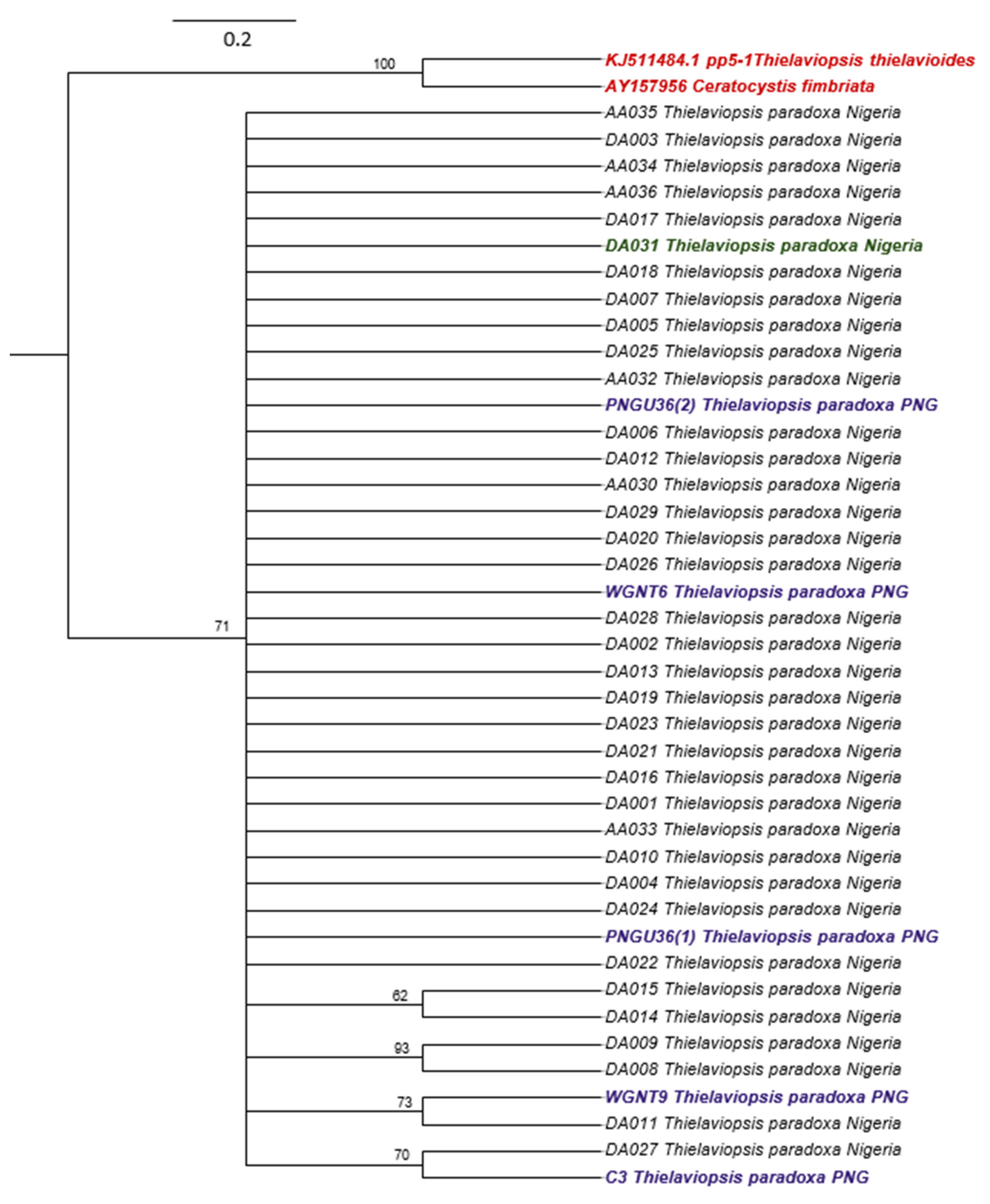
3.2. Phylogenetic Analysis of ITS Sequences

A total of 41 ITS rDNA sequences were generated from the PCR-amplification of the genomic DNA of the isolates with ITS4 primers. The length of the sequences varied between 516 and 528 bp, and the percentage identity shared with T. paradoxa, according to the gene bank, varied between 99.02 and 100%. The 41 ITS sequences of T. paradoxa isolates and the two of Ceratocystis fimbriata and Thielaviopsis thielavioides were used to generate a Bayesian tree. The credibility values (percentage posterior probabilities (PP)) of the branching pattern of the phylogenetic tree are greater than 60% which indicates more than average level of reliability of the expressed relationship among the isolates (Figure 2). There was no definite clustering pattern indicative of origin, hosts, and substrates in the formation of the clades. The isolates formed five clusters: a large clade consisting of 30 Nigerian (oil palm, date fruit and sugar cane) and three PNG oil palm isolates and four small clades consisting of two members each. The clustering pattern evinced by the phylogram failed to address the temperature–isolate relationship observed in this study. While the large clade accommodated different categories of isolates with varied growth responses to temperatures, the four small clades represent members with similar temperature tolerances.

4. Discussion

The behavior of certain fungal species under laboratory conditions tends to correlate with their geographical distribution, seasonal occurrence and evolutional history [57]. This study was designed to evaluate the growth behavior of T. paradoxa isolates under different temperature regimes, characterize available isolate collections using rDNA ITS marker and establish a relationship between the temperature, growth and molecular data of the isolates.
Many studies from different parts of the world have reported various temperatures as optimum for growth of T. paradoxa. However, discrepancies exist in these reports. Our study has clearly demonstrated that a certain level of variability exists among the T. paradoxa isolates with respect to optimum temperature requirement for growth. These differential growth abilities found in some of the tested temperatures varied with source of isolates and geographical distribution. All the temperatures encouraged mycelial growth of the fungal isolates from Nigeria and PNG isolated from oil palm and date palm. While all isolates from PNG and a few from Nigeria grew optimally between 22 °C and 32 °C, the majority had their best growth performance between 25 °C and 32 °C. Meanwhile, some reports documented temperature ranges of 28–32 °C [58,59], 25–31 °C [57] and 25–30 °C [60] as optimal for growth of the fungus. Temperatures between 28 and 32 °C have been linked with high level of pathogenicity of the fungus in sugarcane [47]. We found the growth to be at the lowest rate between 34 °C and 35 °C and no visible mycelial proliferation and spore germination in the sugar cane isolates at these temperatures. Our finding, therefore, differs to some extent from a recent study which reported that T. paradoxa requires a temperature range of 15–35 °C for optimal vegetative growth [61]. Although this present study is only limited to the temperature range tested, it could be imperfect to compare other similar studies that reported temperature 12 °C and 36 °C as minimum and maximum [57] and ranges of 21–22 °C and <10 °C as optimal and inert, respectively, [62] for active mycelial growth and spore germination. Oil palm and date palm isolates performed relatively better compared to sugar cane isolates, with no observable growth in the latter at 35 °C. The oil palm isolate DA029 is obviously the most resilient at this temperature. Such occurrence of higher temperature tolerance in oil palm isolates could be due to a host–pathogen co-evolutionary relationship as these hosts are usually found in tropical regions characterized with high temperature. The physiological information provided in this study might be essential for efficient prediction of the possible effects of climate change on the epidemiology of diseases caused by T. paradoxa [63]. Additionally, the significant variabilities in thermal resistance exhibited by the isolates can influence the choice of strain selection in the formulation of biological control measures, although the relationship between the in vitro temperature–vegetative growth relationship and disease epidemiology must be first investigated [49].
As the findings in this study are based on laboratory observations, the results may not correspond perfectly to what may be observed under natural conditions [64], and further experiments in the presence of the host may be advisable. Nevertheless, our observation that T. paradoxa isolates from oil palm and date palm exhibited some tolerance to higher temperature compared to the sugarcane isolates could provide explanations to an unresolved mechanism that the pathogen attacks certain host species in the presence of others under suitable environmental conditions especially in tropical countries where temperature may reach a favorable level for the fungus to exhibit its pathogenicity. So far, most of the reported cases of the dry basal rot and neck bending disease in West African countries including Nigeria occurred when the atmospheric temperatures were close to the optimum temperature for vegetative growth of the fungus observed in this study. Therefore, regulating the temperature of the environment to an unfavorable level for the survival of the pathogen during the host susceptibility period may be applicable in the prevention of certain diseases such as the neck bending disease of oil palm caused by T. paradoxa. This could be through provision of temporary shading trees such as Gliricidia sepium used for shade management in Theobroma cacao [65]. This system will not only check excessive sunlight radiation which may increase the atmospheric temperature of the plant site but also contribute to soil nitrogen fixation. Soil nitrogen has recently been linked with T. paradoxa disease suppression [30]. The reported cases of T. paradoxa diseases within and across the oil palm growing countries so far vary with respect to severity. This phenomenon could be attributed to variation in temperature in these regions as supported by our observation that all the PNG isolates attained the maximum growth rate at 22 °C compared to only few isolates from Nigeria. Thus, considering such environments where the prevailing temperatures are unfavorable for Thielaviopsis paradoxa disease occurrence for cultivation of any susceptible host species may also be a good management strategy to curtail the disease spread. Even though, other factors such as host population density, genetic constitution and tolerance may also have a significant impact on pathogenicity [15]. This study will be a source of significant data on T. paradoxa disease forecast models for predicting the population dynamics of the pathogen as well as its disease incidence and severity.
The polymorphic nature and massive reference sequences of ITS markers in gene sequence banks have been variously explored in resolving taxonomic issues and gene population studies especially in fungi [42,66]. In this study, information provided by the ITS sequence BLAST on the NCBI interface showed a high-percentage identity of the isolates being shared with the existing T. paradoxa data in the gene bank which further confirms the presence of the pathogen in all the diseased tissues and soil samples where isolations were carried out. This molecular identification thus provides easier and more reliable alternative for identifying the pathogen [67] compared to an in vivo test which is more difficult to accomplish because successful inoculation of the pathogen requires wounding of tissue and aseptic isolation of the pathogen to avoid contamination. The phylogenetic analysis of the ITS sequence data confirmed that all the isolates were T. paradoxa, and limited variation was observed. The genetic closeness is obvious irrespective of origin, host, tissue or substrate from where the fungi were isolated, and the isolates could therefore be inferred to have emerged from a single population which reproduces primarily by vegetative means. Apart from the mode of reproduction, the cultural methods adopted for disease control, which has also been implicated with the spread, may be responsible for the lack of geographical structuring in the phylograms. A recent genetic diversity study carried out in South America using ITS and Random Amplified Polymorphic DNA (RAPD) markers has also suggested T. paradoxa isolates to be a clonal population [47]. However, the present study shows there may be at least five ITS alleles in worldwide populations of this pathogen. This ITS variability should be taken into consideration when designing molecular assays for the diagnosis of the pathogen.
A wider and more robust sampling capturing more sources of the pathogen as well as investigation with more genetic markers could improve understanding of the temperature–isolate growth relationship and the ITS region of T. paradoxa. This is especially important to determining the isolates of the pathogen that are resistant to higher temperature and can withstand climatic change. Additionally, research work to establish the relationship between in vitro growth response of the isolates to temperature changes and pathogenicity as well as disease epidemiology may be warranted to provide insights on development of effective management and control strategies for disease eradication.

Author Contributions

Conceptualization, F.O.A.; Methodology, A.A.A., W.R., A.O. and F.O.A.; Software, A.A.A. and A.A.L.; Validation, A.A.A.; Formal analysis, A.A.A. and A.A.L.; Investigation, Daniel Ofeoritse Esiegbuya and E.J.; Resources, D.O.E. and E.J.; Data curation, A.A.A.; Writing—original draft, A.A.A.; Writing—review and editing, D.O.E., E.J., W.R., A.O. and F.O.A.; Visualization, A.A.A. and A.A.L.; Supervision, F.O.A. and D.O.E.; Project administration, F.O.A.; Funding acquisition, F.O.A. All authors have read and agreed to the published version of the manuscript.

Funding

This research was funded by Ministry of Agriculture and Forestry grant number 4400T-2002 and the APC was funded by Helsinki University Library.

Institutional Review Board Statement

Not applicable.

Informed Consent Statement

Not applicable.

Data Availability Statement

All the data for this research are available on request from the corresponding author. The ITS sequence data can be found at NCBI website (https://www.ncbi.nlm.nih.gov/) (accessed on 24 December 2022).

Acknowledgments

Open access funding provided by University of Helsinki.

Conflicts of Interest

The authors declare no conflict of interest.

References

  1. Keane, P.; Kerr, A. Factors affecting disease development. In Plant Pathogens and Plant Diseases; Brown, J.F., Ogle, H.J., Eds.; Rockvale Publications: Armidale, NSW, Australia, 1997; pp. 287–298. [Google Scholar]
  2. Brader, G.; Compant, S.; Vescio, K.; Mitter, B.; Trognitz, F.; Ma, L.-J.; Sessitsch, A. Ecology and genomic insights into plant-pathogenic and plant-nonpathogenic endophytes. Annu. Rev. Phytopathol. 2017, 55, 61–83. [Google Scholar] [CrossRef] [PubMed]
  3. Agrios, G.N. Plant Pathology; Elsevier: Amsterdam, The Netherlands, 2005; ISBN 978-0-08-047378-9. [Google Scholar]
  4. Kredics, L.; Antal, Z.; Manczinger, L.; Szekeres, A.; Kevei, F.; Nagy, E. Influence of environmental parameters on Trichoderma strains with biocontrol potential. Food Technol. Biotechnol. 2003, 41, 37–42. [Google Scholar]
  5. Almaguer, M.; Rojas, T.I.; Dobal, V.; Batista, A.; Aira, M.J. Effect of temperature on growth and germination of conidia in Curvularia and Bipolaris species isolated from the air. Aerobiologia 2013, 29, 13–20. [Google Scholar] [CrossRef]
  6. Li, Y.; Wadsö, L.; Larsson, L.; Bjurman, J. Correlating two methods of quantifying fungal activity: Heat production by isothermal calorimetry and ergosterol amount by gas chromatography–tandem mass spectrometry. Thermochim. Acta 2007, 458, 77–83. [Google Scholar] [CrossRef]
  7. Freire, S.V.P.; Paiva, L.M.; Lima, E.A.D.L.-A.; Maia, L.C. Morphological, cytological, and cultural aspects of Curvularia pallescens. Rev. Microbiol. 1998, 29, 197–201. [Google Scholar] [CrossRef]
  8. Webster, J.; Weber, R. Introduction to Fungi; Cambridge University Press: Cambridge, UK, 2007; ISBN 1-139-46150-8. [Google Scholar]
  9. Kerry, E. Effects of temperature on growth rates of fungi from subantarctic Macquarie Island and Casey, Antarctica. Polar Biol. 1990, 10, 293–299. [Google Scholar] [CrossRef]
  10. Doohan, F.M.; Brennan, J.; Cooke, B.M. Influence of climatic factors on Fusarium species pathogenic to cereals. Eur. J. Plant Pathol. 2003, 109, 755–768. [Google Scholar] [CrossRef]
  11. Cheng, C.; Gao, X.; Feng, B.; Sheen, J.; Shan, L.; He, P. Plant immune response to pathogens differs with changing temperatures. Nat. Commun. 2013, 4, 2530. [Google Scholar] [CrossRef]
  12. Brasier, C. The effect of light and temperature on reproduction in vitro in two tropical species of Phytophthora. Trans. Br. Mycol. Soc. 1969, 52, 105–113.IN12. [Google Scholar] [CrossRef]
  13. Grove, G.G.; Madden, L.V.; Ellis, M.; Schmitthenner, A. Influence of temperature and wetness duration on infection of immature strawberry fruit by Phytophthora cactorum. Phytopathology 1985, 75, 165–169. [Google Scholar] [CrossRef]
  14. Granke, L.; Hausbeck, M. Effects of temperature, concentration, age, and algaecides on Phytophthora capsici zoospore infectivity. Plant Dis. 2010, 94, 54–60. [Google Scholar] [CrossRef] [PubMed]
  15. Shelley, B.A.; Luster, D.G.; Garrett, W.M.; McMahon, M.B.; Widmer, T.L. Effects of temperature on germination of sporangia, infection and protein secretion by Phytophthora kernoviae. Plant Pathol. 2018, 67, 719–728. [Google Scholar] [CrossRef]
  16. Trecate, L.; Sedláková, B.; Mieslerová, B.; Manstretta, V.; Rossi, V.; Lebeda, A. Effect of temperature on infection and development of powdery mildew on cucumber. Plant Pathol. 2019, 68, 1165–1178. [Google Scholar] [CrossRef]
  17. Sabburg, R.; Obanor, F.; Aitken, E.; Chakraborty, S. Changing fitness of a necrotrophic plant pathogen under increasing temperature. Glob. Chang. Biol. 2015, 21, 3126–3137. [Google Scholar] [CrossRef] [PubMed]
  18. De Beer, Z.W.; Duong, T.; Barnes, I.; Wingfield, B.D.; Wingfield, M.J. Redefining Ceratocystis and allied genera. Stud. Mycol. 2014, 79, 187–219. [Google Scholar] [CrossRef]
  19. Mbenoun, M.; de Beer, Z.W.; Wingfield, M.J.; Wingfield, B.D.; Roux, J. Reconsidering species boundaries in the Ceratocystis paradoxa complex, including a new species from oil palm and cacao in Cameroon. Mycologia 2014, 106, 757–784. [Google Scholar] [CrossRef]
  20. Paulin-Mahady, A.E.; Harrington, T.C.; McNew, D. Phylogenetic and taxonomic evaluation of Chalara, Chalaropsis, and Thielaviopsis anamorphs associated with Ceratocystis. Mycologia 2002, 94, 62–72. [Google Scholar] [CrossRef]
  21. Py, C.; Lacoeuilhe, J.-J.; Teisson, C. The Pineapple: Cultivation and Uses; G.P. Maisonneuve & Larose: Paris, France, 1987; ISBN 978-2-7068-0948-4. [Google Scholar]
  22. Sánchez, V.; Rebolledo, O.; Picaso, R.M.; Cárdenas, E.; Córdova, J.; González, O.; Samuels, G.J. In vitro antagonism of Thielaviopsis paradoxa by Trichoderma longibrachiatum. Mycopathologia 2007, 163, 49–58. [Google Scholar] [CrossRef]
  23. Dade, H.A. Ceratostomella paradoxa, the perfect stage of Thielaviopsis paradoxa (de Seynes) von Höhnel. Trans. Br. Mycol. Soc. 1928, 13, 184–194. [Google Scholar] [CrossRef]
  24. McMartin, A. Pathological conditions affecting growth of sugarcane plant cuttings from Natal. S. Afri. Sugar J. 1937, 21, 353–359. [Google Scholar]
  25. Kile, G. Plant diseases caused by species of Ceratocystis Sensu Stricto and Chalara. In Ceratocystis and Ophiostoma: Taxonomy, Ecology and Pathogenicity; Michael, J.W., Keith, A.S., Joan, F.W., Eds.; APS Press: St. Paul, MN, USA, 1993; Volume 39, pp. 125–131. [Google Scholar]
  26. Abdullah, S.; Asensio, L.; Monfort, E.; Gomez-Vidal, S.; Salinas, J.; Lorca, L.; Jansson, H. Incidence of the two date palm pathogens, Thielaviopsis paradoxa and T. punctulata in soil from date palm plantations in Elx, South-East Spain. J. Plant Prot. Res. 2009, 49, 276–279. [Google Scholar] [CrossRef]
  27. Al-Raisi, Y.M.; B’Chir, M.M.; Al-Mandhari, A.M.; Deadman, M.L.; Gowen, S.R. First report of Ceratocystis radicicola associated with date palm disease in Oman. New Dis. Rep. 2011, 23, 23. [Google Scholar] [CrossRef]
  28. Al-Sadi, A.M. Phylogenetic and population genetic analysis of Ceratocystis radicicola infecting date palms. J. Plant Pathol. 2013, 95, 49–57. [Google Scholar]
  29. Esiegbuya, D.; Ikuenobe, C.; Ghansah, B.; Ojieabu, A. First report of Thielaviopsis ethacetica causing neck bending/inclination of the upper region of oil palms in Nigeria. Agrikul CRI J. 2022, 2, 67–75. [Google Scholar]
  30. Esiegbuya, D.O.; Ikuenobe, C.E.; Ghansah, B.; Ojieabu, A.; Omoregie, K.; Adeh, S.A. Chemotyping of Oil Palm (Elaeis guineensis) Seedlings defense in response to Thielaviopsis ethacetica infection. Vegetos 2022, 1–11. [Google Scholar] [CrossRef]
  31. Johnson, J.A.; Harrington, T.C.; Engelbrecht, C.J.B. Phylogeny and taxonomy of the North American clade of the Ceratocystis fimbriata complex. Mycologia 2005, 97, 1067–1092. [Google Scholar] [CrossRef]
  32. Harrington, T.C. Host specialization and speciation in the American wilt pathogen Ceratocystis fimbriata. Fitopatol. Bras. 2000, 25, 262–263. [Google Scholar]
  33. Engelbrecht, C.J.B.; Harrington, T.C. Intersterility, morphology and taxonomy of Ceratocystis fimbriata on sweet potato, cacao and sycamore. Mycologia 2005, 97, 57–69. [Google Scholar] [CrossRef]
  34. Heath, R.N.; Wingfield, M.J.; Van Wyk, M.; Roux, J. Insect associates of Ceratocystis albifundus and patterns of association in a native savanna ecosystem in South Africa. Environ. Entomol. 2009, 38, 356–364. [Google Scholar] [CrossRef]
  35. Thorpe, D.J.; Harrington, T.C.; Uchida, J.Y. Pathogenicity, Internal Transcribed Spacer-RDNA Variation, and Human Dispersal of Ceratocystis fimbriata on the Family Araceae. Phytopathology 2005, 95, 316–323. [Google Scholar] [CrossRef] [PubMed]
  36. Li, Q.; Harrington, T.C.; McNew, D.; Li, J. Ceratocystis Uchidae, a New Species on Araceae in Hawaii and Fiji. Mycoscience 2017, 58, 398–412. [Google Scholar] [CrossRef]
  37. Baker, C.J.; Harrington, T.C.; Krauss, U.; Alfenas, A.C. Genetic variability and host specialization in the Latin American clade of Ceratocystis fimbriata. Phytopathology 2003, 93, 1274–1284. [Google Scholar] [CrossRef]
  38. Wingfield, B.; Wyk, M.; Roos, H.; Wingfield, M. Ceratocystis: Emerging evidence for discrete generic boundaries. CBS Biodivers. Ser. 2013, 12, 57–64. [Google Scholar]
  39. Fajarningsih, N.D. Internal Transcribed Spacer (ITS) as DNA barcoding to identify fungal species: A review. Squalen Bull. Mar. Fish. Postharvest Biotechnol. 2016, 11, 37–44. [Google Scholar] [CrossRef]
  40. Ali, M.A.L.; Xi, J.; Coudray, A.J. Comparisons of molecular methods in the diagnosis of pathogenic fungi. Int. J. Sci. Technol. Res. 2016, 4, 234–236. [Google Scholar]
  41. Pang, K.-L.; Mitchell, J.I. Molecular approaches for assessing fungal diversity in marine substrata. Bot. Mar. 2005, 48, 332–347. [Google Scholar] [CrossRef]
  42. Samson, R.A.; Houbraken, J.; Thrane, U.; Frisvad, J.C.; Andersen, B. Food and Indoor Fungi; CBS Laboratory Manual Series; CBS-KNAW Fungal Biodiversity Centre: Utrecht, The Netherlands, 2010; Volume 2. [Google Scholar]
  43. Chase, M.W.; Fay, M.F. Barcoding of Plants and Fungi. Science 2009, 325, 682–683. [Google Scholar] [CrossRef]
  44. Das, S.; Deb, B. DNA barcoding of fungi using ribosomal ITS Marker for genetic diversity analysis: A review. Int. J. Pure Appl. Biosci. 2015, 3, 160–167. [Google Scholar]
  45. Seifert, K.A. Progress towards DNA barcoding of fungi. Mol. Ecol. Resour. 2009, 9, 83–89. [Google Scholar] [CrossRef] [PubMed]
  46. Schoch, C.L.; Seifert, K.A.; Huhndorf, S.; Robert, V.; Spouge, J.L.; Levesque, C.A.; Chen, W.; Fungal Barcoding Consortium; Fungal Barcoding Consortium Author List; Bolchacova, E.; et al. Nuclear Ribosomal Internal Transcribed Spacer (ITS) Region as a Universal DNA Barcode Marker for Fungi. Proc. Natl. Acad. Sci. USA 2012, 109, 6241–6246. [Google Scholar] [CrossRef]
  47. Alvarez, E.; Llano, G.A.; Loke, J.B.; Chacon, M.I. Characterization of Thielaviopsis paradoxa Isolates from oil palms in Colombia, Ecuador and Brazil. J. Phytopathol. 2012, 160, 690–700. [Google Scholar] [CrossRef]
  48. Borges, A.F.; de Alcântara Neto, F.; da Silva Matos, K.; Júnior, J.E.A.B.; Júnior, N.S.M.; Moreira, S.I.; de Melo, M.P. Thielaviopsis ethacetica the etiological agent of sugarcane pineapple sett rot disease in Brazil. Trop. Plant Pathol. 2019, 44, 460–467. [Google Scholar] [CrossRef]
  49. Ouedraogo, A.; Fargues, J.; Goettel, M.S.; Lomer, C.J.; Ouedraogo, A.; Fargues, J.; Goettel, M.S.; Lomer, C.J. Effect of temperature on vegetative growth among isolates of Metarhizium anisopliae and M. flavoviride. Mycopathologia 1997, 137, 37–43. [Google Scholar] [CrossRef] [PubMed]
  50. Alwakeel, S.S. Indoor fungal and bacterial contaminations on household environment in Riyadh, Saudi Arabia. Saudi J. Biol. Sci. 2008, 15, 113–119. [Google Scholar]
  51. Terhonen, E.; Marco, T.; Sun, H.; Jalkanen, R.; Kasanen, R.; Vuorinen, M.; Asiegbu, F. The effect of latitude, season and needle age on the mycota of Scots Pine (Pinus sylvestris) in Finland. Silva Fenn. 2011, 45, 301–317. [Google Scholar] [CrossRef]
  52. Thermo Scientific “Phusion” High–Fidelity DNA Polymerase Product Information Sheet. Thermofisher Scientific Inc. 2021. Available online: https://assets.thermofisher.com/TFS-Assets/LSG/manuals/MAN0012393_Phusion_HighFidelity_DNAPolymerase_UG.pdf (accessed on 16 September 2022).
  53. White, T.J.; Bruns, T.; Lee, S.; Taylor, J. Amplification and direct sequencing of fungal ribosomal RNA genes for phylogenetics. In PCR Protocols; Innis, M.A., Gelfand, D.H., Sninsky, J.J., White, T.J., Eds.; Academic Press: San Diego, CA, USA, 1990; pp. 315–322. ISBN 978-0-12-372180-8. [Google Scholar]
  54. Ronquist, F.; Teslenko, M.; Van Der Mark, P.; Ayres, D.L.; Darling, A.; Höhna, S.; Larget, B.; Liu, L.; Suchard, M.A.; Huelsenbeck, J.P. MrBayes 3.2: Efficient Bayesian phylogenetic inference and model choice across a large model space. Syst. Biol. 2012, 61, 539–542. [Google Scholar] [CrossRef]
  55. Miller, M.A.; Pfeiffer, W.; Schwartz, T. Creating the CIPRES Science Gateway for inference of large phylogenetic trees. In Proceedings of the 2010 Gateway Computing Environments Workshop (GCE), New Orleans, LA, USA, 14 November 2010; pp. 1–8. [Google Scholar]
  56. Davis, C.T.; Ebel, G.D.; Lanciotti, R.S.; Brault, A.C.; Guzman, H.; Siirin, M.; Lambert, A.; Parsons, R.E.; Beasley, D.W.C.; Novak, R.J.; et al. Phylogenetic analysis of North American West Nile virus isolates, 2001–2004: Evidence for the emergence of a dominant genotype. Virology 2005, 342, 252–265. [Google Scholar] [CrossRef] [PubMed]
  57. Liu, L.-J.; Cortés-Monllor, A. Effect of temperature and moisture on various aspects of development, growth, and pathogenicity of Thielaviopsis paradoxa from sugarcane in Puerto Rico. J. Agric. Univ. P. R. 1972, 56, 162–170. [Google Scholar] [CrossRef]
  58. Kiryu, T. Studies on the physiological characters of Ceralostomella paradoxa. Rept. Govt. Sugar Expt. Sta. 1939, G, 21–37. [Google Scholar]
  59. Chi, C.C. A preliminary report on the study of pineapple disease of sugarcane in Taiwan. J. Sugarcane Res. 1949, 3, 71–102. [Google Scholar]
  60. Bachiller, N.C.S.J. Effect of environmental factors on the growth and sporulation of Thielaviopsis paradoxa (de Seynes) von Hohnel in culture. Philipp. J. Crop Sci. 1998, 23, 43. [Google Scholar]
  61. Sikder, M.; Ahmmed, M.; Sultana, A.; Alam, N. First report on black spot disease of Phyllanthus emblica L. fruits caused by Thielaviopsis paradoxa in Bangladesh. Int. J. Agril. Res. Innov. Technol. 2021, 10, 38–46. [Google Scholar] [CrossRef]
  62. Hewajulige, I.G.N.; Wijesundera, R.L.C. Thielaviopsis paradoxa, Thielaviopsis basicola (Black Rot, Black Root Rot). In Postharvest Decay; Elsevier: Amsterdam, The Netherlands, 2014; pp. 287–308. ISBN 978-0-12-411552-1. [Google Scholar]
  63. Paterson, R.R.M.; Sariah, M.; Lima, N. How will climate change affect oil palm fungal diseases? Crop Prot. 2013, 46, 113–120. [Google Scholar] [CrossRef]
  64. McConnell, M.; Balci, Y. Fine root dynamics of oak saplings in response to Phytophthora cinnamomi infection under different temperatures and durations. For. Pathol. 2015, 45, 155–164. [Google Scholar] [CrossRef]
  65. Kaba, J.S.; Zerbe, S.; Agnolucci, M.; Scandellari, F.; Abunyewa, A.A.; Giovannetti, M.; Tagliavini, M. Atmospheric nitrogen fixation by Gliricidia trees (Gliricidia sepium (Jacq.) Kunth Ex Walp.) intercropped with cocoa (Theobroma cacao L.). Plant Soil 2019, 435, 323–336. [Google Scholar] [CrossRef]
  66. Op De Beeck, M.; Lievens, B.; Busschaert, P.; Declerck, S.; Vangronsveld, J.; Colpaert, J.V. Comparison and validation of some ITS primer pairs useful for fungal metabarcoding studies. PLoS ONE 2014, 9, e97629. [Google Scholar] [CrossRef]
  67. Saeed, E.E.; Sham, A.; El-Tarabily, K.; Abu Elsamen, F.; Iratni, R.; AbuQamar, S.F. Chemical Control of Black Scorch Disease on Date Palm Caused by the Fungal Pathogen Thielaviopsis punctulata in United Arab Emirates. Plant Dis. 2016, 100, 2370–2376. [Google Scholar] [CrossRef] [PubMed]
Figure 1. A dry basal rot-infected oil palm showing fracture of the lower fronds (A). A cross section of a dry basal rot-infected oil palm showing fruit bunch rotting symptom (B). A pure culture of Thielaviopsis paradoxa (C). Microscopic view of two ascospore types produced by T. paradoxa (D).
Pathogens 12 00727 g001
Figure 2. Phylogenetic tree constructed by Bayesian analysis of ITS rDNA sequences of 41 Thielaviopsis paradoxa isolates and two related species, Ceratocystis fimbriata and T. thielavioides (serving as outgroup; written in red colour). Numbers indicated above the branches are the relevant %PP values for the consensus tree. The scale bar represents the number of nucleotide substitutions per site. Isolates are written with black (oil palm isolates from Nigeria), purple (oil palm isolates from PNG), green (date fruit isolate from Nigeria) and blue (sugarcane isolates from Nigeria) colours.
Pathogens 12 00727 g002
Table 1. Sources of isolates used.
S/NIsolate IDHostSubstrateOriginGPS CoordinateName of CollectorsHambi Reference NumberGenebank Accession
Number
1DA001Oil palmTrunkUhiere, Nigeria6°46′05.9″ N, 6°29′45.5″ ED. O Esiegbuya and A. Ojieabu2737OQ422120
2DA002Oil palmTrunkUhiere, Nigeria6°46′05.9″ N, 6°29′45.5″ ED. O Esiegbuya and A. Ojieabu2736OQ422121
3DA003Oil palmTrunkUhiere, Nigeria6°46′05.9″ N, 6°29′45.5″ ED. O Esiegbuya and A. Ojieabu2738OQ422122
4DA004Oil palmTrunkUhiere, Nigeria6°46′05.9″ N, 6°29′45.5″ ED. O Esiegbuya and A. Ojieabu2739OQ422123
5DA005Oil palmTrunkUhiere, Nigeria6°46′05.9″ N, 6°29′45.5″ ED. O Esiegbuya and A. Ojieabu2740OQ422124
6DA006Oil palmTrunkUhiere, Nigeria6°46′05.9″ N, 6°29′45.5″ ED. O Esiegbuya and A. Ojieabu2741OQ422125
7DA007Oil palmTrunkUhiere, Nigeria6°46′0.01″ N, 5°51′39.5″ ED. O Esiegbuya and A. Ojieabu2742OQ422126
8DA008Oil palmTrunkUhiere, Nigeria6°46′0.01″ N, 5°51′39.5″ ED. O Esiegbuya and A. Ojieabu2743OQ422127
9DA009Oil palmTrunkUhiere, Nigeria6°46′0.01″ N, 5°51′39.5″ ED. O Esiegbuya and A. Ojieabu2744OQ422128
10DA010Oil palmTrunkUhiere, Nigeria6°46′0.01″ N, 5°51′39.5″ ED. O Esiegbuya and A. Ojieabu2745OQ422129
11DA011Oil palmTrunkUhiere, Nigeria6°46′0.01″ N, 5°51′39.5″ ED. O Esiegbuya and A. Ojieabu2746OQ422130
12DA012Oil palmTrunkUhiere, Nigeria6°46′0.01″ N, 5°51′39.5″ ED. O Esiegbuya and A. Ojieabu2747OQ422131
13DA013Oil palmTrunkUhiere, Nigeria6°46′08.7″ N, 5°51′04.3″ ED. O Esiegbuya and A. Ojieabu2748OQ422132
14DA014Oil palmTrunkUhiere, Nigeria6°46′08.7″ N, 5°51′04.3″ ED. O Esiegbuya and A. Ojieabu2749OQ422133
15DA015Oil palmTrunkUhiere, Nigeria6°46′08.7″ N, 5°51′04.3″ ED. O Esiegbuya and A. Ojieabu2750OQ422134
16DA016Oil palmTrunkUhiere, Nigeria6°46′08.7″ N, 5°51′04.3″ ED. O Esiegbuya and A. Ojieabu2751OQ422135
17DA017Oil palmTrunkUhiere, Nigeria6°46′14.5″ N, 5°50′45.1″ ED. O Esiegbuya and A. Ojieabu2752OQ422136
18DA018Oil palmTrunkUhiere, Nigeria6°46′14.5″ N, 5°50′45.1″ ED. O Esiegbuya and A. Ojieabu2753OQ422137
19DA019Oil palmTrunkUhiere, Nigeria6°46′14.5″ N, 5°50′45.1″ ED. O Esiegbuya and A. Ojieabu2754OQ422138
20DA020Oil palmTrunkUhiere, Nigeria6°46′14.5″ N, 5°50′45.1″ ED. O Esiegbuya and A. Ojieabu2755OQ422139
21DA021Oil palmTrunkUhiere, Nigeria6°46′14.5″ N, 5°50′45.1″ ED. O Esiegbuya and A. Ojieabu2756OQ422140
22DA022Oil palmSoilUgbowo, Nigeria6°39′69.3″ N, 5°60′92.02″ ED. O Esiegbuya and A. Ojieabu2757OQ422141
23DA023Oil palmSoilUgbowo, Nigeria6°39′69.3″ N, 5°60′92.02″ ED. O Esiegbuya and A. Ojieabu2758OQ422142
24DA024Oil palmSoilUgbowo, Nigeria6°39′69.3″ N, 5°60′92.02″ ED. O Esiegbuya and A. Ojieabu2759OQ422143
25DA025Oil palmSoilUgbowo, Nigeria6°39′69.3″ N, 5°60′92.02″ ED. O Esiegbuya and A. Ojieabu2760OQ422144
26DA026Oil palmSoilUgbowo, Nigeria6°39′69.3″ N, 5°60′92.02″ ED. O Esiegbuya and A. Ojieabu2761OQ422145
27DA027Oil palmSoilUgbowo, Nigeria6°39′69.3″ N, 5°60′92.02″ ED. O Esiegbuya and A. Ojieabu2762OQ422146
28DA028Oil palmSoilUgbowo, Nigeria6°39′69.3″ N, 5°60′92.02″ ED. O Esiegbuya and A. Ojieabu2763OQ422147
29DA029Oil palmSoilUdo, Nigeria5°28′39.3″ N, 8°05′07.2″ ED. O Esiegbuya and A. Ojieabu2764OQ422148
30AA030Oil palmSoilUdo, Nigeria5°28′39.3″ N, 8°05′07.2″ EA. A Azeez2765OQ422149
31DA031Date palmFruitDutse, Nigeria11°18′79.3″ N, 9°55′17.2″ ED. O Esiegbuya and A. Ojieabu.2735OQ422150
32AA032Sugar caneStemAsaba, Nigeria5°29′21.32″ N, 6°00′14..42″ EA. A Azeez2766OQ422151
33AA033Sugar caneStemAsaba, Nigeria5°29′18.32″ N, 6°00′04.62″ EA. A Azeez2767OQ422152
34AA034Sugar caneStemAsaba, Nigeria5°29′54.37″ N, 6°00′17.64″ EA. A Azeez2734OQ422153
35AA035Sugar caneStemAsaba, Nigeria5°29′33.72″ N, 6°00′28.01″ EA. A Azeez2735OQ422154
36AA036Sugar caneStemAsaba, Nigeria5°29′26.11″ N, 6°00′33.34″ EA. A Azeez2768OQ422155
37C3Oil palmTrunkMBE, Papua New GuineaUnknownB. Ritchie and PNGOPRA team2769OQ422156
38WGNT6Oil palmTrunkMBE, Papua New GuineaUnknownB. Ritchie and PNGOPRA team2771OQ422160
39WGNT9Oil palmTrunkMBE, Papua New GuineaUnknownB. Ritchie and PNGOPRA team2772OQ422159
40PNGU36(1)Oil palmTrunkMBE, Papua New GuineaUnknownB. Ritchie and PNGOPRA team2773OQ422157
41PNGU36(2)Oil palmTrunkMBE, Papua New GuineaUnknownB. Ritchie and PNGOPRA team2774OQ422158
Table 2. Growth rates (cm/day) of Thielaviopsis paradoxa isolates at different temperature three days after inoculation.
Isolate22 °C25 °C30 °C32 °C34 °C35 °C
DA0012.93 a2.93 a2.93 a2.75 abc1.08 BC0.17 OP
DA0022.17 jklmn2.93 a2.93 a2.93 a1.57 tuvwxy0.30 KLMNO
DA0032.93 a2.93 a2.93 a2.93 a1.70 rstuvw0.40 GHIJKLMNO
DA0042.47 bcdef2.93 a2.93 a2.93 a1.73 qrstu0.67 GHIJKLMN
DA0052.75 ab2.93 a2.93 a2.93 a1.63 rstuvwxy0.33 JKLMNO
DA0062.53 cdefgh2.93 a2.93 a2.93 a1.73 rstuv0.67 EFG
DA0072.93 a2.93 a2.93 a2.93 a1.73 rstuv0.70 DEFGH
DA0082.80 abc2.93 a2.93 a2.93 a1.83 pqrst0.50 FGHIJK
DA0092.93 a2.93 a2.93 a2.93 a1.77 qrstu0.37 HIJKLMN
DA0102.32 ghijkl2.93 a2.93 a2.93 a1.07 BC0.23 MNOP
DA0112.93 a2.93 a2.93 a2.93 a1.80 pqrst0.50 FGHIJKLM
DA0122.77 abc2.93 a2.93 a2.93 a1.90 nopqr0.67 EFG
DA0132.93 a2.93 a2.93 a2.93 a1.5 uvwxyz0
DA0142.53 cdefgh2.93 a2.93 a2.93 a1.67 rstuvwx0.67 EFGH
DA0152.8 abc2.93 a2.93 a2.93 a1.67 rstuvwx0.37 IJKLMNO
DA0162.93 a2.93 a2.93 a2.93 a1.70 rstuvw0.27 LMNOP
DA0172.36 fghijk2.93 a2.93 a2.93 a1.70 rstuvw0.30 KLMNO
DA0182.20 ijklm2.93 a2.93 a2.93 a1.67 rstuvwx0.40 GHIJKLMNO
DA0192.16 jklmn2.93 a2.93 a2.93 a2.03mnopq0.27 LMNOP
DA0202.93 a2.93 a2.93 a2.93 a1.87 opqrs0.63 EFGHI
DA0212.40 efghij2.93 a2.93 a2.93 a1.70 rstuvw0.67 EFG
DA0222.30 ghijklm2.93 a2.93 a2.93 a1.77 qrstu0.37 IJKLMNO
DA0232.16 jklmn2.93 a2.93 a2.60 bcdef1.23 zAB0
DA0241.80 pqrst2.93 a2.93 a2.93 a1.43 wxyzA0.43 GHIJKLMNO
DA0252.53 cdefgh2.93 a2.93 a2.93 a1.63 rstuvwxy0.63 EFGHI
DA0262.67 abcde2.93 a2.93 a2.93 a1.70 rstuvw0.47 GHIJKLMN
DA0272.53 cdefg2.93 a2.93 a2.93 a1.77 qrstu0.43 GHIJKLMNO
DA0282.40 efghij2.93 a2.93 a2.93 a1.70 rstuvw0.60 EFGHIJ
DA0292.70 abcd2.93 a2.93 a2.93 a1.60 stuvwxy0.97 BCD
AA0302.12 klmno2.93 a2.93 a2.93 a1.47 vwxyzA0.23 MNOP
DA0312.60 bcdef2.93 a2.93 a2.93 a1.37 yzA0.47 GHIJKLMN
AA0321.85 opqrs2.93 a2.46 defghi1.36 yzA0.20 NOP0
AA0331.63 rstuvwxy2.75 abc2.93 a2.93 a1.67 rstuvwx0
AA0341.47 vwxyzA2.07 lmnop1.47 vwxyzA1.40 xyzA0.77 DEF0
AA0352.26 hijklm2.93 a2.93 a2.93 a1.40 xyzA0
AA0360.53 FGHIJKL1.83 pqrst2.23 ijklm1.20 AB0.87 CDE0
C32.93 a2.93 a2.93 a2.93 a1.03 BCD0.53 FGHIJKL
WGNT62.93 a2.93 a2.93 a2.93 a0.77 DEF0.33 JKLMNO
WGNT92.93 a2.93 a2.93 a2.93 a1.41 xyzA0.57 FGHIJK
PNGU36(1)2.93 a2.93 a2.93 a2.93 a1.47 vwxyzA0.57 FGHIJK
PNGU36(2)2.93 a2.93 a2.93 a2.93 a1.37 yzA0.57 FGHIJK
Growth rate values designated with the same lower and/or upper case letter(s) are not significantly different (p < 0.05) according to Tukey-test.
Disclaimer/Publisher’s Note: The statements, opinions and data contained in all publications are solely those of the individual author(s) and contributor(s) and not of MDPI and/or the editor(s). MDPI and/or the editor(s) disclaim responsibility for any injury to people or property resulting from any ideas, methods, instructions or products referred to in the content.

