Article

The Effect of Patellar Tendon Release on the Characteristics of Patellofemoral Joint Squat Movement: A Simulation Analysis

School of Mechanical and Power Engineering, Henan Polytechnic University, Jiaozuo 454000, China
*
Author to whom correspondence should be addressed.
Appl. Sci. 2019, 9(20), 4301; https://doi.org/10.3390/app9204301
Submission received: 7 August 2019 / Revised: 16 September 2019 / Accepted: 11 October 2019 / Published: 14 October 2019
(This article belongs to the Special Issue Engineering for Surgery)

Abstract

:
Objectives: This paper studies the patellar tendon release’s effect on the movement characteristics of the artificial patellofemoral joint squat to provide reference data for knee joint surgery. Methods: Firstly, the dynamic finite element model of the human knee joint under squatting was established. Secondly, in the above no-release models, the release of 30% of the attachment area at the upper end, the lower end, or both ends of the patellar tendon were conducted, respectively. Then the simulations of all above four models were conducted. Finally, the results of the simulation were compared and analyzed. Results: The simulation results show that, after releasing the patellar tendon (compared with the no-release simulation’s results), the relative flexion, medial-lateral rotation, medial-lateral tilt, and superior-inferior shift of the patella relative to the femur increased; the medial-lateral shift and anterior-posterior shift of the patella relative to the femur decreased. Conclusion: In this paper, the maximum flexion angle of the patella increased after the patellar tendon being released (compared with the no-release model), which indicated that the mobility of knee joint was improved after the patellar tendon release. The simulation data in this paper can provide technical reference for total knee arthroplasty.

1. Introduction

With the application of prostheses in total knee arthroplasty (TKA), post-operative complications concomitantly usually occur [1]. One of these complications, often being ignored, is patella baja [2,3,4]. At the same time, relevant studies [5,6,7,8,9] show that, after TKA, the patellar tendon suffers certain reductions. Other studies [6,10] indicate that the patellar tendon can be shortened, the position of the patella lowered, reducing the knee joint movement and variation. A slightly low patella does not, however, affect the function of the knee joint. Check points during surgery must include patella contracture syndrome [11] and true patella baja [10], which can be treated by release of scar operation, patellar tendon lengthening, and patellar ligament insertion. Patients with pseudo-patella baja and slight joint line up, and those that do not affect knee function, are treated with follow-up observation. However, excessive upward movement of the joint line can result in serious symptoms [12], such as the need for surgery to replace a thin pad [13] and/or patellar ligament release. This paper is for studying the patellar tendon release’s effect on the movement characteristics of the artificial patellofemoral joint squat to provide reference data for knee joint surgery.

2. Materials and Methods

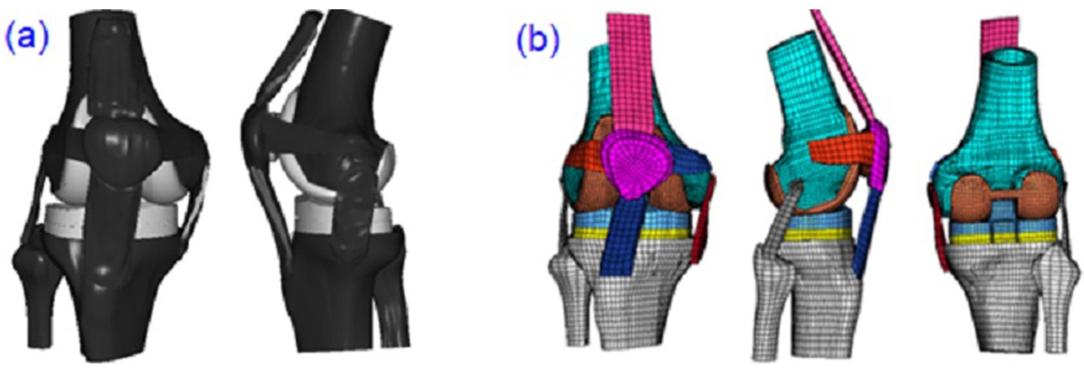
2.1. The Establishment of a Finite Element Model

In this paper, a healthy male volunteer with a height of 173 cm and a weight of 60 kg was scanned by medical instrument computed tomography (CT) in the range of 10 cm above and below the knee joint center. The correlative parameter was set to 120 kVp and 150 mA, with a scanning interval of 1 mm. The volunteer was scanned with magnetic resonance imaging (MRI). MR (GE Sigma HD1.5T, Boston, MA, USA) was used for proton density weighting (PDWI) and chemical shift fat suppression weighted scanning, and the image coordinates of each layer of different weighted phases were consistent. Flux density is 1.5 T, and slide thickness is 1 mm [14]. Based on the CT and MRI images of the healthy volunteer, a three-dimensional geometric anatomical model including all bone tissues and main soft tissues was established (as shown in Figure 1). A three-dimensional solid model of press fit condylar (PFC) femoral prosthesis (PFC sigma, DePuy orthopaedics, Warsaw, IN, USA.) commonly used in TKA was established. Then the total knee replacement operation was simulated, and the prosthesis was assembled on the three-dimensional geometric anatomical model of the knee joint (as shown in Figure 2a). The model of the knee joint was divided into finite element meshes after TKA. In the finite element model of the knee joint, most of the unit types were hexahedrons (c3d8r, an eight-node linear brick element with reduced integration), and very few were pentahedrons (C3D6, a six-node linear triangular prism element) and tetrahedrons (C3D4, a four-node linear tetrahedron element), with a total unit number of 31,767, and 37,031 nodes (as shown in Figure 2b). The volunteer was provided details of the study and signed an informed consent form.
Hypermesh software, version 11.0 (Altair Engineering Corp, MI, USA) was used to simulate the release at ends of the patellar tendon. The width of the patellar tendon at the upper end was 6.84 mm before release (Figure 3a) and 4.788 mm after release (Figure 3b). The height of the patellar tendon at the lower end was 8.09 mm before release (Figure 3c) and 5.633 mm after release (Figure 3d). Figure 3 shows the finite element models before and after release the 30% of the patellar tendon at both ends (clinically, one-third of the central patellar tendon is often transplanted to repair damaged ligaments [15,16,17,18]. Therefore, 30% of the patellar tendon was used as an example). Figure 3a represents the model before release of the patellar tendon at the upper end. Figure 3b represents the model released the patellar tendon at the upper end. Figure 3c represents the model before releasing the patellar tendon at the lower end. Figure 3d represents the model releasing the patellar tendon at the lower end. The establishment of the model was based on the model built by Wang Jian-ping, and the validity of the model was verified by in vitro cadaveric experiments [19].

2.2. The Establishment of a Coordinate System for the Knee Joint’s Motion

The corresponding reference coordinate systems of femur, tibia and patella are established by the following methods: (1) The femur posterior condylar cortical bone was contour-fitted to two laps, with one line through the center of two circles as the X axis of the femur. Parallel with lines of force and through the centerline of the femoral medial condyle represent the Z axis of the femur. Regarding the line, this passes through the center of the femoral medial condyle and perpendicular to the X-axis and Z-axis, as the Y-axis. (2) The intersection point of the Z-axis and the plane of the tibial joint line serves as the origin of the tibial coordinate system, and the femoral coordinate system moves to this point to obtain the tibial coordinate system. (3) The center of the patellar anterior surface moves inward 10 mm, then the center serves as the origin of the patellar coordinate system. The coordinate system of the femur was relocated to the origin, and the patellar coordinate system can be obtained [20]. The bone coordinate systems are shown in Figure 4.

2.3. Loading Conditions and the Setting of Material Properties

For this study, 400 N [19,21] of force was applied to the quadriceps, and the direction of the force was parallel with the femoral shaft, pointing to the starting point of the quadriceps. At the same time, half of the body’s weight (0.5BW) was applied along the force line of the knee joint, that is, a force of 300 N perpendicular to the ground was applied to the center of the femoral head. The boundary conditions were defined: the reference point of the ankle corresponding to the rotation center was fully fixed with six degrees of freedom. The material properties of bone tissue and femoral prosthesis were defined as isotropic and linear elastic, respectively. The material properties of polymer polyethylene pad were defined as non-linear elastic-plastic deformability [22,23]. The material properties of soft tissue were defined as nonlinear elastic [24]. The friction coefficient of polymer polyethylene and cobalt chromium molybdenum material was defined as 0.04 [22]. The penalty function with weighted factor was adopted [25,26]. For the femur, polymer polyethylene pad, tibial plateau, patellar prosthesis, and other soft tissues, seven surface contact pairs were defined in the finite element model after TKA. Moreover, nine surface contact pairs were defined in the finite element model of normal human.

3. Results

The four different knee joint finite element models were imported into the finite element analysis ABAQUS software, version 6.10 (Dassault SIMULIA Corp., Paris, France). Within 0–135 degrees, the data of motion characteristics at different flexion degrees were calculated and analyzed, as shown in Figure 5.

3.1. Medial-Lateral Shift and Flexion of Patella Along the Inner and Outer Axes

Figure 6a shows the medial-lateral shift data of the patellofemoral joint within 0–135 degrees of knee flexion. As the figure shows, from 0 to 30 degrees of flexion, the patella relative to the femur shifted outward. After releasing the patellar tendon, the patella shifted outward greatly. The lateral shift of the patellar tendon released at the lower end was larger than that of release at the upper end. The lateral shift was the largest when both ends were released simultaneously. From 40 degrees to 135 degrees of flexion, the patella relative to the femur shifted inward. After releasing the patellar tendon, the patella medial shift was smaller than that of no-release. The medial shift of the patellar tendon released at different ends was basically balanced. After the upper end of the patellar tendon was released, the maximum of the patella medial shift was 2.62 mm at 110 degrees of flexion, which was 11% lower than that of 2.94 mm at 100 degrees of flexion for the no-release model. After the lower end of the patellar tendon was released, the maximum medial shift of the patella was 2.63 mm at 110 degrees of flexion, and decreased 11% by compared with the no-release model. After both ends of the patellar tendon were released, the maximum medial shift of the patella was 2.61 mm at 110 degrees of flexion, and decreased 11% by compared with the no-release model.
Figure 6b shows the changes of patella flexion before and after the release. At the same flexion degree of the knee, the release of the patellar tendon at the upper end and lower end was compared with the no-release model, the flexion of the patella relative to femur was slightly larger. The patella flexion after both ends release (compared with the no-release model), in the same flexion degree of the knee, showed a smaller degree of patella flexion (within 130–135 degrees of flexion, being slightly larger than the no-release model). At 135 degrees of knee flexion, for the three releasing models: the upper end release, lower end release, and both ends release models, the patellar flexion reached maximums of 92, 92.73, and 93.17 degrees, respectively. Compared with the no-release model of 89 degrees, this shows an increase of 3%, 4%, and 5%, respectively.

3.2. Superior-Inferior Shift and Medial-Lateral Rotation of the Patella Along Its Upper and Lower Axes

As Figure 6c shows, during the knee joint’s squatting movement, the patella relative to the femur shifted downward. The patella inferior shift of no-release model was slightly smaller than that of the upper end release. For the lower end release simulation, the patella inferior shift was slightly smaller within early 0–20 degrees of flexion and slightly larger after the 20-degree flexion than that of the no-release model. Within all flexion degrees, the patella inferior shift of the both ends release was slightly larger than that of no-release model, other than 20-degree flexion. After releasing the upper end of the patellar tendon, the maximum inferior shift of the patella was 43.80 mm at 110-degree flexion, which increased 6% compared with 41.42 mm at 100-degree flexion of the no-release model. For the lower end release model, the maximum inferior shift of the patella was 43.49 mm at 100-degree flexion, which increased 5% compared with the no-release model. After releasing the patellar tendon at both ends, the maximum inferior shift of the patellar was 43.43 mm at 100-degree flexion, which increased 5% compared with the no-release model.
Figure 6d shows the data of medial-lateral rotation. The data of the patella relative to the femur showed lateral rotation first and then medial rotation. Compared to the no-release model and the upper end model, the knee flexion degree of the initial patella medial rotation was greater for the lower end model and both-ends model. Meanwhile, it is shown that the patella medial-rotation for all three release models as larger than that of the no-release model. At 60 degrees of knee flexion, the patella medial-rotation reached the maximum of 5.41 degrees, 5.70 degrees, and 5.72 degrees, respectively, for the upper-end, lower-end and both-ends models, which increased 1%, 7%, and 7%, respectively, compared with the 5.34 degree value of the no-release model.

3.3. Anterior-Posterior Shift and Medial-Lateral Tilt of Patella Along the Anteroposterior Axis

Figure 6e shows the patella translated backward relative to the femur. During the knee joint’s squatting movement, the patella relative to the femur translated backward. Within all flexion degrees, the patella backward translations of all three release models were smaller than that of no-release model, other than the 30-degree flexion, and the patella posterior shift was the least when both ends were released. At 135 degrees of knee flexion, the patella posterior shift reached the maximum values of 61.82 mm, 62.57 mm, and 61.07 mm, respectively, for the upper-end, lower-end, and both-ends models, which decreased 5%, 4%, and 6% compared with the value 64.97 mm of the no-release model.
Figure 6f shows the data of medial-lateral tilt. Before and after patellar tendon release, within all flexion degrees, the patella showed an inward movement pattern relative to the femur other than 30-degree flexion. At the same degree of knee flexion, after the release of the patella tendon, the medial tilt angle of the patella relative to the femur was larger than that of no release. After 80 degrees of knee flexion, when both ends of the patellar tendon were released at the same time, the patella medial tilt angle was the largest. At 135 degrees of knee flexion, the patella medial tilt angle reached the maximum values of 5.51 degrees, 5.01 degrees, and 5.92 degrees, respectively, for the upper-end, lower-end, and both-ends models, which increased 21%, 10%, and 30%, respectively, compared with the 4.55 degree value of the no-release model.

4. Discussion

This paper investigates the patellar tendon release’s effect on the movement characteristics of the artificial patellofemoral joint squat. The simulation results show that, after the patellar tendon being released, respectively, at the upper end, the lower end, and both ends, being compared with the no-release simulation results, the relative flexion, medial-lateral rotation, medial-lateral tilt, and superior-inferior shift of the patella relative to the femur increased, and the medial-lateral shift and anterior-posterior shift of the patella relative to the femur decreased. Different release models have different effects on the squatting motion characteristics of the patellofemoral joint. In this study, the data of patellofemoral joint squatting after TKA of the no-release model was analyzed and compared with those of all three release models and other related research results of the natural knee joint.

4.1. Medial-Lateral Shift and Flexion of the Patella

For the medial-lateral shift of the patella relative to the femur the maximum patella medial-shift of all three release models were balanced, and the maximum medial-shift was decreased compared with that of the no-release model (the maximum reduction was about 11% for the both-ends model). The patella shifted outward first and then inward relative to the femur, which is similar to the related studies [27,28].
For the flexion movement of the patella, relative to the femur, previous studies [27,28] showed that the flexion of patella changes linearly with the flexion of the patellofemoral joint. It is consistent with the results of this study. In this study, the maximum flexion angle of the patella increased after the patellar tendon being released (compared with the no-release model, the maximum of the both-ends model increased by 5%), which indicated that the mobility of the knee joint was improved after the patellar tendon release.

4.2. Superior-Inferior Shift and Medial-Lateral Rotation of Patella

For the superior-inferior shift of the patella, relative to the femur, after the patellar tendon was released in different ends, the patella shifted downward relative to the femur. In the high flexion, the patella was moved up slightly, and the maximum inferior shift (about 43.5 mm at 110 degrees) was increased 5% compared with that of no-release model. The maximum increase occurred in the upper-end release model and the minimum one occurred at both-ends release model. Related studies [27,29] also showed that the patella, with the knee flexion, shifted downward relative to the femur, and the maximum flexion angle of the knee was 110 degrees in their studies (the maximum inferior shift was about 50 mm), which was basically similar to this paper (in this paper, the maximum flexion angle of the knee was 135 degrees, and the maximum inferior shift was 43.8 mm).
For the medial-lateral rotation of the patella, relative to the femur, after the patellar tendon was released at different ends, the motion pattern of the patella relative to the femur was lateral rotation first and then medial rotation, which is consistent with that of the no-release model. For all the above three release models, the maximum medial rotation of the patella increased; among them, the increase of the upper end release was the minimum (1%), and the increase of the both-ends release were the maximum (7%). Heegaard et al. [30] showed that within 0–110 degrees of knee flexion, the patellar rotation pattern changed from medial to lateral rotation, and then lateral rotation within 110–135 degrees of knee flexion. This phenomenon indicates that the patellar motion changes during the high flexion, which might be related to the way of femoral fixation and passive loading.

4.3. Anterior-Posterior Shift and Medial-Lateral Tilt of the Patella

For the anterior-posterior shift of the patella, relative to the femur, the results of Azmy et al.’s [31] in vitro experiments showed that with the flexion of the knee joint, the patella shifted backward; after 30 degree knee flexion, posterior movement rate of the patella increased, and the maximum posterior movement of the patella reached 32 mm. The results of Baldwin et al. [29], based on the experimental dynamic finite element model, also show that the patella continues to shift backward with the knee flexion, and the maximum posterior movement of the patella is about 30 mm. The motion analysis results of patella anterior-posterior shift in this paper are consistent with those of the above studies; with knee flexion, patella continued to shift backward. Meanwhile, the analysis results of this paper gave the data of the posterior shift of the patella during high knee flexion. At 135 degree knee flexion, the posterior shift of the patella after the upper end, the lower end, and both ends of the patellar tendon were released to reach the maximum values of 61.82 mm, 62.57 mm, and 61.07 mm, respectively, which was 5%, 4%, and 6% lower than the no-release values of 64.97 mm.
For the medial-lateral tilt of the patella relative to the femur with the flexion of the knee joint, the patella relative to the femur exhibits an inward motion pattern before and after release. At the same degree of flexion, the medial tilt angle of the patella relative to the femur was larger than that of the no-release model, the increase of the both ends release was the maximum (30%), and the increase of the lower end release was the minimum (10%). The relevant findings [27,29,31,32] also showed that the patella, with the flexion of knee, tilted inward relative to the femur, which is consistent with the results of this study. At the same time, there are different research results for the medial-lateral tilt motion of the patella [33]. The results of Amis et al. [33] showed that the patella continued to tilt outward with knee flexion, which may be related to the loading method that the quadriceps were divided into six bundles. There are differences in related studies, which may be related to the fact that the patella was less constrained by the femoral articular surface in the direction of patella medial-lateral tilt.
Several limitations of the present study should be noted. In this study, only 30% of the patella was released, and others were not analyzed. The finite element model of the human knee joint after TKA was established in this paper, based on the consideration of all bone tissue and the main soft tissue of the knee; although the transverse patellar ligaments, which play an important role in the movement of the patella, are added, the effect of the joint capsule, hamstring, or gastrocnemius on knee joint activity was not considered. In the subsequent analysis of the knee joint, these tissues could be taken into account to make the model more in line with human physiology.
The anterior cruciate ligament (ACL) has the functions of restricting the forward movement of the tibia and controlling the internal and external valgus of the knee [34,35,36]. The ACL is extremely vulnerable during exercise and it is difficult to repair itself. If not treated in time, the patient’s daily activities will be seriously affected [37]. The posterior cruciate ligament (PCL), aside from providing restraint to posterior tibial translation, is reported to have a considerable role in providing rotational stability to the knee [38]. Injuries to the PCL are far less common than injuries to other knee structures, such as the ACL or the menisci [39]. In addition, it has been found that the clinical outcomes of PCL reconstruction are less satisfactory and less predictable relative to those of ACL reconstruction [39]. The medial patellofemoral ligament (MPFL) is one of the most important restraints to lateral displacement of the patella from 0° to 30° of flexion, providing up to 60% of lateral patellofemoral stability [40]. In the last decade, the understanding of the pathoanatomic mechanisms involved in lateral patellar dislocation (LPD) has evolved tremendously and reconstruction of the medial patellofemoral ligament (MPFL), as the most important passive restraint against LPD, has evolved to become an established operative procedure [41]. This paper mainly investigates the patellar tendon release’s effect on the movement characteristics of the artificial patellofemoral joint squat. However, different knee ligaments play different roles. Based on the models used in this study, we will further study the ligaments’ reconstruction and other related issues.

5. Conclusions

This paper investigates the patellar tendon release’s effect on the movement characteristics of the artificial patellofemoral joint squat. The post-TKA patellofemoral joint’s motion characteristics in different patellar tendon release pattern were obtained. Compared with the no-release model, the flexion, medial-lateral rotation, medial-lateral tilt, and superior-inferior shift of the patella relative to the femur increased; and the medial-lateral shift and anterior-posterior shift of the patella relative to the femur decreased. In this paper, the maximum flexion angle of the patella increased after the patellar tendon being released (compared with the no-release model), which indicated that the mobility of knee joint was improved after the patellar tendon release. Different release models have different effects on the squatting motion characteristics of patellofemoral joint. In this paper, the simulation results can be used to moderately release the ends of the patellar tendon in the TKA surgery, in order to improve the motility of the patella relative to the femur. The results of this paper can provide a reference for the study of the pathology and rehabilitation of the knee joint, as well as related operations.

Author Contributions

Conceptualization: J.W.; data curation: L.F. and Y.L.; investigation: J.W. and L.F.; methodology: J.W. and Y.L.; resources: J.W.; software: J.W., Y.Y., and D.G.; supervision: J.W. and Y.L.; validation: Y.Y., D.G., and S.W.; writing—original draft: J.W.; writing—review and editing: Y.Y., D.G., S.W. and Y.L.

Funding

This research was funded by National Natural Science Foundation of China, grant number 31370999.

Conflicts of Interest

The authors declare no conflict of interest.

References

  1. Adravanti, P.; Tecame, A.; De Girolamo, L.; Ampollini, A.; Papalia, R. Patella Resurfacing in Total Knee Arthroplasty: A Series of 1280 Patients at Midterm Follow-Up. J. Arthroplast. 2018, 33, 696–699. [Google Scholar] [CrossRef] [PubMed]
  2. Chonko, D.J.; Lombardi, A.V.; Berend, K.R. Patella baja and total knee arthroplasty (TKA): Etiology, diagnosis, and management. Surg. Technol. Int. 2004, 12, 231–238. [Google Scholar] [PubMed]
  3. Ali, S.A.; Helmer, R.; Terk, M.R. Patella Alta: Lack of Correlation Between Patellotrochlear Cartilage Congruence and Commonly Used Patellar Height Ratios. J. Am. Roentgenol. 2009, 193, 1361–1366. [Google Scholar] [CrossRef] [PubMed]
  4. Nakagawa, S.; Arai, Y.; Inoue, H.; Atsumi, S.; Ichimaru, S.; Ikoma, K.; Fujiwara, H.; Kubo, T. Two Patients with Osteochondral Injury of the Weight-Bearing Portion of the Lateral Femoral Condyle Associated with Lateral Dislocation of the Patella. Case Rep. Orthop. 2014, 2014, 876410. [Google Scholar] [CrossRef] [PubMed]
  5. Chougule, S.S.; Stefanakis, G.; Stefan, S.C.; Rudra, S.; Tselentakis, G. Effects of fat pad excision on length of the patellar tendon after total knee replacement. J. Orthop. 2015, 12, 197–204. [Google Scholar] [CrossRef] [Green Version]
  6. Weale, A.E.; Murray, D.W.; Newman, J.H.; Ackroyd, C.E. The length of the patellar tendon after unicompartmental and total knee replacement. Bone Jt. J. 1999, 81, 790–795. [Google Scholar] [CrossRef]
  7. Davies, G.S.; Van Duren, B.; Shorthose, M.; Roberts, P.G.; Morley, J.R.; Monk, A.P. Changes in patella tendon length over 5 years after different types of knee arthroplasty. Knee Surg. Sports Traumatol. Arthrosc. 2016, 24, 3029–3035. [Google Scholar] [CrossRef]
  8. FlORen, M.; Davis, J.; Peterson MG, E.; Laskin, R.S. A mini-midvastus capsular approach with patellar displacement decreases the prevalence of patella baja. J. Arthroplast. 2007, 22, 51–57. [Google Scholar] [CrossRef]
  9. Meneghini, R.M.; Ritter, M.A.; Pierson, J.L.; Meding, J.B.; Berend, M.E.; Faris, P.M. The effect of the Insall-Salvati ratio on outcome after total knee arthroplasty. J. Arthroplast. 2006, 21, 116–120. [Google Scholar] [CrossRef]
  10. Kazemi, S.M.; Daftari, B.L.; Eajazi, A.; Miniator Sajadi, M.R.; Okhovatpoor, M.A.; Farhang, Z.R. Pseudo-patella baja after total knee arthroplasty. Med. Sci. Monit. Int. Med. J. Exp. Clin. Res. 2011, 17, CR292. [Google Scholar] [CrossRef]
  11. Krishnan, S.G.; Steadman, J.R.; Millett, P.J.; Hydeman, K.; Close, M. Lysis of Pretibial Patellar Tendon Adhesions (Anterior Interval Release) to Treat Anterior Knee Pain After ACL Reconstruction; Springer: London, UK, 2006. [Google Scholar]
  12. Seo, J.G.; Moon, Y.W.; Kim, S.M.; Park, S.H.; Lee, B.H.; Chang, M.J.; Jo, B.C. Prevention of pseudo-patella baja during total knee arthroplasty. Knee Surg. Sports Traumatol. Arthrosc. 2015, 23, 3601–3606. [Google Scholar] [CrossRef] [PubMed]
  13. Bugelli, G.; Ascione, F.; Cazzella, N.; Franceschetti, E.; Franceschi, F.; Dell’Osso, G.; Giannotti, S. Pseudo-patella baja: A minor yet frequent complication of total knee arthroplasty. Knee Surg. Sports Traumatol. Arthrosc. 2017, 26, 1831–1837. [Google Scholar] [CrossRef] [PubMed]
  14. Wang, J.-P.; Qian, L.-W.; Wang, C.-T. Simulation of Geometric Anatomy Model of Human Knee Joint. J. Syst. Simul. 2009, 21, 2806–2809. [Google Scholar]
  15. Matava, M.J.; Hutton, W.C. A biomechanical comparison between the central one-third patellar tendon and the residual tendon. Br. J. Sports Med. 1995, 29, 178–184. [Google Scholar] [CrossRef]
  16. Mansson, O.; Sernert, N.; Rostgard-Christensen, L.; Kartus, J. Long-term clinical and radiographic results after delayed anterior cruciate ligament reconstruction in adolescents. Am. J. Sports Med. 2015, 43, 138–145. [Google Scholar] [CrossRef]
  17. George, D.S.L.M.; Pampolha, A.G.M.; Orlando Junior, N. Functional results from reconstruction of the anterior cruciate ligament using the central third of the patellar ligament and flexor tendons. Rev. Bras. Ortop. (Engl. Ed.) 2015, 50, 705–711. [Google Scholar]
  18. Haviv, B.; Yassin, M.; Rath, E.; Bronak, S. Prevalence and clinical implications of nerve injury during bone patellar tendon bone harvesting for anterior cruciate ligament reconstruction. J. Orthop. Surg. 2017, 25, 230949901668498. [Google Scholar] [CrossRef]
  19. Wang, J.; Tao, K.; Li, H.; Wang, C. Modelling and analysis on biomechanical dynamic characteristics of knee flexion movement under squatting. Sci. World J. 2014, 2014, 321080. [Google Scholar]
  20. Grood, E.S.; Suntay, W.J. A joint coordinate system for the clinical description of three-dimensional motions: Application to the knee. J. Biomech. Eng. 1983, 105, 136–144. [Google Scholar] [CrossRef]
  21. Li, G.; Most, E.; Otterberg, E.; Sabbag, K.; Zayontz, S.; Johnson, T.; Rubash, H. Biomechanics of Posterior-Substituting Total Knee Arthroplasty: An In Vitro Study. Clin. Orthop. Relat. Res. 2002, 404, 214–225. [Google Scholar] [CrossRef]
  22. Godest, A.C.; Beaugonin, M.; Haug, E.; Taylor, M. Simulation of a knee joint replacement during a gait cycle using explicit finite element analysis. J. Biomech. 2002, 35, 267–275. [Google Scholar] [CrossRef]
  23. Taylor, M.; Barrett, D.S. Explicit finite element simulation of eccentric loading in total knee replacement. Clin. Orthop. Relat. Rec 2003, 414, 162–171. [Google Scholar] [CrossRef] [PubMed]
  24. Pena, E.; Calvo, B.; Martinez, M.A.; Doblare, M. A three-dimensional finite element analysis of the combined behavior of ligaments and menisci in the healthy human knee joint. J. Biomech. 2006, 39, 1686–1701. [Google Scholar] [CrossRef] [PubMed]
  25. Halloran, J.P.; Petrella, A.J.; Rullkoetter, P.J. Explicit finite element modeling of total knee replacement mechanics. J. Biomech. 2005, 38, 323–331. [Google Scholar] [CrossRef]
  26. Bustince, H.; Fernandez, J.; Burillo, P. Penalty Function in Optimization Problems: A Review of Recent Developments; Springer: Cham, Switzerland, 2018. [Google Scholar]
  27. Tang, T.S.; MacIntyre, N.J.; Gill, H.S.; Fellows, R.A.; Hill, N.A.; Wilson, D.R. Accurate assessment of patellar tracking using fiducial and intensity-based fluoroscopic techniques. Med. Image Anal. 2004, 8, 343–351. [Google Scholar] [CrossRef] [PubMed]
  28. Fellows, R.A.; Hill, N.A.; Gill, H.S.; MacIntyre, N.J.; Harrison, M.M.; Ellis, R.E.; Wilson, D.R. Magnetic resonance imaging for in vivo assessment of three-dimensional patellar tracking. J. Biomech. 2005, 38, 1643–1652. [Google Scholar] [CrossRef]
  29. Baldwin, M.A.; Clary, C.; Maletsky, L.P.; Rullkoetter, P.J. Verification of predicted specimen-specific natural and implanted patellofemoral kinematics during simulated deep knee bend. J. Biomech. 2009, 42, 2341–2348. [Google Scholar] [CrossRef]
  30. Heegaard, J.; Leyvraz, P.F.; Curnier, A.; Rakotomanana, L.; Huiskes, R. The biomechanics of the human patella during passive knee flexion. J. Biomech. 1995, 28, 1265–1279. [Google Scholar] [CrossRef] [Green Version]
  31. Azmy, C.; Guerard, S.; Bonnet, X.; Gabrielli, F.; Skalli, W. EOS (R) orthopaedic imaging system to study patellofemoral kinematics: Assessment of uncertainty. Orthop. Traumatol. Surg. Res. 2010, 96, 28–36. [Google Scholar] [CrossRef]
  32. McWalter, E.J.; Hunter, D.J.; Wilson, D.R. The effect of load magnitude on three-dimensional patellar kinematics in vivo. J. Biomech. 2010, 43, 1890–1897. [Google Scholar] [CrossRef]
  33. Amis, A.A.; Senavongse, W.; Bull, A.M.J. Patellofemoral kinematics during knee flexion-extension: An in vitro study. J. Orthop. Res. 2006, 24, 2201–2211. [Google Scholar] [CrossRef] [PubMed]
  34. Testa, R. Knee rotational laxity and proprioceptive function 2A years after partial ACL reconstruction. Knee Surg. Sports Traumatol. Arthrosc. 2012, 20, 762–766. [Google Scholar]
  35. Gokeler, A.; Benjaminse, A.; Hewett, T.E.; Lephart, S.M.; Engebretsen, L.; Ageberg, E.; Dijkstra, P.U. Proprioceptive deficits after ACL injury: Are they clinically relevant? Br. J. Sports Med. 2012, 46, 180–192. [Google Scholar] [CrossRef] [PubMed]
  36. Smith, C.K.; Howell, S.M.; Hull, M.L. Anterior laxity, slippage, and recovery of function in the first year after tibialis allograft anterior cruciate ligament reconstruction. Am. J. Sports Med. 2011, 39, 78. [Google Scholar] [CrossRef]
  37. Tianqu, H. Finite Element Analysis of the Influence of Femoral Tunnel Locating Points on the Isometricity of Grafts in Anterior Cruciate Ligament Reconstruction. Master’s Thesis, Central South University, Changsha, China, 24 May 2014. [Google Scholar]
  38. Laprade, C.M.; Civitarese, D.M.; Rasmussen, M.T.; Laprade, R.F. Emerging Updates on the Posterior Cruciate Ligament: A Review of the Current Literature. Am. J. Sports Med. 2015, 0363546515572770. [Google Scholar] [CrossRef]
  39. Narvy, S.J.; Pearl, M.; Vrla, M.; Yi, A.; Hatch, G.F.R. Anatomy of the Femoral Footprint of the Posterior Cruciate Ligament: A Systematic Review. Arthrosc. J. Arthrosc. Relat. Surg. 2015, 31, 345–354. [Google Scholar] [CrossRef]
  40. Tucker, A.; Mcmahon, S.; Mcardle, B.; Rutherford, B.; Acton, D. Synthetic versus autologous reconstruction (Syn-VAR) of the medial patellofemoral ligament: A study protocol for a randomised controlled trial. Trials 2018, 19, 268. [Google Scholar] [CrossRef]
  41. Balcarek, P.; Rehn, S.; Howells, N.R.; Eldridge, J.D.; Kita, K.; Dejour, D.; Friede, T. Results of medial patellofemoral ligament reconstruction compared with trochleoplasty plus individual extensor apparatus balancing in patellar instability caused by severe trochlear dysplasia: A systematic review and meta-analysis. Knee Surg. Sports Traumatol. Arthrosc. 2016, 25, 3869–3877. [Google Scholar] [CrossRef]
Figure 1. (a) Contour tracing of bone from CT; (b) MRI image; (c) the geometric anatomy model of human knee.
Applsci 09 04301 g001
Figure 2. (a) 3D geometric anatomical model of knee joint after TKA; (b) finite element model of knee joint after TKA.
Applsci 09 04301 g002
Figure 3. The finite element model of patellar tendon: (a) the upper before release; (b) the upper released; (c) the lower before release; (d) the lower released.
Applsci 09 04301 g003
Figure 4. Coordinate system of bone. (a) femur; (b) tibia; (c) patella.
Applsci 09 04301 g004
Figure 5. A sample of simulation results after TKA. Flexion degrees: (a) 0°; (b) 30°; (c) 90°; (d) 135°.
Applsci 09 04301 g005
Figure 6. Patellofemoral movement data at different flexion degrees before and after the patellar tendon was released. (a) Medial-lateral shift; (b) flexion of patella relative to femur; (c) superior-inferior shift; (d) medial-lateral rotation; (e) anterior-posterior shift; (f) medial-lateral tilt.
Applsci 09 04301 g006

