Article

Endophytic Fungi Isolated from Plants Growing in Central Andean Precordillera of Chile with Antifungal Activity against Botrytis cinerea

Faculty of Chemistry and Biology, University of Santiago of Chile, Bernardo O’Higgins Avenue 3363, Estación Central, Santiago 9160000, Chile
*
Authors to whom correspondence should be addressed.
J. Fungi 2020, 6(3), 149; https://doi.org/10.3390/jof6030149
Submission received: 20 June 2020 / Revised: 7 August 2020 / Accepted: 12 August 2020 / Published: 26 August 2020

Abstract

:
Botrytis cinerea is an important phytopathogenic fungus affecting the fruit production around the world. This fungus is controlled mainly by using synthetic fungicides, but many resistant isolates have been selected by the indiscriminate use of fungicides. Endophytic fungi or secondary metabolites obtained from them become an alternative method of control for this fungus. The aim of this work was to identify endophytic fungi with antifungal activity against the plant pathogenic fungus B. cinerea isolated from plants from Central Andean Precordillera of Chile. Three endophytic fungi (Ac1, Lc1 and Ec1) with antifungal activity against B. cinerea were isolated from native and endemic plants growing in Central Andean Precordillera of Chile. The isolates Lc1 (isolated from Lithraea caustica) and Ac1 (isolated from Acacia caven) were identified as Alternaria spp. and the isolate Ec1 (isolated from Echinopsis chiloensis) was identified as Aureobasidium spp. The isolated endophytic fungi would inhibit B. cinerea through the secretion of diffusible and volatile compounds affecting the mycelial growth, conidia germination and interestingly, it was also shown that the volatile compounds produced by the three isolated endophytic fungi suppressed the sporulation of B. cinerea.

1. Introduction

Botrytis cinerea is a filamentous necrotrophic fungus that infects more than 250 plant species around the world, and it is the causal agent of “gray mould” disease [1]. Gray mold causes great economic losses in fruit production due to decay of crops and it produces biochemical changes that alter the organoleptic quality of fruits [1]. The estimated lost ranging from USD 10 billion to USD 100 billion [1]. Traditional control of this disease is realized through cultural management, elimination of remains of infected plants, and by the use of synthetic fungicides and biofungicides, including biological control [2]. Currently, the management of the disease is affected by the selection of B. cinerea isolates resistant to fungicides [3]. In a study realized in table grapes of Thompson Seedless variety in the Central Valley of Chile, it was demonstrated that only 1% of the B. cinerea isolates was sensitive to all of fungicide families [4]. Therefore, new alternatives for the control of this disease are necessary. An alternative is the use of endophytic fungi or secondary metabolites obtained from them [5]. Endophytic fungi are organisms that can live inside plants without causing symptoms of disease in their host. These fungi can have a great impact on host plants, because they can increase the plant tolerance to biotic and abiotic stress [6]. Plant tolerance to biotic stress can be increased because some endophytic fungi produce compounds with antimicrobial activity [5]. Numerous endophytic fungi, among them, species of the genera Aspergillus, Aureobasidium, Alternaria, Phoma, Fusarium, Trichoderma, Penicillium, that significantly suppress the growth of B. cinerea have been isolated from different plant species [7]. There are different criteria for selection of plants as a source of isolation of fungal endophytes with a considerable inhibition effect to B. cinerea growth; one of them is to look for endemic plants, because these plant species are distributed in an exclusive reduced geographical location; therefore, it is expected that these plants possess specific endophytes [8]. Central Andean Precordillera of Chile has a Mediterranean climate called “Chilean scrub”. This climate is characterized by rainy winters and long dry summers. Mediterranean climate is only present in five regions of the world [9,10]. Therefore, plants growing in these regions encounter in conditions of abiotic stress [11].
The aim of this work was to identify endophytic fungi obtained from different tissue types of various plant families from Central Andean Precordillera of Chile producing volatile or diffusible compounds with antifungal activity against the plant pathogenic fungus B. cinerea.

2. Materials and Methods

2.1. Collection of Plant Material

Samples of different tissue types (stem, leaves or flowers) from healthy plants growing in El Ingenio, Cajón del Maipo in central Andean Precordillera of Chile (Latitude: −33°46′8.76″; Longitude: −70°16′35.03″ and Altitude 1200 m) was collected in August 2017. The study material was obtained from endemic and native species of different plant families (Calceolaria polifolia (Cp), Lithraea caustica (Lc), Colliguaja odorifera (Co), Echinopsis chiloensis (Ec), Tropaeolum tricolor (Tt), Puya chilensis (Pc) and Acacia caven (Ac)). Stems, leaves, or flowers were excised from one plant of each plant species with a sterile scalpel and placed in sterile Falcon tubes at 4 °C until use. In the case of E. chiloensis, stem pieces of 5 cm were collected for endophyte isolation.

2.2. Isolation of Endophytic Fungi

Stems, leaves, and flowers were surface sterilized by immersing them in 0.1% Tween 20 for 30 s followed by treatment with 2% sodium hypochlorite for 5 min; they were washed in sterile water for 5 min. Subsequently, they were immersed in 70% ethanol for 5 min and then washed with sterile distilled water [12]. The stems, leaves, and flowers were cut into small pieces of 0.5 cm approximately and these sterilized tissue segments were inoculated on potato dextrose agar plates (PDA) (Difco™) supplemented with 50 μg/mL Kanamycin Sulfate (PanReac AppliChem, Darmstadt, Germany). The pieces of tissues of each plant were inoculated in triplicate in a Petri dish. A total of three plates were utilized by tissue. The plates were incubated for fourteen days in dark at 22 °C. Then, the fungi that emerged from the vegetal fragments were transferred to new plates with PDA medium and incubated at 22 °C. Then, each fungal species was subcultured several times to obtain a pure culture [12].
As surface sterilization control, 100 μL of water from the last wash was inoculated in a PDA plates. The plates were incubated at 22 °C. The absence of fungal growth on the culture media indicated that the sterilization of the plant segment surface was effective to eliminate the surface fungi [12]. The control was realized in triplicate.
The growth type of the obtained fungi was analyzed macroscopically by observing the colony on PDA medium and microscopically by observation of vegetative structures such as hyphae or yeasts by light microscopy (Optical Motic microscope, Hong Kong, China).

2.3. Antifungal Activity Determination against B. cinerea

In this study, the G29 isolate of B. cinerea was used. This isolate was originally obtained from a naturally infected grape (Vitis vinifera) [13].

2.3.1. Dual Confrontation Assay

To evaluate the inhibitory effect on growth of B. cinerea of the endophytic fungi, dual confrontation assays were performed in PDA Petri dishes supplemented with 50 μg/mL Kanamycin Sulfate [14]. For the assays, mycelium discs (0.5 cm of diameter) of the endophytic fungus and B. cinerea were inoculated in a Petri dish. The mycelium discs of both fungi were placed 6 cm apart on culture medium in opposite sides of the plate. In the control, B. cinerea mycelium was inoculated in both sides of the plates [15]. After 10 days of incubation in the dark at 22 °C, the radial mycelial growth of B. cinerea towards the endophytic fungus (Ri) and radial mycelial growth of B. cinerea on a control plate (Rc) were measured and the percentage of inhibition was calculated according to the formula (Rc−Ri)/Rc × 100. The assays were performed four times with three replicates.

2.3.2. Effect of Volatile Compounds on the Growth of B. cinerea

The effect of volatile compounds produced by the endophytic fungi on the growth of B. cinerea was examined using Sandwiched Petri plates assay; this culture technique prevents any direct contact between endophytic fungi and B. cinerea [16]. Endophyte fungi were inoculated in Petri plates containing PDA and were allowed to grow for 7 days at 22 °C. Then, B. cinerea was inoculated into another Petri plate containing PDA, and the base of Petri dish containing the endophytic fungus was put on top of the base of Petri dish containing B. cinerea. This system was completely sealed with parafilm to avoid the release of the volatile compounds. The effect on the volatile compounds on B. cinerea mycelial growth was evaluated by comparing the radial growth of the pathogen with the control corresponding to B. cinerea inoculated in the same conditions, but in the absence of the endophyte fungi. Inhibition percent was established as (R1 − R2/R1) × 100, where R1 is the distance covered by the pathogen in the control treatments, and R2 is the distance covered by the pathogen in the presence of the endophyte. All experiments were carried out in triplicate.

2.3.3. Effect of Volatile Compounds on the Sporulation of B. cinerea

To determine if the volatile compounds secreted by endophytic fungi had an effect on sporulation of B. cinerea, the isolate Ec1 (isolated from E. chiloensis) was used. For this purpose, B. cinerea and the endophytic fungus were grown in Petri dishes containing PDA as it was described in Section 2.3.2. As negative control, only B. cinerea was used. After 7 days of incubation in the dark at 22 °C, B. cinerea conidia were removed from the Petri dishes by adding 3 mL of 0.9% NaCl and conidia were counted in a Neubahuer chamber. In this experiment, three independent experiments were performed, each one in triplicate.

2.3.4. Effect of Diffusible Compounds on Growth and Germination of B. cinerea

Additionally, the effect of diffusible compounds produced by the isolate Ec1 on B. cinerea was analyzed by using the diffusion test assay or a cell-free filtrate.
For the diffusion test assay, the isolate Ec1 was inoculated in a Petri dish with PDA on a cellophane layer and incubated for 3 days at 22 °C. After this incubation time, cellophane containing the mycelium of the endophytic fungus was removed from the Petri dish and a 5-mm disc of B. cinerea mycelium was inoculated at the center of this Petri dish [17]. The culture was incubated for 5 days at 22 °C and radial growth was recorded and compared with that of the control. As control, a disc of B. cinerea mycelium from a 3-day old culture was inoculated in fresh culture medium. The inhibition of mycelial growth was evaluated by measuring the radial growth of B. cinerea. The percentage of inhibition was calculated as follows: % Inhibition = (R1 − R2) × 100/R1, where R1 is the growth distance of the B. cinerea in the control, and R2 corresponds to the distance of the growth of the pathogen in the diffusion test. The test was carried out in triplicate.
On the other hand, the effect of cell-free filtrates obtained from isolate Ec1 on germination of B. cinerea conidia was evaluated. To obtain cell-free filtrates, the isolate Ec1 was inoculated into a 250 mL flask containing 100 mL of PDB (Potato Dextrose Broth). The culture was incubated at 22 °C with agitation (140 rpm) for 12 days. The culture was centrifuged at 5200× g for 30 min at 4 °C to collect the supernatant and, the supernatant was filtered with a 0.22-µm filter [18]. To determine the effect of the cell-free filtrate on conidial germination, the assay was executed as described [19]. A conidial suspension of B. cinerea at a concentration of 1 × 10³ conidia mL−1 was inoculated in 24-well plates containing PDB medium and the cell-free filtrate at 50 or 100% supplemented with 1% glucose. As germination control, the suspension of conidia of B. cinerea was inoculated in PDB medium. The cultures were incubated for 4 h at 22 °C. After this time of incubation, percentage of germination was determined as cg/ct × 100, where cg are the germinated conidia and ct are the total conidia. The conidia were considered germinated when germ tube length was equal or greater than conidial diameter. The rate of germ tube growth (measured in µm/h) after 4 h of incubation was also determined. In both experiments at least 100 conidia were photographed for each concentration of cell-free filtrate and for the control. The images were captured with a digital camera Moticam 2300 (Hong Kong, China) coupled to a light Motic Microscope and the images were analyzed with Motic Image Plus 2.0 software. These experiments were carried out in triplicate.
To detect the diffusible compounds with antifungal activity produced by the isolate Ec1, thin-layer chromatography (TLC) coupled with bioautographic was performed [20]. For this purpose, the isolate Ec1 was inoculated in PDB medium and was incubated for 12 days at 22 °C with agitation (140 rpm). Then, the culture was centrifuged at 5200× g for 30 min at 4 °C to separate the fungal biomass, the pellet was discarded and the supernatant was filtered with a 0.22-µm filter. Then, filtrated supernatant was subjected to liquid–liquid partition with ethyl acetate three times [20]. As control, an ethyl acetate extraction was carried out from PDB culture medium without inoculation with the fungus. The extracts were dried using a rotary evaporator.
Subsequently, compounds of the extracts were separated on aluminum-backed TLC plates (silica gel 60 F254, Merck, Santiago, Chile). The TLC plates were developed under saturated conditions with the system chloroform/methanol (95:5) as eluent. Compounds on the plates were visualized under UV light at 254 nm. To detect the compounds with antifungal activity on the TLC plate, a bioautography was conducted [20]. For this, the developed plates were dried to eliminate the solvent. The dry plate was placed in a Petri dish on PDA medium supplement with 50 µg/mL Kanamycin Sulfate. Then, 3 mL of a conidial suspension of B. cinerea (1 × 10⁶ conidia/mL) was poured on the plate. Cultures were incubated for 7 days at 22 °C.

2.4. Identification of Endophytic Fungi

For the identification of the fungi, morphological and molecular techniques were used. For morphological identification, microcultures were performed. Sterile pieces 2 cm2 of PDA were placed in a slide and were inoculated with the isolated endophytic fungi and then the PDA pieces were covered with a coverslip. Then, the slides containing the inoculated solid culture medium were put inside sterile Petri dishes containing filter paper with glycerol 30% to maintain humidity. The fungal cultures were incubated for 7 to 10 days at 22 °C until the formation of the aerial structures. The visualization of the structures was performed using a light Motic microscope Hong Kong, China.
For the identification at genus level of the endophyte fungi, comparison of the ITS regions of the rDNA was used. For this, mycelia obtained from axenic cultures were inoculated on a cellophane paper layer placed on Petri dishes with PDA. The culture was incubated for 3 days at 22 °C. Then, the fungal mycelium was recovered and the genomic DNA was obtained using the CTAB method [21] with some modifications. For this, approximately 200 mg of fungal tissue was deposited in a falcon tube, then 800 µL of CTAB buffer (3% CTAB, 100 mM Tris-HCl pH 8.0, 1.4 M NaCl, 20 mM EDTA) was added to the tube and then the tissue was ground with glass balls of 4 mm diameter and vortex for 3 min. The ground tissue was incubated at 60 °C for 30 min. Then, the cellular debris was centrifuged at 5000× g for 10 min at room temperature and the supernatant was recovered. Subsequently, an equal volume of chloroform: isoamyl alcohol (24: 1) was added. The mixture was centrifuged at 7000× g for 10 min at 4 °C and the aqueous phase was recovered, an equal volume of cold isopropanol was added and, the solution was incubated at −20 °C for 2 h. Then, 500 µL of absolute ethanol was added to the pellet and incubated for 2 h at −20 °C, the suspension was centrifuged at 7000× g for 10 min at 4 °C and the supernatant was discarded. Finally, 50 µL of DNAse free H2O was added and the DNA was stored at −20 °C until use.
The ITS sequences were amplified using the ITS-1 and ITS-4 primers forward primer was ITS-1 5′-TCCGTAGGTGAACCTGCGG-3′and reverse primer 5′-TCCTCCGCTTATTGATATGC-3′ [22]. The PCR reaction was performed in a volume of 50 μL containing; 2 μL of genomic DNA, 1 μL of ITS-1 primer (10 μM), 1 μL of ITS-4 primer (10 μM), 25 μL of GoTaq® Green Master Mix 2x (Promega, Madison WI, USA) and 21 μL of nuclease-free H2O. The PCR program consisted of an initial denaturation at 94 °C for 3 min, then 38 cycles consisting of 94 °C for 40 s, followed by 57 °C for 40 s and 72 °C for 40 s, finally an elongation at 72 °C for 5 min. The PCR products were sequenced by Macrogen Inc. (Seoul, Korea) in both directions
Fungal ITS region sequences from endophytes were manually edited with Geneious Prime® 2020.0.4 software. Final nucleotide sequence of the isolates Lc1, Ac1 and Ec1 were submitted to GenBank NCBI using the Basic Local Alignment Search Tool (BLAST) [23]. Sequences were aligned against fungi ITS sequences from NCBI, and BLASTn tool was used to search sequences that presented similarity with them. Consensus sequences tree construction was achieved with Geneious Prime® 2020.0.4 software, and the closest species sequences obtained from GenBank were selected to perform the alignment. The sequences used are detailed in Table S1. Clustal Ω algorithms were used, and dendograms were constructed using Tamura-Nei genetic distance model with neighbor-joining method. The resampling for each dendrogram was 1000 bootstrap and the support threshold was of 50% [24].

2.5. Statistical Analysis

All the statistical analyses were conducted using GraphPad Prism 5.0. To determine the effects of the different treatments, Analysis of variance (ANOVA) with Tukey’s test was carried out. Means were separated with the least significant difference test (p < 0.05).

3. Results

3.1. Isolation of Endophytic Fungi

From all of endemic and native plants studied, it was possible to isolate endophytic fungus. A total of eleven endophyte isolates were obtained (Table 1). Among these, eight corresponded to filamentous fungi and three to yeast-like fungi. In general, one endophyte was isolated from most of the plants, with exception of T. tricolor.
To evaluate the antifungal activity against B. cinerea of isolated endophytic fungi, a confrontation assay was used. From these fungi, only the isolates Lc1, Ac1 and Ec1 inhibited the mycelial growth of B. cinerea (Table 2). These isolates exhibited a similar antifungal effect on the mycelial growth of B. cinerea.
In addition, the antifungal effect against B. cinerea of volatile compounds produced by the endophytic fungi was evaluated (Table 2). The three fungi inhibited the mycelial growth of B. cinerea through the production of volatile compounds. The antifungal effect against B. cinerea produced by the volatile compounds from the three endophytic fungi was similar. In addition, phenotypical differences in the B. cinerea mycelium were observed when the fungus was exposed to volatile compounds produced by the endophytic fungi (Figure 1). The B. cinerea mycelium without treatment with volatile compounds showed a gray coloration due to the presence of conidia, on the other hand, when B. cinerea was exposed to the volatile compounds produced by the isolates Lc1, Ec1 or, Ec1, the mycelia remained white, suggesting that the volatile compounds would affect the sporulation of B. cinerea.
To quantify the effect of volatile compounds on B. cinerea sporulation, the fungus was cultivated in the presence or absence of volatile compounds produced by the isolate Ec1. Then, conidia were extracted from the culture media of B. cinerea and were quantified. The volatile compounds produced by isolate Ec1 reduced the sporulation of B. cinerea by 98.4% (Figure 2a).
To determine if the effect of the endophytic fungi on the mycelial growth of B. cinerea in the confrontation assays was due to the secretion of diffusible compounds, diffusion test assays and a study using a cell-free filtrate were realized.
For the diffusion test assay, the isolate Ec1 was inoculated in a Petri dish with PDA on a cellophane layer and incubated for 3 days. After this incubation time, cellophane containing the mycelium of the endophytic fungus was discarded and the effect on B. cinerea of the compounds secreted by the endophytic fungus to the culture medium was determined (Figure 2b). The compounds secreted by the isolate Ec1 to the culture medium inhibited the mycelial growth in about 42.1%. Correspondingly, an antifungal assay using a cell-free filtrate obtained from liquid medium culture of the isolate Ec1 was performed. This filtrate was used to evaluate their effect on conidial germination of B. cinerea (Figure 2c). The results show that the cell-free filtrate obtained from isolate Ec1 at 50 or 100% inhibited the germination in 25.8% and 42.3%, respectively. Moreover, the effect of the cell-free filtrate on the elongation of the germ tube was evaluated (Figure 2d). A decrease in the germ tube growth with respect to control was observed. The growth rate of germ tube in the absence cell-free filtrate was 5 µm/h, and in the presence of cell-free filtrate at 50 or 100% was 4.19 µm/h or 4.11 µm/h, respectively. Therefore, the isolate Ec1 was able to secrete diffusible compounds affecting the mycelia growth, conidia germination and elongation of germ tube.
Figure 3a,b show chromatograms of the compounds secreted by the isolate Ec1. Additionally, chromatograms of the compounds extracted from the culture medium without inoculating the fungus are shown. The isolate Ec1 secreted several compounds which were different to those extracted from culture medium without inoculation with this fungus. It was also determined, through a bioautography analysis, that only some compounds secreted by the isolate Ec1 had antifungal activity against B. cinerea. An inhibition halo on the chromatogram is observed in the zone corresponding to migration of the more polar compounds (Figure 3c).

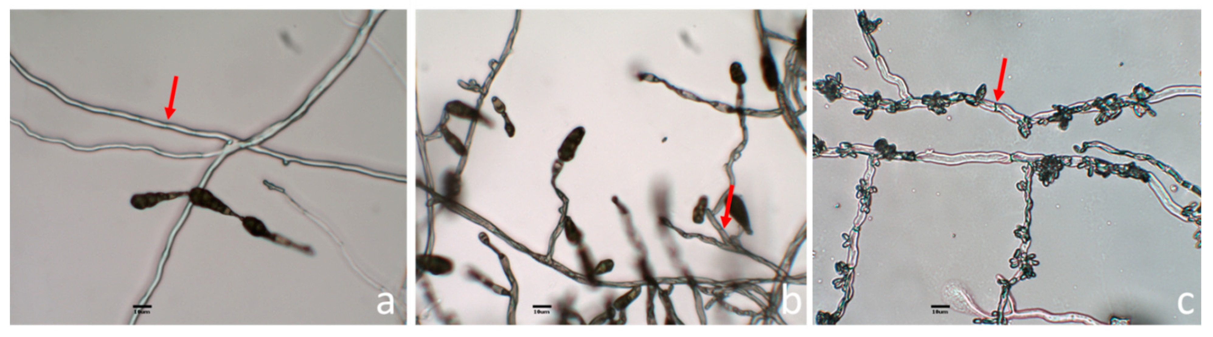
3.2. Morphological Identification of Endophytic Fungi with Antifungal Activity

For the identification of the fungi, morphological and molecular techniques were used. According to the microscopic observation, the isolate Ac1 presented branched septate hyphae and the conidia were ovoid or ellipsoidal with horizontal and vertical septa and were produced in chains (Figure 4a). The isolate Lc1 also presented septate hyphae and the conidia were similar to those observed in Ac1, but concatenated conidia were not observed (Figure 4b). The Ec1 isolate showed septate hyphae. The hyphae contained conidia in clusters which were located along the hyphae (Figure 4c).
Morphological analysis of the isolates was complemented by phylogenetic analysis of ITS markers, the Neighbor-joining tree generated from the data is presented in the Figure 5 for Lc1 and Ac1 sequences and, in the Figure 6 for Ec1 sequence. The trees generated were made by Neighbor-joining using BLAST alignment with NCBI database.
The isolates Lc1 and Ac1 showed conidia morphology similar to Alternaria genus [25,26]. Neighbor-joining tree of these fungi were clustered with Alternaria species which includes A. alternata and A. tenuissima [27,28]. Therefore, the conidia morphology and the phylogenetic analysis support the assignation of these fungi to this genus.
On the other hand, phylogenetic analyses based on internal transcribed spacer (ITS) allowed the identification of the isolate Ec1 as Aureobasidium spp. (Figure 6). This isolate showed morphology similar to yeast-like fungus A. pullulans; this fungus changes between its filamentous and yeast form [29].

4. Discussion

In this work, three endophytic fungi (Ac1, Lc1 and Ec1) with antifungal activity against B. cinerea obtained from native and endemic plants growing in Central Andean Precordillera of Chile were isolated. This region has a Mediterranean climate [9,10,30]. It has been described that the plant colonization by endophytic fungus is dependent on the ecosystem. Colonization rate was from less than 1 to 44% in arctic and boreal ecosystems to more than 90% in tropical ecosystems [31]. Endophyte communities along a latitudinal gradient from the Canadian arctic to the tropical forest of central Panama were studied and it was shown that endophytic fungi increase in incidence from arctic to tropical sites [32]. These authors defined the leaves of tropical trees as hotspots for fungal species diversity [32]. Little is known about the abundance of endophytic fungi in Mediterranean ecosystems. Some studies have been conducted in oaks and pines [33,34,35,36]. Additionally, a total of 319 fungal species were obtained from roots of 24 plant species from the Province of Alicante (Spain). Fusarium and Phoma species were the most abundant genera, followed by Aspergillus, Alternaria and Acremonium [37]. These results coincide with those found in this work since Alternaria was found as endophytic fungus from two different plants. However, in this work low numbers of the isolates of endophytic fungi were obtained. The rigorous conditions of disinfection of the plant surface to eliminate the epiphytic fungi could explain this result.
This is the first report of endophytic fungi isolated from plants located in the studied area and it is also the first time that endophytic fungi with antifungal activity against B. cinerea have been isolated from L. caustica, A. caven and E. chiloensis. L. caustica (litre) is an endemic tree of Chile and E. chiloensis is an endemic columnar cactus species widely distributed in north-central Chile that inhabits mainly equatorial-facing slopes in semiarid environments [30,38]. A. caven is a native tree found in several countries in South America [39]. Although endemic or native plants of this region were used in this work, the isolated fungi Alternaria spp. (Lc1 from L. caustica), Alternaria spp. (Ac1 from A. caven) and Aureobasidium spp. (Ec1 from E. chiloensis) have been commonly found as endophytes in different types of plants around the world [12,40]. Alternaria and Aureobasidium genera have been isolated as endophytes from different cacti species in Arizona [12] and from grapevine cultivars in Canary Islands [40].
In Chile, some studies have reported the presence of endophytic fungi in native, endemic and introduced plants [41,42,43,44]. The endophytic fungi Microsphaeropsis olivacea and Penicillium janczewskii isolated from native gymnosperms collected in southern Chile showed antifungal activity on B. cinerea [43].
Species from genus Alternaria have been described as saprobic, endophytic, and pathogenic [45]. Many species of Alternaria isolated as endophytic produce antifungal compounds [39]. Some of these species have been reported with activity against B. cinerea [40,46,47]. On the other hand, it has been reported that A. pullulans is an important antagonist of B. cinerea [15,48,49]. Alternaria and Aureobasidium species are able to inhibit the mycelial growth of B. cinerea by antibiosis mediated by diffusible compounds [50,51,52] and, in the case of A. pullulans also through the production of volatile compounds [15,49,53]. Indeed, A. pullulans is the active ingredient of fungicides that are currently commercialized against B. cinerea [7].
In this work, the results suggest that the isolated endophytic fungi would inhibit to B. cinerea through the secretion of diffusible and volatile compounds affecting the mycelial growth. In the case of the isolate Ec1 corresponding to Aureobasidium spp., it was also demonstrated that extracts obtained from this fungus inhibited the mycelial growth, the conidia germination and the elongation of the germ tube. The compounds secreted by Aureobasidium spp. were not identified, but it was shown that the most polar compounds of this extract exhibited the antifungal activity.
Interestingly, in this work it was also shown that the volatile compounds produced by the three isolated endophytic fungi suppressed the sporulation of B. cinerea. There are no works published in the literature describing the effect of volatiles produced by Alternaria or Aureobasidium species on the sporulation of B. cinerea. Reduction of sporulation is a crucial step for controlling B. cinerea disease cycle in plants.
A. pullulans presents great phenotypical variability. In some varieties nine secondary metabolite biosynthetic clusters were identified. However, in other varieties 32 or 37 biosynthetic clusters have been identified [54] therefore, it is possible that the isolate Ec1 might produce new compounds with antifungal activity that have not been previously described.

5. Conclusions

Three endophyte fungi with antifungal activity against B. cinerea were isolated form endemic and native plants of Chile. The antifungal activity is mediated by diffusible and volatile compounds which reduced mycelial growth, sporulation and conidia germination of B. cinerea.

Supplementary Materials

The following are available online at https://www.mdpi.com/2309-608X/6/3/149/s1.

Author Contributions

Conceptualization, L.M. and M.C.; methodology, A.V.; software, R.P.; investigation, A.V. and R.P.; writing—original draft preparation, A.V. and R.P.; writing—review and editing, M.C. and L.M.; project administration, L.M.; funding acquisition, M.C. All authors have read and agreed to the published version of the manuscript.

Funding

This research was funded by “USA 1899—Vridei 021943-CT PAP Universidad de Santiago de Chile”.

Acknowledgments

The authors thank to Universidad de Santiago de Chile Grant USA 1899—Vridei 021943-CT PAP for their financial support.

Conflicts of Interest

The authors declare no conflict of interest.

References

  1. Dean, R.; van Kan, J.A.; Pretorius, Z.A.; Hammond-Kosack, K.E.; Di Pietro, A.; Spanu, P.D. The Top 10 fungal pathogens in molecular plant pathology. Mol. Plant. Pathol. 2012, 13, 414–430. [Google Scholar] [CrossRef] [PubMed] [Green Version]
  2. Jacometti, M.A.; Wratten, S.D.; Walter, M. Review: Alternatives to synthetic fungicides for Botrytis cinerea management in vineyards. Aust. J. Grape Wine Res. 2010, 16, 154–172. [Google Scholar] [CrossRef]
  3. Hahn, M. The rising threat of fungicide resistance in plant pathogenic fungi: Botrytis as a case study. J. Biol. Chem. 2014, 7, 133–141. [Google Scholar] [CrossRef] [PubMed] [Green Version]
  4. Esterio, M.; Copier, C.; Román, A.; Araneda, M.; Rubilar, M.; Pérez, I.; Auger, J. Frequency of fungicide-resistant Botrytis cinerea populations isolated from ‘Thompson Seedless’ table grapes in the Central Valley of Chile. Cienc. Investig. Agrar. 2017, 44, 295–306. [Google Scholar] [CrossRef] [Green Version]
  5. Deshmukh, S.; Gupta, M.; Prakash, V.; Saxena, S. Endophytic Fungi: A source of potential antifungal compounds. J. Fungi 2018, 4, 77. [Google Scholar] [CrossRef] [PubMed] [Green Version]
  6. Jia, M.; Chen, L.; Xin, H.; Zheng, C.; Rahman, K.; Han, T.; Qin, L. A Friendly relationship between endophytic fungi and medicinal plants: A systematic review. Front. Microbiol. 2016, 7, 1–14. [Google Scholar] [CrossRef] [Green Version]
  7. Bolívar-Anillo, H.J.; Garrido, C.; Collado, I.G. Endophytic microorganisms for biocontrol of the phytopathogenic fungus Botrytis cinerea. Phytochem. Rev. 2019, 19, 721–740. [Google Scholar] [CrossRef]
  8. Strobel, G. Endophytes as sources of bioactive products. Microbes Infect. 2003, 5, 535–544. [Google Scholar] [CrossRef]
  9. Castri, F.; Mooney, H.A. Mediterranean Type Ecosystems: Origin and Structure; Springer: Berlin/Heidelberg, Germany, 1973. [Google Scholar]
  10. Dallman, P.R. Plant Life in the World’s Mediterranean Climates: California, Chile, South Africa, Australia, and the Mediterranean Basin; Oxford University Press: Oxford, UK, 1998. [Google Scholar]
  11. Nardini, A.; Lo Gullo, M.A.; Trifilò, P.; Salleo, S. The challenge of the Mediterranean climate to plant hydraulics: Responses and adaptations. Env. Exp. Bot. 2014, 103, 68–79. [Google Scholar] [CrossRef]
  12. Suryanarayanan, T.S.; Wittlinger, S.K.; Faeth, S.H. Endophytic fungi associated with cacti in Arizona. Mycol. Res. 2005, 109, 635–639. [Google Scholar] [CrossRef] [Green Version]
  13. Muñoz, G.; Hinrichsen, P.; Brygoo, Y.; Giraud, T. Genetic characterization of Botrytis cinereal populations in Chile. Mycol. Res. 2002, 106, 594–601. [Google Scholar] [CrossRef]
  14. Montealegre, J.; Ochoa, F.; Besoain, X.; Herrera, R.; Pérez, L. In Vitro and glasshouse biocontrol of Rhizoctonia solani with improved strains of Trichoderma spp. Cienc. Investig. Agrar. 2014, 41, 197–206. [Google Scholar] [CrossRef] [Green Version]
  15. Chen, P.H.; Chen, R.Y.; Chou, J.Y. Screening and evaluation of yeast antagonists for biological control of Botrytis cinerea on strawberry fruits. Mycobiology 2018, 46, 33–46. [Google Scholar] [CrossRef] [PubMed] [Green Version]
  16. Li, N.; Alfiky, A.; Wang, W.; Islam, M.; Nourollahi, K.; Liu, X.; Kang, S. Volatile compound-mediated recognition and inhibition between Trichoderma biocontrol agents and Fusarium Oxysporum. Front. Microbiol. 2018, 9, 2614. [Google Scholar] [CrossRef] [PubMed]
  17. Rubio, M.B.; Hermosa, R.; Reino, J.L.; Collado, I.G.; Monte, E. Thctf1 transcription factor of Trichoderma harzianum is involved in 6-pentyl-2H-pyran-2-one production and antifungal activity. Fungal Genet. Biol. 2009, 46, 17–27. [Google Scholar] [CrossRef] [PubMed]
  18. Gao, P.; Qin, J.; Li, D.; Zhou, S. Inhibitory effect and possible mechanism of a Pseudomonas strain QBA5 against gray mold on tomato leaves and fruits caused by Botrytis cinerea. PLoS ONE 2018, 13, e0190932. [Google Scholar] [CrossRef] [Green Version]
  19. Morales, J.; Mendoza, L.; Cotoras, M. Alteration of oxidative phosphorylation as a possible mechanism of the antifungal action of p-coumaric acid against Botrytis cinerea. J. Appl. Microbiol. 2017, 123, 969–976. [Google Scholar] [CrossRef]
  20. Dissanayak, M.L.M.C.; Ito, S.I.; Yoshihiko, A. TLC Bioautography guided detection and biological activity of antifungal compounds from medicinal plant Acorus calamus Linn. Asian J. Plant. Pathol. 2015, 9, 16–26. [Google Scholar] [CrossRef] [Green Version]
  21. Doyle, J.J.; Doyle, J.L. Isolation of plant DNA from fresh tissue. Focus 1990, 12, 13–15. [Google Scholar]
  22. White, T.; Bruns, T.; Lee, S.; Taylor, J. Amplification and Direct Sequencing of Fungal Ribosomal Rna Genes for Phylogenetics. Pcr Protocols: A Guide to Methods and Applications; New York Academic Press Inc.: New York, NY, USA, 1990; pp. 315–322. [Google Scholar]
  23. Altschul, S.F.; Gish, W.; Miller, W.; Myers, E.W.; Lipman, D.J. Basic local alignment search tool. J. Mol. Biol. 1990, 215, 403–410. [Google Scholar] [CrossRef]
  24. Saitou, N.; Nei, M. The neighbor-joining method: A new method for reconstructing phylogenetic trees. Mol. Biol. Evol. 1987, 4, 406–425. [Google Scholar] [PubMed]
  25. Agrios, G.N. Plant Pathology, 5th ed.; Elsevier Acad. Press: Boston, MA, USA, 2005. [Google Scholar]
  26. Deacon, J.W. Fungal Biology, 4th ed.; Blackwell Publishing Ltd.: Oxford, UK, 2006. [Google Scholar]
  27. Hoog, G.; Horre, R. Molecular taxonomy of the Alternaria and Ulocladium species from humans and their identification in the routine laboratory. Mycoses 2002, 45, 259–276. [Google Scholar] [CrossRef] [PubMed] [Green Version]
  28. Zhu, X.; Xiao, C. Phylogenetic, morphological, and pathogenic characterization of Alternaria Species associated with fruit rot of blueberry in California. Phytopathology 2015, 105, 1555–1567. [Google Scholar] [CrossRef] [Green Version]
  29. Zalar, P.; Gostinčar, C.; de Hoog, G.S.; Uršič, V.; Sudhadham, M.; Gunde-Cimerman, N. Redefinition of Aureobasidium pullulans and its varieties. Stud. Mycol. 2008, 61, 21–38. [Google Scholar] [CrossRef] [PubMed] [Green Version]
  30. Peña-Rojas, K.; Donoso, S.; Pacheco, C.; Riquelme, A.; Gangas, R.; Guajardo, A.; Durán, S. Respuestas morfo-fisiológicas de plantas de Lithraea caustica (Anacardiaceae) sometidas a restricción hídrica controlada. Bosque (Valdivia) 2018, 39, 27–36. [Google Scholar] [CrossRef]
  31. Higgins, K.L.; Arnold, A.E.; Miadlikowska, J.; Sarvate, S.D.; François, L. Phylogenetic relationships, host affinity, and geographic structure of boreal and arctic endophytes from three major plant lineages. Mol. Phylogenet. Evol. 2007, 42, 543–555. [Google Scholar] [CrossRef]
  32. Arnold, A.E.; Lutzoni, F. Diversity and host range of foliar fungal endophytes: Are tropical leaves biodiversity hotspots? Ecology 2007, 88, 541–549. [Google Scholar] [CrossRef]
  33. Moricca, S.; Ragazzi, A. Fungal endophytes in Mediterranean oak forests: A lesson from Discula quercina. Phytopathology 2008, 98, 380–386. [Google Scholar] [CrossRef] [Green Version]
  34. Linaldeddu, B.T.; Sirca, C.; Spano, D.; Franceschini, A. Variation of endophytic cork oak-associated fungal communities in relation to plant health and water stress. Pathology 2011, 41, 193–201. [Google Scholar] [CrossRef] [Green Version]
  35. Martinez-Alvarez, P.; Martin-Garcia, J.; Rodriguez-Ceinos, S.; Diez, J.J. Monitoring endophyte populations in pine plantations and native oak forests in Northern Spain. For. Syst. 2012, 21, 373–382. [Google Scholar] [CrossRef] [Green Version]
  36. Moricca, S.; Ginetti, B.; Ragazzi, A. Species and organ specificity in endophytes colonizing healthy and declining Mediterranean oaks. Phytopathol. Mediterr. 2012, 51, 587–598. [Google Scholar]
  37. Maciá-Vicente, J.G.; Jansson, H.B.; Abdullah, S.K.; Descals, E.; Salinas, J.; Lopez-Llorca, L.V. Fungal root endophytes from natural vegetation in Mediterranean environments with special reference to Fusarium spp. Fems Microbiol. Ecol. 2008, 64, 90–105. [Google Scholar] [CrossRef] [PubMed] [Green Version]
  38. Cares, R.A.; Medel, R.; Botto-Maham, C. Frugivory in Echinopsis chiloensis (Caryophyllales: Cactaceae). Rev. Chil Hist Nat. 2013, 86, 489–491. [Google Scholar] [CrossRef] [Green Version]
  39. Root-Bernstein, M.; Valenzuela, R.; Huerta, M.; Armesto, J.; Jaksic, F. Acacia caven nurses endemic sclerophyllous trees along a successional pathway from silvopastoral savanna to forest. Ecosphere 2017, 8, e01667. [Google Scholar] [CrossRef]
  40. Cosoveanu, A.; Cabrera, Y.; Hernandez, G.; Cabrera, R. Endophytic fungi from grapevine cultivars in Canary Islands and their activity against phytopatogenic fungi. Int. J. Agric. Crop. Sci. 2014, 7, 1497–1503. [Google Scholar]
  41. González-Teuber, M.; Vilo, C.; Bascuñán-Godoy, L. Molecular characterization of endophytic fungi associated with the roots of Chenopodium quinoa inhabiting the Atacama Desert, Chile. Genom. Data 2017, 11, 109–112. [Google Scholar] [CrossRef]
  42. Oses, R.; Valenzuela, S.; Freer, J.; Sanfuentes, E.; Rodríguez, J. Fungal endophytes in xylem of healthy Chilean trees and their possible role in early wood decay. Fungal Divers. 2008, 33, 77–86. [Google Scholar]
  43. Hormazabal, E.; Piontelli, E. Endophytic fungi from Chilean native gymnosperms: Antimicrobial activity against human and phytopathogenic fungi. World J. Microb. Biot. 2009, 25, 813–819. [Google Scholar] [CrossRef]
  44. Schmeda-Hirschmann, G.; Hormazabal, E.; Astudillo, L.; Rodriguez, J.; Theoduloz, C. Secondary metabolites from endophytic fungi isolated from the Chilean gymnosperm Prumnopitys Andin. (Lleuque). World J. Microbiol. Biotechnol. 2005, 21, 27–32. [Google Scholar] [CrossRef]
  45. Eram, D.; Arthikala, M.; Melappa, G.; Santoyo, G. Alternaria species: Endophytic fungi as alternative sources of bioactive compounds. Ital. J. Mycol. 2018, 47, 40–54. [Google Scholar]
  46. Chowdhary, K.; Kaushik, N. Fungal endophyte diversity and bioactivity in the Indian medicinal plant Ocimum sanctum Linn. PLoS ONE 2015, 10, e0141444. [Google Scholar] [CrossRef] [PubMed]
  47. Park, Y.H.; Kim, Y.; Mishra, R.C.; Bae, H. Fungal endophytes inhabiting mountain-cultivated ginseng (Panax ginseng Meyer): Diversity and biocontrol activity against ginseng pathogens. Sci. Rep. 2017, 7, 1–10. [Google Scholar] [CrossRef] [PubMed]
  48. Bozoudi, D.; Tsaltas, D. The multiple and versatile roles of Aureobasidium pullulans in the vitivinicultural sector. Fermentation 2018, 4, 85. [Google Scholar] [CrossRef] [Green Version]
  49. Di Francesco, A.; Ugolini, L.; D’Aquino, S.; Pagnotta, E.; Mari, M. Biocontrol of Monilinia laxa by Aureobasidium pullulans strains: Insights on competition for nutrients and space. Int. J. Food Microbiol. 2017, 248, 32–38. [Google Scholar] [CrossRef] [PubMed]
  50. Lou, J.; Fu, L.; Peng, Y.; Zhou, L. Metabolites from Alternaria fungi and their bioactivities. Molecules 2013, 18, 5891–5935. [Google Scholar] [CrossRef]
  51. Feng, C.; Ma, Y. Isolation and anti-phytopathogenic activity of secondary metabolites from Alternaria sp. FL25, an endophytic fungus in Ficus carica. Chin. J. Appl. Environ. Biol. 2010, 16, 76–78. [Google Scholar] [CrossRef]
  52. Prasongsuk, S.; Lotrakul, P.; Ali, I.; Bankeeree, W.; Punnapayak, H. The current status of Aureobasidium pullulans in biotechnology. Folia Microbiol. 2018, 63, 129–140. [Google Scholar] [CrossRef]
  53. Di Francesco, A.; Ugolini, L.; Lazzer, L.; Mari, M. Production of volatile organic compounds by Aureobasidium pullulans as a potential mechanism of action against postharvest fruit pathogens. Biol. Control. 2015, 81, 8–14. [Google Scholar] [CrossRef]
  54. Gostinčar, C.; Ohm, R.A.; Kogej, T.; Sonjak, S.; Turk, M.; Zajc, J.; Gunde-Cimerman, N. Genome sequencing of four Aureobasidium pullulans varieties: Biotechnological potential, stress tolerance, and description of new species. BMC Genom. 2014, 15, 549. [Google Scholar] [CrossRef] [Green Version]
Figure 1. Macroscopic morphology of B. cinerea mycelia after 7 days of incubation in the presence of volatile compounds produced by the isolates Lc1 (a), Ac1 (b) and Ec1 (c). B. cinerea growth in the absence of volatile compounds (d).
Jof 06 00149 g001
Figure 2. Antifungal activity of volatile and diffusible compounds secreted by the isolate Ec1 against B. cinerea. (a) Effect of volatile compounds secreted by Ec1 on sporulation of B. cinerea after 7 days of incubation at 22 °C. (b) Effect of diffusible compounds on mycelial growth of B. cinerea after 3 days of incubation at 22 °C. Gray bars represent B. cinerea control and black bars represent isolate Ec1 test treatment. (c) Effect of the cell-free filtrate obtained from Ec1 at different concentrations on germination of B. cinerea conidia after 4-h incubation. (d) Growth rate (µm/h) of the germ tube of B. cinerea after 4 h of incubation at different concentration of cell-free filtrates obtained from Ec1. Values represent the mean ± SD. a, b: A Student’s t-test and Mann Whitney test were completed. c, d: Analysis of variance (ANOVA) with Tukey’s test was carried out. Columns with different letters are significantly different (p < 0.05).
Jof 06 00149 g002
Figure 3. Compounds extracted from Ec1 isolate and their antifungal activity on B. cinerea. (a) Chromatogram of the extract visualized under UV light at 254 nm. (b) Chromatogram of the extract revealed with 25% H2SO4. Lane 1, compounds obtained from the culture medium in the absence of isolate Ec1. Lane 2, compounds secreted by the isolate Ec1. Mobile phase CHCl3/MeOH 95: 5. (c) Bioautography of the extract obtained from Ec1 after 7 days of B. cinerea incubation at 22 °C.
Jof 06 00149 g003
Figure 4. Morphological examination of microscopic aerial structures from Ac1 (a), Lc1 (b) and, Ec1 (c). The aerial structures of the three fungi were observed with the 40× objective. The septa are indicated by a red arrow.
Jof 06 00149 g004
Figure 5. Neighbor-joining tree based on fungal internal transcribed spacer (ITS) sequences of Ac1, Lc1, 15 Alternaria spp. and Pleospora welwitschiae as outgroup. Numbers labeled at each node indicate bootstrap value (%) from 1000 replicated.
Jof 06 00149 g005
Figure 6. Neighbor-joining tree based on fungal internal transcribed spacer (ITS) sequences of Ec1, Aureobasidium spp. and Selenophoma mahoniae as outgroup. Numbers labeled at each node indicate bootstrap value (%) from 1000 replicated.
Jof 06 00149 g006
Table 1. Endophytic fungi isolated from plants growing in Central Andean Precordillera of Chile.
PlantIsolation OrganFungal IsolateGrowth Type
C. polifoliaLeafCp1Filamentous
L. causticaLeafLc1Filamentous
C. odoriferaLeafCo1Filamentous
E. chiloensisStemEc1Filamentous
T. tricolorFlowerTt1Filamentous
StemTt2Filamentous
FlowerTt3Yeast
StemTt4Yeast
P. chilensisFlowerPc1Filamentous
StemPc2Yeast
A. cavenStemAc1Filamentous
Table 2. Effect of endophytic fungus on B. cinerea mycelial growth in confrontation assays and by volatile compounds.
Fungal EndophyteInhibition of Mycelial Growth in the Confrontation Test (%)Inhibition of Mycelial Growth Produced by Volatile Compounds (%)
Lc136.0 ± 4.022.4 ± 3.5
Ac141.9 ± 2.818.0 ± 3.8
Ec136.6 ± 4.517.0 ± 1.5
Mean of four independent experiments ± the standard deviation.

