Article

Scedosporium spp. from Clinical Setting in Argentina, with the Proposal of the New Pathogenic Species Scedosporium americanum

1
Departamento Micología, Instituto Nacional de Enfermedades Infecciosas, Administración Nacional de Laboratorios e Institutos de Salud ‘Dr. C. G. Malbrán’, C1282 AFF Buenos Aires, Argentina
2
Hospital San Bernardo de la provincia de Salta, A4400 Salta, Argentina
3
Hospital Italiano de Buenos Aires, C1199 CABA Buenos Aires, Argentina
4
Hospital Alemán de Buenos Aires, C1118 AAT Buenos Aires, Argentina
5
Mycology Unit, Faculty of Medicine and IISPV, Universitat Rovira i Virgili, 43201 Reus, Spain
*
Author to whom correspondence should be addressed.
J. Fungi 2021, 7(3), 160; https://doi.org/10.3390/jof7030160
Submission received: 12 January 2021 / Revised: 7 February 2021 / Accepted: 11 February 2021 / Published: 24 February 2021
(This article belongs to the Section Fungal Evolution, Biodiversity and Systematics)

Abstract

:
Species of the genus Scedosporium (family Microascaceae, phylum Ascomycota) are responsible for a wide range of opportunistic human infections, and have a low susceptibility to most antifungal drugs. It is well known that the pattern of Scedosporium species distribution varies according to geographic region. To assess the diversity of Scedosporium species in Argentina involved in human infections, we carried out a retrospective study reviewing 49 strains from clinical samples sent for diagnosis to the National Clinical Mycology Reference Laboratory between 1985 and 2019. Then, a phenotypic characterization, a phylogenetic study and and in vitro susceptibility test to antifungals were carried out. An analysis of combined nucleotide sequences dataset of the internal transcribed spacer of the ribosomal DNA (ITS) and of a fragment of the β-tubulin gene (BT2) demonstrated that 92% of the strains belonged to the species S. boydii, S. apiospermum and S. angustum, all them pertaining to S. apiospermum species complex. However, two strains (4%) were identified as S. aurantiacum, a species never reported in clinical settings in the Americas’. Surprisingly, one of them displayed a polycytella-like conidiogenesis, up to date only reported for S. apiospermum. In addition, the strain DMic 165285 was phylogenetically located far away from the rest of the species, so is proposed as the novel species Scedosporium americanum. On the other hand, from all seven antifungals tested, voriconazole and posaconazole were the most active drugs against Scedosporium spp.

1. Introduction

The Microascaceae is a monophyletic family of highly polymorphic fungi that includes the genera Acaulium, Cephalotrichum, Fairmania, Fuscoannellis, Gamsia, Kernia, Lomentospora, Lophotrichus, Microascus, Parascedosporium, Petriella, Petriellopsis, Pithoascus, Pseudoscopulariopsis, Scedosporium, Scopulariopsis, Wardomyces, Wardomycopsis and Yunnania [1,2,3]. Some of these genera are well-known human opportunistic human pathogens. The best known of these fungi is probably Scopulariopsis brevicaulis, a non-dermatophyte mostly causing onychomycosis. However, over the last two decades Scedosporium spp. have become increasing prevalent in immunocompromised patients, despite being better known for causing post-traumatic infections in healthy people [4]. The genus comprises species that are highly polymorphic: all them produce the scedosporium-like asexual morph, characterized by the production of solitary conidia from annelidic conidiogenous cells; a graphium-like synnematous synanamorph; unicellular sessile conidia; a polycytella-like synanamorph, a sort of “macroconidia” only seen in certain strains of S. apiospermum [5,6]; and a sexual morph, comprising dark, non-ostiolate globose ascomata with a thin peridial wall of textura epidermoidea, 8-spored, soon evanescent asci, and unicellular, yellowish or reddish brown, broadly fusiform to ellipsoidal ascospores with (usually) a germ pore at each end. The generic name was proposed by Saccardo (1911) [7], who described Monosporium apiospermum (isolated by Radaeli in 1911) from a case of eumycetoma) [8] that had morphological similarities with Monosporium acremonioides (actually Harzia acremonioides), the former producing non-verticillate, vaguely branched decumbent conidiophores (which are erect and verticillate in H. acremonioides) [9]. The generic name was validated by Castellani and Chalmers (1919), who accepted Scedosporium apiospermum instead of M. apiospermum. In 1943, Negroni and Fisher [10] isolated a fungus, which had asexual and sexual reproductive structures, from purulent material from a case of knee arthritis. Despite its great similarity to Allescheria boydii (isolated from a foot eumycetoma by Shear 1922) [11], they erected a new genus and species Pseudallescheria shearii. Consequently, the holomorph was given a different scientific name from the most common asexual morph (Scedosporium). In 2014, in order to remove the dual nomenclature that had been based on the anamorph/teleomorph concept, Pseudallescheria was accepted as a synonym of Scedosporium [12]. Ten species of Scedosporium are currently recognized (S. aurantiacum, S. cereisporum, S. desertorum, S. dehoogii and S. minutisporum, and S. apiospermum species complex including S. angustum, S. apiospermum, S. boydii, S. ellipsoideum, and S. fusoideum), which are easily distinguishable phylogenetically by comparing the sequences of a fragment of the β-tubulin gene (TUB2) rather than by phenotype [4,13]. Scedosporium species have been linked to various ecological niches, mostly being soils and waters contaminated with aliphatic and aromatic compounds, the consequence of anthropogenic industrial activity [14,15]. Infections by Scedosporium spp. cause a wide range of clinical entities, such as osteomyelitis, keratitis, lymphocutaneous infection and sinusitis [4,16,17,18] and is the primary etiological agent of white grain mycetoma worldwide [19]. Scedosporium spp. can also colonize the respiratory tract of cystic fibrosis patients (CF), causing allergic bronchitis, and has produced disseminated diseases and central nervous system (CNS) infections in immunocompetent patients after near-drowning incidents. Scedosporium spp. infections occur by traumatic implantation of contaminated materials (e.g., wood chips), or can be acquired by inhalation of contaminated aerosols [16,17].
The most common systemic antifungals display high minimum inhibitory concentration (MIC) values for Scedosporium spp., making successful treatment difficult. The main clinically relevant species, S. apiospermum and S. boydii, show variable, strain-dependent in vitro antifungal susceptibility [20,21]. Another clinically important species, S. aurantiacum, also has high MIC values against every antifungal apart from voriconazole (VZ), which is the first-line antimycotic drug against Scedosporium spp. infections [21].
Several reports show that S. apiospermum species complex is found worldwide [4]. However, Scedosporium aurantiacum has been reported to colonise airways of patients primarily in Europe and Australia, where there is a larger number of patients with CF [22,23,24]. It has also been isolated once in Japan [25] but so far has not been reported in clinical cases in the Americas.
The aim of this work is to increase knowledge of the diversity of Scedosporium spp. involved in human infections in Argentina by presenting the results of a retrospective study of 49 clinical isolates that includes phylogenetic analyses, phenotypic characterizations and in vitro susceptibility assays to antifungals. Scedosporium aurantiacum and S. dehoogii are reported for the first time in clinical samples for the Americas, and Scedosporium americanum is described as a novel species.

2. Materials and Methods

2.1. Fungal Strains

We studied 49 fungal strains that were received for identification between 1985 and 2019 at the laboratory of the Mycology Department of the National Institute of Infectious Diseases, ‘Dr. Carlos G. Malbrán’. The strains were sent from public and private hospitals of eight Argentinian provinces (Table 1).

2.2. DNA Extraction, Amplification and Sequencing

Total DNA was extracted and purified from fungal colonies according to Refojo et al. [26]. The internal transcribed spacer (ITS) region of the nuclear ribosomal DNA and a fragment of the β-tubulin (BT2) gene were amplified with the primers ITS5 and ITS4 and Bt2a and Bt2b, respectively [27,28]. The PCR reaction mixture (50 μL) included 10 to 20 ng of fungal DNA template, MgCl2 1.5 mM, dNTPs 250 mM, TRIS-HCl 20 mM (pH 8.4), KCl 50 mM, 2.5 U Taq DNA polymerase (Invitrogen, Life Technologies, Carlsbad, CA, USA) and 0.2 μM of each primer. The amplification programme included an initial denaturation step at 94 °C for 5 min followed by 35 cycles of denaturation at 95 °C for 45 s, annealing for 1 min at 55 °C (ITS) or 59 °C (BT2), and extension for 1 min at 72 °C. A final extension step at 72 °C for 10 min was added at the end of the amplification. After PCR, the products were purified with Accuprep® PCR Purification Kit (Bioneer, Oakland, CA, USA) and stored at −20 °C until they were used. PCR products were sequenced in both directions using the primers described above, using the Big Dye ™ Terminator v3.1 Cycle Sequencing kit (Applied Biosystems, Foster City, CA, USA) according to the manufacturer’s instructions, on a 3500 ABI genetic analyzer (Applied Biosystems). The sequences obtained were edited using Bioedit Sequence Alignment Editor v7.0.4.1 [29].

2.3. Molecular Identification and Phylogeny

A preliminary identification was carried out using the Basic Local Alignment Search Tool (BLAST; https://blast.ncbi.nlm.nih.gov/Blast.cgi). For species-level identification, an identity ≥ 98% (and a coverage ≥99%) was considered when the nucleotide sequences of our strains were compared with those of type or reference strains. ITS and BT2 sequences were aligned using Muscle in MEGA v6.0 software followed by manual adjustment. The best-fit model of sequence evolution was determined by MODELTEST v2.3 [30,31]. For phylogenetic analysis, sequences of ex-type strains of Scedosporium spp. and related taxa of the Microascaceae were taken from GenBank (https://www.ncbi.nlm.nih.gov/genbank/). All sequences generated in this study were deposited in GenBank and their accession numbers are listed in Table 1. Gene sequences of Parascedosporium tectoneae (CBS 120338) were used as outgroup. The computer program MEGA v6.0 [30] was used to build the phylogenetic tree based on the maximum-likelihood (ML) algorithm with a Kimura-2-parameters model with a gamma distribution, treating gaps as complete deletions. A 1000 bootstrap replicates was used, and bootstrap support (BS) values ≥ 75% were considered significant. Other phylogenetic analyses were carried out by MrBayes v3.1.2 [32]. Two parallel runs of four chains were performed for 10,000,000 generations, and trees were sampled every 100 generations. TRACER version 1.5 was used to verify that the mean likelihood value, effective sample size (ESS), and other parameters reached a plateau. For each run, 25% of the trees were discarded as they were obtained during the burning phase. The posterior probability (PP) is shown with a cut-off of 0.95. Trees were viewed and edited with FIGTREE v1.1.2 (http://tree.bio.ed.ac.uk/software/figtree/) and MEGA v6.0 [30] software.

2.4. Morphological Characterization

The strains were cultured at 25, 37, 40, and 45 °C in the dark onto potato dextrose agar (PDA; 75 g potatoes, 20 g dextrose, 15 g agar-agar, 1 L tap water; home made) and oatmeal agar (OA; 30 g oat flakes, 1 g MgSO4·7H2O, 1.5 g H2PO4, 15 g agar, 1 L tap water; home made). Also, the fungal strains were grown at 15, 38, 39, 42 and 43 °C when it was considered necessary to gather such information. Cultures were characterized after 7 days and 14 days of incubation, color notations in parentheses are from Kornerup and Wanscher [33], reproductive structures were observed from slide cultures using 65% lactic acid under a Leica DMF2500 (Leica Microsystems, Concord, ON, Canada) bright field microscope. The measurements of fungal structures and the micrographs were taken by a Leica DMC 2900 camera with LAS v4.8 software (Leica Microsystems, Concord, ON, Canada).

2.5. Antifungal Susceptibility Testing

MIC values were determined according to the reference document M38-3rd ed. from the Clinical and Laboratory Standards Institute (CLSI) [34]. The antifungal drugs tested were: amphotericin (AMB), itraconazole (ITR) and voriconazole (VOR) (Sigma-Aldrich, Buenos Aires, Argentina), caspofungin (CAS) and posaconazole (POS) (Merck, Co., Argentina), anidulafungin (ANI) (Pfizer, Buenos Aires, Argentina) and micafungin (MICA) (Astellas, Tokio, Japan). The strains were cultured on PDA at 35 °C for 5–7 days to induce sporulation. Because strains DMic 165285 and CBS 218.35 showed little growth on PDA, both were cultured on OA. For AMB and triazoles, the MIC endpoint was considered at 100% of growth inhibition, while for echinocandins the minimal effective concentration (MEC) was used, which is the concentration of drug at which hyphae are rounded and compact, and stopped growing (CLSI M38) when compared with the growth control well. Geometric mean, mode, range, MIC50 and MIC90 were calculated. Neither clinical breakpoint nor epidemiological cut-off values have been defined for Scedosporium spp., therefore the susceptible/resistant and wild type/not-wild type categories were not used. For analytical purposes, we assigned a cut-off MIC value ≥2 mg/L to interpret results. Candida parapsilosis ATCC 22019, C. krusei ATCC 6258 and Aspergillus flavus ATCC 204304 were used as controls in all assays.

3. Results

3.1. Molecular Presumptive Identification of the Strains

A BLAST search using ITS sequences identified most of our fungal strains as S. boydii (33/49 strains), followed by S. apiospermum (12/49), S. aurantiacum (2/49), and S. dehogii and S. ellipsoideum (one strain each). However, when BLAST was performed using TUB2 sequences, five of the strains re-identified as S. angustum, and two others as S. ellipsoideum (Tables S1 and S2). On the other hand, one of our strains (DMic 165285) identified as S. boydii by ITS BLAST could not be located at species level using TUB2 sequence.

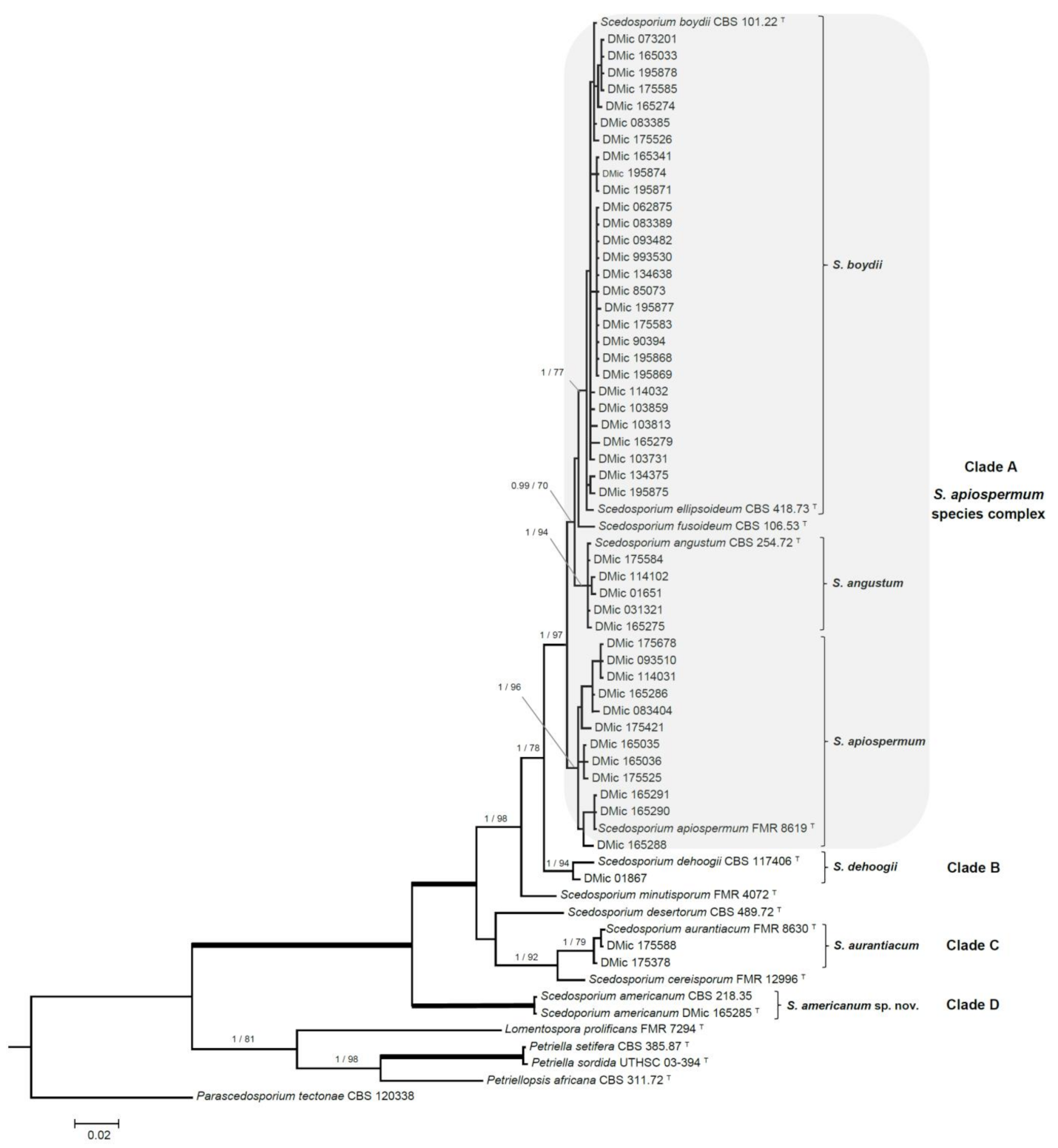
3.2. Phylogenetic Analyses
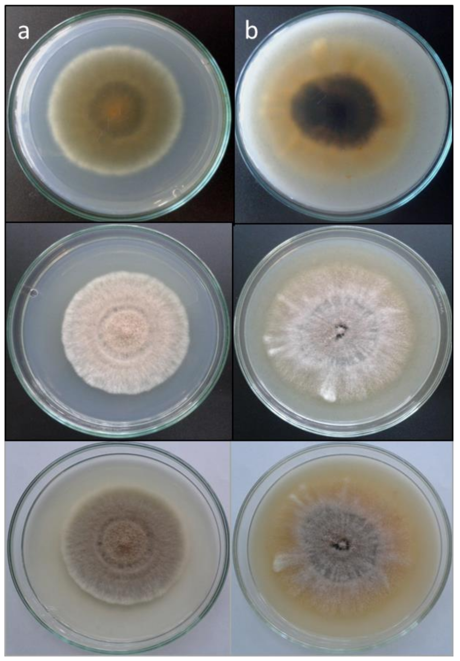
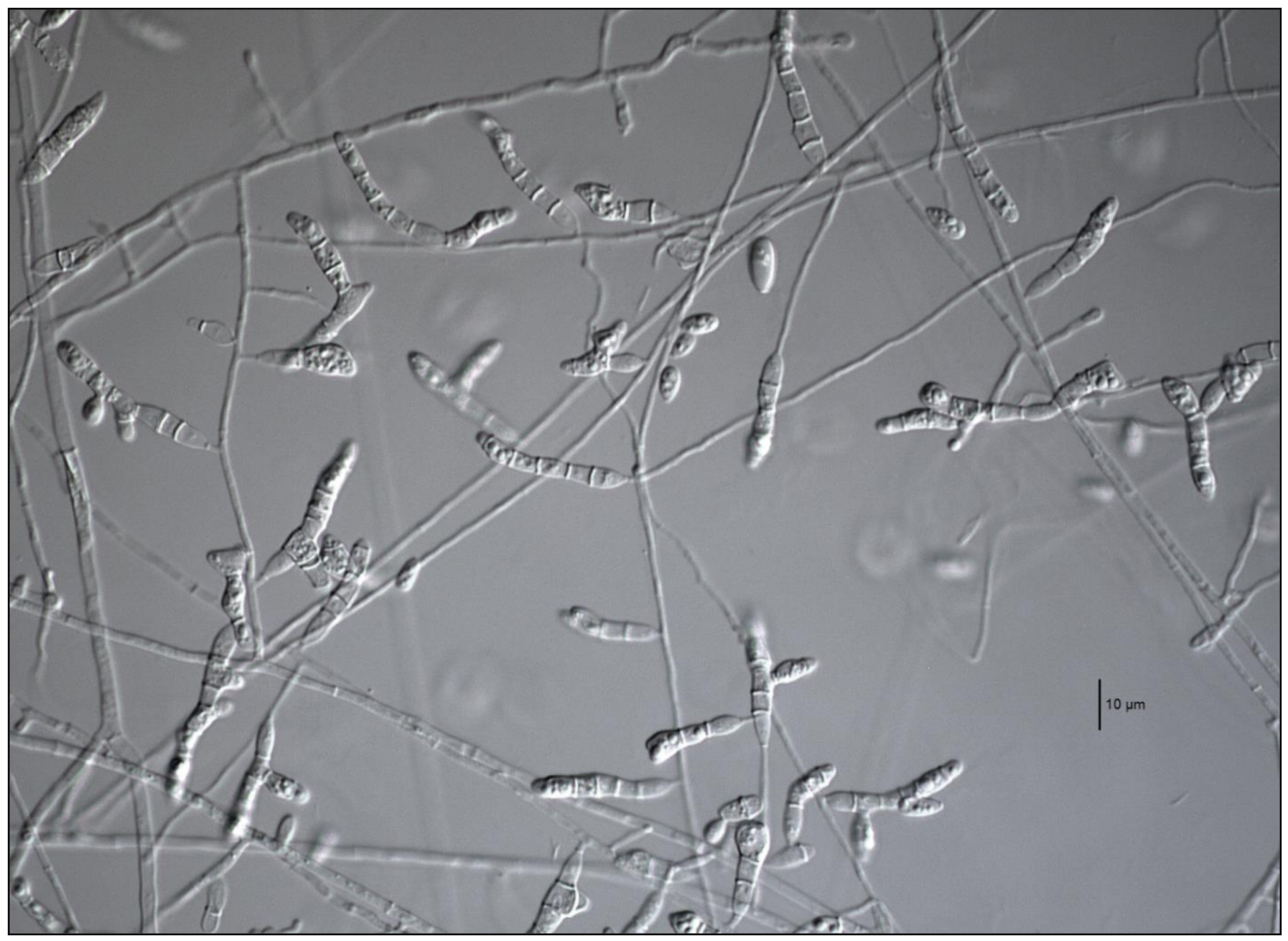
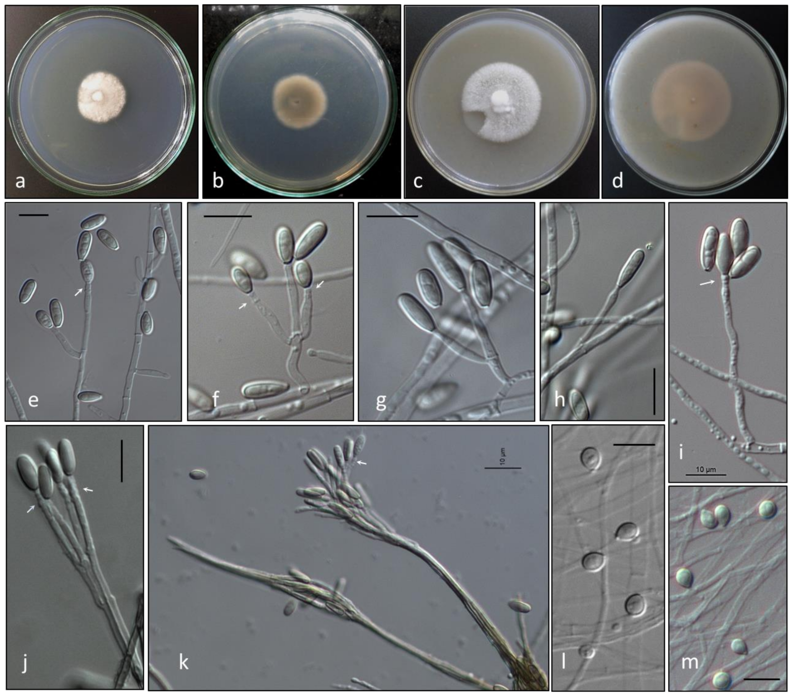
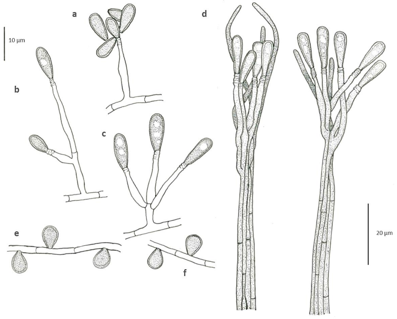
The phylogenetic study included 65 ITS and TUB2 sequences, with a total of 989 characters including gaps, from which 244 were parsimony informative. The ML analysis was congruent with the BI analysis, both displaying a similar topology. In the combined ITS-TUB2 tree, our strains were distributed across four clades, into a well-supported main clade (1 PP/100 BS) grouping all Scedosporium species (Figure 1). Forty-five of the isolates (92%, 45/49) were placed into the S. apiospermum species complex clade A (1 PP/97% BS), and distributed across several terminal clades: A terminal clade (1 PP/77% BS) containing the type strains of S. boydii and S. ellipsoideum, included 28 of our strains, displaying a relatively high intraspecific genetic variation; another twelve strains were placed into a terminal clade (1 PP/96% BS) containing the type strain of S. apiospermum, also with a relatively high intraspecific genetic variation, and a third terminal clade (1 PP/94% BS) included the type strain of S. angustum and four of our strains. Clade B (1 PP/94% BS) included only the type strain of S. dehoogii and one of our strains. Clade C (1 PP/79% BS) included two Argentinian strains and the type strain of S. aurantiacum (Figure 1, Table 1). And clade D (1 PP/100% BS), placed in a basal position and phylogenetically distant from the rest of Scedosporium spp., included the strain DMic 165285 and CBS 218.35. The latter strain was included in the phylogeny because it displayed the highest identity in the BLAST search (ITS: 546/551 bp, 99.1%, 5 gaps). The estimation of the evolutionary distance showed a greater proximity within Scedosporium spp. than those from other related genera belonging to the Microascaceae (data not shown).

3.3. Morphology

All strains formed the typical scedosporium-like asexual morph, and were able to grow up to 37 °C. Also, 42/45 strains of the S. apiospermum species complex (clade A) were able to grow up to 40 °C. Three of our strains that failed to grow at 40 °C were in the S. boydii/S. ellipsoideum terminal clade. In clade A, 23 of our strains produced ascomata on PDA and OA, 18 corresponding to S. boydii and 5 to S. angustum. None of our S. apiospermum strains formed ascomata in either culture media tested. Strain DMic 01867, placed in clade B (S. dehoogii), did not produce a synnematous asexual morph or a sexual morph, nor grew at 40 °C. Two of the strains placed in clade C (S. aurantiacum) produced the characteristic yellowish pigment in both culture media tested (Figure 2), did not manifest the sexual morph either, but both produced the synnematous asexual morph [35], and grew up to 42 °C, despite it being described that some strains can grow up to 45 °C. Strain DMic 175588 formed, in addition, a polycytella-like asexual morph (Figure 3), so far only reported for S. apiospermum [6]. Strains DMic 165285 and CBS 218.35, in the clade D (not matching any of previously known species of the genus) failed to grow at 40 °C, and their growing rates on OA and PDA at 25 °C and at 37 °C were significantly lower than the rest of the species [36] (Table S3).

3.4. Taxonomy

Given the phylogenetic distance between clade D and the rest of those representing the species of Scedosporium, and due to the distinctiveness of their phenotype, we erect Scedosporium americanum, selecting DMic 165285 as its ex-type strain, due to the greatest diversity of reproductive structures.
Scedosporium americanum Abrantes, Refojo, Hevia, J. Fernández et Stchigel, sp. nov. (Figure 4 and Figure 5). MycoBank MB 836850.
Etymology: Due to the geographical origin of the fungus.
Typus: Argentina, Salta, from hand subcutaneous lesions of a man, 5 Febr. 2016, M. F. Dávalos (holotype DMic-H 165285, culture ex-type DMic 165285).
Additional material examined: USA, unknown origin (cited as probably mycetoma pedis), April 1935, Fred D. Weidmann (culture CBS 218.35).
Mycelium composed by septate, hyaline to pale brown, smooth-, thin- to thick-walled hyphae of 1.3–2.9 μm wide. Conidiophores solitary, often reduced to a single conidiogenous cell arising laterally from undifferentiated hypha, or short-stalked (5–15 µm long), bearing two or three conidiogenous cells at the top. Conidiogenous cells annellidic, lateral or terminal, hyaline, smooth- and thin-walled, cylindrical to flask-shaped, 5–16 µm long, 1.5–2.5 µm wide, with several, distinct annellations at the top with the age. Conidia enteroblastic, one-celled, solitary, arranged in slimy masses, hyaline to sub-hyaline, ovoid when young, 7.5–8.8 × 3.8–4.5 μm, then becoming brown, smooth- and thick-walled, gutulate, ellipsoidal to claviform with a truncated base, 10–13.6 × 3.1–4.8 μm. Conidiomata synnematous, more abundantly produced on PDA after 4 weeks at 25 °C, consisting in 3–5 adpressed elongated conidiophores, attaining up to 210 μm in length at first determined, then increasing its length with age; conidiogenous cells annellidic, indistinguishable from those of the conidiophores; conidia claviform with a truncate base, 11–17 × 3.3–3.8 μm. Sessile conidia holoblastic, one-celled, solitary, scattered and scarcely produced, ovoid, 5.4–6.8 × 4.2–4.8 μm, attached directly to the hyphae or on short lateral branches, more abundant on OA than on PDA. Sexual morph not observed.
Colonies on PDA attaining 30–32 mm diam after 14 days at 25 °C, cottony, slightly elevated, woolly at centre, nearly white, margins irregular and fimbriated; reverse brown at centre (4D2) and olive brown (4E8) to colourless towards the periphery. Colonies on OA attaining 40–45 mm diam after 14 d at 25 °C, slightly elevated at the centre, woolly to floccose, white, with slightly greyish patches (4C1), margins lobed; reverse yellowish grey (4B2) at the centre and greyish to colourless towards the periphery. Colonies on PDA and OA at 37 °C after 7 days attaining 5–6 mm and 9–10 mm, respectively. The fungus does not produce diffusible pigment on PDA or OA. The minimum, optimum and maximum temperatures of growth are: 15 °C, 25–30 °C and 39 °C, respectively.
Case report: DMic 165285 was recovered in 2016 from a 76-year-old man who lives in Salta city, a blacksmith by trade, with type II diabetes and no other previous history of disease. He reported not remembering any major trauma, only small skin punctures by metal shavings. He had a hand lesion of 18 years of evolution, and presented fistulae draining pus and local bone involvement. The fungus was recovered from two clinical samples (a biopsy of subcutaneous tissues and a pus discharge from a fistulated tract) by culturing on Sabouraud’s dextrose agar. On direct examination with KOH 30%, only septate hyaline hyphae were seen, and the drainage material did not contain eumycotic grains (Figure 6). Unfortunately, the patient did not return for medical consultation, so it was not possible to follow its clinical evolution.

3.5. Antifungal Susceptibility Testing

The MIC and MEC values obtained are given in Table 2. Overall, 66% of the tested strains (33/50) showed a MIC value ≥2 mg/L for AMB, followed by 54% (27/50) for ANI, 50% (25/50) for CAS, 28% (14/50) for ITR, and 16% (8/50) for MICA. However, VOR and POS were the most active antifungals, and only two isolates exhibited MIC values ≥2 mg/L for either drug.

4. Discussion

Our results demonstrate that the most frequently recovered species of Scedosporium from clinical specimens in Argentina belonged to the S. apiospermum species complex (clade A; Figure 1), which is clearly monophyletic and presents an intraspecific genetic variability consistent with its definition [13]. This variation is mainly manifested in the S. boydii and S. apiospermum subclades, as described in the review by Ramírez-García et al. [4]. In our phylogenetic analysis, clade A was divided into two robust subclades, where the homothallic lineages (corresponding to S. boydii and to S. angustum) are separated from the heterothallic ones (S. apiospermum). Scedosporium aurantiacum (clade B) is represented by two strains from sputum of patients with CF. The strain DMic 175378 was from an 11-year-old patient (already deceased) from Santa Fé province, who never left the country, so we consider this as the first recorded autochthonous case of infection by S. aurantiacum. The second strain (DMic 175588) was recovered from a 27-year-old diabetic patient in Buenos Aires city, who had a history of travelling to Egypt and Europe [14]. It is remarkable that this isolate displays a polycytella-like asexual morph previously reported only in an “aberrant” strain of S. apiospermum [5,6]. Both cases of infection by S. aurantiacum represent the first report in the Americas. Furthermore, we report for the first time an autochthonous case of infection by S. dehoogii, although this fungus has been isolated from environment in Chile and Mexico [37,38]. Surprisingly, our strain DMic 165285 was found to represent a novel species, Scedosporium americanum (clade D). In 2008, Gilgado et al. [39] carried out a comprehensive study of the genus Scedosporium and its sexual counterpart, Pseudallescheria. These authors included the strain CBS 218.35, highlighting that, despite it showing morphological similarities with the asexual reproductive structures of other Scedosporium spp., it was phylogenetically placed at distance from the other species studied. This suggested that further studies were required to determine whether or not it is a different species, which we believe has been resolved during the development of our work. We have erected the novel species Scedosporium americanum since it produces a greater diversity of reproductive structures and because CBS 218.35 has a dubious geographical and clinical origin, and we have chosen DMic-H 165285 as its type material.
In the present study, MIC values of antifungals tested against S. boydii, S. apiospermum and S. angustum are in agreement with those reported by other authors [21,37,40,41]. In addition, we have confirmed VOR and POS show the highest activity in vitro against these species (MIC values ≤1 mg/L against 48/50 strains) and that the in vitro behaviour of antifungal drugs against the fungi is intra- and interspecies-dependent. Interestingly, among echinocandins, micafungin was slightly more active against all Scedosporium strains tested, our results being in agreement with those of others researchers [20,37].
In conclusion, the present study demonstrates the diversity of Scedosporium species from clinical samples in Argentina, expanding the currently available epidemiological information. Despite there being an increasing number of reports of infections due to Scedosporium spp. [18] in recent years, their identification is, in many countries, based on conventional morphological characterization and mostly limited at genus-level. We highlight the importance of supporting continuous scrutiny of not only possible changes in distribution of the species but also in the emergence of other species that are poorly or not susceptible to the most common antifungal drugs.

Supplementary Materials

Table S1. Preliminary molecular identification of Scedosporium Argentinian strains based on TUB2 sequences. Table S2. Preliminary molecular identification of Scedosporium Argentinian strains based on ITS sequences. Table S3. Growing rates of S. americanum on OA and PDA at 25 °C.

Author Contributions

Conceptualization: R.A.A. and N.R.; methodology: R.A.A., A.I.H., J.F., G.I. and S.C.; software: J.F.; formal analysis: A.M.S.; investigation: R.A.A. and A.M.S.; resources: G.O.D.; data curation: J.F., M.F.D., S.L. and I.M.; writing—original draft preparation: R.A.A.; writing—review and editing: R.A.A., N.R., A.I.H.and A.M.S.; supervision: N.R. and A.M.S.; Project administration: N.R. and G.O.D.; All authors have read and agreed to the published version of the manuscript.

Funding

This research received no external funding.

Institutional Review Board Statement

Not applicable.

Informed Consent Statement

Not applicable.

Data Availability Statement

The data presented in this study are available in “Scedosporium spp. from clinical setting in Argentina, with the proposal of the new pathogenic species Scedosporium americanum”.

Acknowledgments

We thank the National Mycology Laboratory Network of Argentina for the collaborative work carried out for decades.

Conflicts of Interest

The authors declare no conflict of interest.

References

  1. Sandoval-Denis, M.; Guarro, J.; Cano-Lira, J.; Sutton, D.; Wiederhold, N.; De Hoog, G.; Abbott, S.; Decock, C.; Sigler, L.; Gené, J. Phylogeny and taxonomic revision of Microascaceae with emphasis on synnematous fungi. Stud. Mycol. 2016, 83, 193–233. [Google Scholar] [CrossRef] [PubMed] [Green Version]
  2. Woudenberg, J.; Meijer, M.; Houbraken, J.; Samson, R. Scopulariopsis and scopulariopsis-like species from indoor environments. Stud. Mycol. 2017, 88, 1–35. [Google Scholar] [CrossRef] [PubMed]
  3. Su, L.; Zhu, H.; Niu, Y.; Guo, Y.; Du, X.; Guo, J.; Zhang, L.; Qin, C. Phylogeny and taxonomic revision of Kernia and Acaulium. Sci. Rep. 2020, 10, 10302. [Google Scholar]
  4. Ramirez-Garcia, A.; Pellon, A.; Rementeria, A.; Buldain, I.; Barreto-Bergter, E.; Rollin-Pinheiro, R.; de Meirelles, J.V.; Xisto, M.I.D.S.; Ranque, S.; Havlicek, V.; et al. Scedosporium and Lomentospora: An updated over-view of underrated opportunists. Med. Mycol. 2018, 56, 102–125. [Google Scholar] [CrossRef] [PubMed]
  5. Campbell, C.K. Polycytella hominisgen. et sp. nov., a cause of human pale grain mycetoma. Med. Mycol. 1987, 25, 301–305. [Google Scholar] [CrossRef]
  6. Borman, A.M.; Campbell, C.K.; Linton, C.J.; Bridge, P.D.; Johnson, E.M. Polycytella Hominis is a mutated form of Scedosporium Apiospermum. Med. Mycol. 2006, 44, 33–39. [Google Scholar] [CrossRef] [Green Version]
  7. Saccardo, P.A. Notae mycologicae. Series XIII. Ann. Mycol. 1911, 9, 249–257. [Google Scholar]
  8. Radaeli, F. Caso singolare di alterazione cutánea e profonda di natura probabilmente micotica in un piede (con pre-sentizioni di culture, di preparati microscopici e di microfotografie). Giornale Italiano Delle Malattie Veneree e Della Pelle 1911, 52, 109–116. [Google Scholar]
  9. Costantin, J. Les Mucédinées Simples; Paul Klincksieck: Paris, France, 1888. [Google Scholar]
  10. Negroni, P.; Fisher, I. Pseudallescheria sheari n. gen., n. sp. aislada de un paramicetoma de la rodilla. Rev. Inst. Bacteriológico “Dr Carlos G Malbrán” 1944, 12, 195–204. [Google Scholar]
  11. Shear, C. Life history of an undescribed ascomycete isolated from a granular mycetoma of man. Mycologia 1922, 14, 239–243. [Google Scholar] [CrossRef]
  12. Lackner, M.; de Hoog, G.S.; Yang, L.; Ferreira Moreno, L.; Ahmed, S.A.; Andreas, F.; Kaltseis, J.; Nagl, M.; Lass-Flörl, C.; Risslegger, B.; et al. Proposed nomenclature for Pseudallescheria, Scedosporium and related genera. Fungal Divers 2014, 67, 1–10. [Google Scholar] [CrossRef]
  13. Chen, M.; Zeng, J.; De Hoog, G.S.; Stielow, B.; Ende, A.H.G.G.V.D.; Liao, W.; Lackner, M. The ‘species complex’ issue in clinically relevant fungi: A case study in Scedosporium apiospermum. Fungal Biol. 2016, 120, 137–146. [Google Scholar] [CrossRef]
  14. Rougeron, A.; Giraud, S.; Alastruey-Izquierdo, A.; Cano-Lira, J.; Rainer, J.; Mouhajir, A.; Le Gal, S.; Nevez, G.; Meyer, W.; Bouchara, J.P. Ecology of Scedosporium Species: Present Knowledge and Future Research. Mycopathologia 2018, 183, 185–200. [Google Scholar] [CrossRef] [PubMed]
  15. Rougeron, A.; Schuliar, G.; Leto, J.; Sitterl, E.; Landry, D.; Bougnoux, M.E.; Kobi, A.; Bouchara, J.P.; Giraud, S. Human-impacted areas of France are environmental reservoirs of the Pseudallescheria boydii/Scedosporium apiospermum species complex. Environ. Microbiol. 2015, 17, 1039–1048. [Google Scholar] [CrossRef]
  16. Guarro, J.; Kantarcioglu, A.S.; Horré, R.; Rodriguez-Tudela, J.L.; Estrella, M.C.; Berenguer, J.; De Hoog, G.S. Scedosporium apiospermum: Changing clinical spectrum of a therapy-refractory opportunist*. Med. Mycol. 2006, 44, 295–327. [Google Scholar] [CrossRef] [Green Version]
  17. Cortez, K.J.; Roilides, E.; Quiroz-Telles, F.; Meletiadis, J.; Antachopoulos, C.; Knudsen, T.; Buchanan, W.; Milanovich, J.; Sutton, D.A.; Fothergill, A.; et al. Infections Caused by Scedosporium spp. Clin. Microbiol. Rev. 2008, 21, 157–197. [Google Scholar] [CrossRef] [Green Version]
  18. Mello, T.P.; Bittencourt, V.C.B.; Liporagi-Lopes, L.C.; Aor, A.C.; Branquinha, M.H.; Santos, A.L. Insights into the social life and obscure side of Scedosporium/Lomentospora species: Ubiquitous, emerging and multidrug-resistant opportunistic pathogens. Fungal Biol. Rev. 2019, 33, 16–46. [Google Scholar] [CrossRef]
  19. van de Sande, W.W.J. Global Burden of Human Mycetoma: A Systematic Review and Meta-analysis. PLoS Negl. Trop. Dis. 2013, 7, e2550. [Google Scholar] [CrossRef] [Green Version]
  20. Lackner, M.; de Hoog, G.S.; Verweij, P.E.; Najafzadeh, M.J.; Curfs-Breuker, I.; Klaassen, C.H.; Meis, J.F. Species-Specific Antifun-gal Susceptibility Patterns of Scedosporium and Pseudallescheria Species. Antimicrob. Agents Chemother. 2012, 56, 2635–2642. [Google Scholar] [CrossRef] [Green Version]
  21. Lackner, M.; Hagen, F.; Meis, J.F.; Gerrits van den Ende, A.H.G.; Vu, D.; Robert, V.; Fritz, J.; Moussa, T.A.A.; de Hoog, G.S. Susceptibility and Diversity in the Therapy-Refractory Genus Scedosporium. Antimicrob. Agents Chemother. 2014, 58, 5877–5885. [Google Scholar] [CrossRef] [Green Version]
  22. Delhaes, L.; Harun, A.; Chen, S.C.; Nguyen, Q.; Slavin, M.; Heath, C.H.; Maszewska, K.; Halliday, C.; Robert, V.; Sorrell, T.C.; et al. Molecular Typing of AustralianScedosporiumIsolates Showing Genetic Variability and NumerousS. aurantiacum. Emerg. Infect. Dis. 2008, 14, 282–290. [Google Scholar] [CrossRef]
  23. Kaur, J.; Kautto, L.; Penesyan, A.; Meyer, W.; Elbourne, L.D.H.; Paulsen, I.T.; Nevalainen, H.K.M. Interactions of an Emerging Fungal Pathogen Scedosporium aurantiacum with Human Lung Epithelial Cells. Sci. Rep. 2019, 9, 5035. [Google Scholar] [CrossRef] [PubMed]
  24. Heath, C.; Slavin, M.; Sorrell, T.; Handke, R.; Harun, A.; Phillips, M.; Nguyen, Q.; Delhaes, L.; Ellis, D.; Meyer, W.; et al. Population-based surveillance for scedosporiosis in Australia: Epidemiology, disease manifestations and emergence of Scedosporium aurantiacum infection. Clin. Microbiol. Infect. 2009, 15, 689–693. [Google Scholar] [CrossRef]
  25. Nakamura, Y.; Suzuki, N.; Nakajima, Y.; Utsumi, Y.; Murata, O.; Nagashima, H.; Saito, H.; Sasaki, N.; Fujimura, I.; Ogino, Y.; et al. Scedosporium auranti-acum brain abscess after near-drowning in a survivor of a tsunami in Japan. Respir. Investig. 2013, 51, 207–211. [Google Scholar] [CrossRef] [PubMed]
  26. Refojo, N.; Minervini, P.; Hevia, A.I.; Abrantes, R.A.; Fernández, J.; Apestey, N.; Garnero, M.; Villada, M.; Davel, G. Keratitis caused by moulds in Santa Lucía Ophthalmology Hospital in Buenos Aires, Argentina. Rev. Iberoam. Micol. 2016, 33, 1–6. [Google Scholar] [CrossRef] [PubMed]
  27. White, T.J.; Bruns, T.; Lee, S.T.J. Amplification and direct sequencing offungal ribosomal RNA genes for phylogenetics. In PCR Protocols; Academic Press, Inc.: Cambridge, MA, USA, 1990. [Google Scholar]
  28. Glass, N.L.; Donaldson, G.C. Development of primer sets designed for use with the PCR to amplify conserved genes from filamentous ascomycetes. Appl. Environ. Microbiol. 1995, 61, 1323–1330. [Google Scholar] [CrossRef] [PubMed] [Green Version]
  29. Hall, T.A. BioEdit: A User-Friendly Biological Secuence Alignament Editor and Analysis Program for Windows 95/98/NT; ScienceOpen, Inc.: Burlington, MA, USA, 1999. [Google Scholar]
  30. Tamura, K.; Stecher, G.; Peterson, D.; Filipski, A.; Kumar, S. MEGA6: Molecular Evolutionary Genetics Analysis Version 6.0. Mol. Biol. Evol. 2013, 30, 2725–2729. [Google Scholar] [CrossRef] [PubMed] [Green Version]
  31. Nylander, J.A.A. MrModeltest v2. Program distributed by the author. Evol. Biol. Cent. Uppsala Univ. 2004, 2, 1–2. [Google Scholar]
  32. Huelsenbeck, J.P.; Ronquist, F. MRBAYES: Bayesian inference of phylogenetic trees. Bioinformatics 2001, 17, 754–755. [Google Scholar] [CrossRef] [Green Version]
  33. Kornerup, A.; Wanscher, J.H. Methuen Handbook of Color, 3rd ed.; Eyre Methuen: London, UK, 1978. [Google Scholar]
  34. CLSI Standars M38. Reference Method for Brtoth Dlution Anti-fungal Susceptibility Testing of Filamentous Fungi, 3rd ed.; Clinical and Laboratory Standars Institute, CLSI: Wayne, PA, USA, 2017. [Google Scholar]
  35. Gilgado, F.; Cano, J.; Gené, J.; Guarro, J. Molecular Phylogeny of the Pseudallescheria boydii Species Complex: Proposal of Two New Species. J. Clin. Microbiol. 2005, 43, 4930–4942. [Google Scholar] [CrossRef] [Green Version]
  36. de Hoog, G.S.; Guarro, J.; Gené, J.; Ahmed, S.; Al-Hatmi, A.M.S.; Figueras, M.J.; Vitale, R.G. Atlas of Clinical Fungi, 3rd ed.; Reus: Utrecht, The Netherlands, 2019. [Google Scholar]
  37. Elizondo-Zertuche, M.; Treviño-Rangel, R.d.J.; Robledo-Leal, E.; Luna-Rodríguez, C.E.; Martínez-Fierro, M.L.; Rodríguez-Sánchez, I.P.; González, G.M. Molecular identification and in vitro antifungal susceptibility of Scedosporium complex isolates from high human activity sites in Mexico. Mycologia 2017, 109, 874–881. [Google Scholar] [CrossRef] [PubMed]
  38. Alvarez, E.; Sanhueza, C. New record of Scedosporium dehoogii from Chile: Phylogeny and susceptibility profiles to classic and novel putative antifungal agents. Rev. Iberoam. Micol. 2016, 33, 224–229. [Google Scholar] [CrossRef]
  39. Gilgado, F.; Cano, J.; Gené, J.; Sutton, D.A.; Guarro, J. Molecular and Phenotypic Data Supporting Distinct Species Statuses for Scedosporium apiospermum and Pseudallescheria boydii and the Proposed New Species Scedosporium dehoogii. J. Clin. Microbiol. 2007, 46, 766–771. [Google Scholar] [CrossRef] [PubMed] [Green Version]
  40. Alastruey-Izquierdo, A.; Cuenca-Estrella, M.; Monzón, A.; Tudela, J.L.R. Prevalence and Susceptibility Testing of New Species of Pseudallescheria and Scedosporium in a Collection of Clinical Mold Isolates. Antimicrob. Agents Chemother. 2006, 51, 748–751. [Google Scholar] [CrossRef] [PubMed] [Green Version]
  41. Rivero-Menendez, O.; Cuenca-Estrella, M.; Alastruey-Izquierdo, A. In vitro activity of APX001A against rare moulds us-ing EUCAST and CLSI methodologies. J. Antimicrob. Chemother. 2019, 74, 1295–1299. [Google Scholar] [CrossRef] [PubMed]
Figure 1. Bayesian phylogenetic tree built using DNA sequences from two loci (ITS and BT2). Bayesian posterior probability values over 0.95 and bootstrap support scores over 70 % are indicated on the nodes. Branches in bold represent 1/100 values. Parascedosporium tectonae was selected as outgroup. T = ex-type strain. Alignment length 989 bp. The sequences not generated by us were retrieved from GenBank.
Jof 07 00160 g001
Figure 2. Scedosporium aurantiacum DMic 175378. (a) Colony on PDA (column, from top to down: reverse and surface on a black background, and surface on a white background) after 14 days at 25 °C. (b) Colony on OA (same explanation than previously) after 14 days at 25 °C.
Jof 07 00160 g002
Figure 3. Scedosporium aurantiacum DMic 175588. Polycytella-like asexual morph. Bar = 10 µm.
Jof 07 00160 g003
Figure 4. Scedosporium americanum DMic 165285 T. Colony on PDA (a, surface; b, reverse), and on OA (c, surface; d, reverse) after 14 days at 25 °C. Conidiogenous cells and conidia (ei). Synnematous conidiomata. (j,k). Sessile conidia (l,m). Arrows indicate annellations at the top of conidiogenous cells. Bars = 10 µm.
Jof 07 00160 g004
Figure 5. Scedosporium americanum DMic 165285 T. Conidiogenous cells and conidia (ac). Synnematous conidiomata. (d). Sessile conidia (e,f).
Jof 07 00160 g005
Figure 6. Subcutaneous infection of the right hand: arrows indicate drain sinuses produced in the lesion (a). X-ray of hand showing bone lesions (b).
Jof 07 00160 g006
Table 1. Strains and sequences used in the study.
TaxonCollection Accession NumberSourceGenBank Accession Number
BT2 ITS
Scedosporium boydiiDMic 85073No data, 1985MT813143MT803004
Scedosporium boydiiDMic 90394No data, 1990MT813144MT803005
Scedosporium boydiiDMic 993530Lung tissue, 1999MT813145MT803006
Scedosporium angustumDMic 01651No data, 2001MT813146MT803007
Scedosporium dehoogiiDMic 01867Corneal abscess, 2001 *MT813147MT803008
Scedosporium angustumDMic 031321No data, 2003MT813148MT803009
Scedosporium boydiiDMic 062875Lung tissue, 2006MT813149MT803010
Scedosporium boydiiDMic 073201Sputum, 2007MT813150MT803011
Scedosporium boydiiDMic 083385Sputum, 2008MT813151MT803012
Scedosporium boydiiDMic 083389Lung tissue, 2008MT813152MT803013
Scedosporium apiospermumDMic 083404Corneal abscess, 2008MT813153MT803014
Scedosporium boydiiDMic 093482Lung tissue, 2009MT813154MT803015
Scedosporium apiospermumDMic 093510Corneal abscess, 2009MT813155MT803016
Scedosporium boydiiDMic 103859Sputum, 2010MT813156MT803017
Scedosporium boydiiDMic 103731Corneal abscess, 2010MT813157MT803018
Scedosporium boydiiDMic 103813Corneal abscess, 2010MT813158MT803019
Scedosporium apiospermumDMic 114031Corneal abscess, 2011MT813159MT803020
Scedosporium boydiiDMic 114032Bronchoalveolar lavage, 2011MT813160MT803021
Scedosporium angustumDMic 114102Subcutaneous mycosis (foot), 2011MT813161MT803022
Scedosporium boydiiDMic 134638Sputum, 2013MT813162MT803023
Scedosporium boydiiDMic 165274Subcutaneous mycosis (leg), 2013MT813163MT803024
Scedosporium boydiiDMic 134375Sputum, 2014MT813164MT803025
Scedosporium angustumDMic 165275Corneal abscess, 2014MT813165MT803026
Scedosporium boydiiDMic 165279Tracheal aspirate, 2014MT813166MT803027
Scedosporium boydiiDMic 165033Bronchoalveolar lavage, 2015MT813167MT803028
Scedosporium apiospermumDMic 165035Subcutaneous mycosis (arm), 2015MT813168MT803029
Scedosporium apiospermumDMic 165036Corneal abscess, 2015MT813169MT803030
Scedosporium apiospermumDMic 165286Subcutaneous mycosis (hand), 2015MT813170MT803031
Scedosporium americanum sp. nov.DMic 165285Subcutaneous mycosis (hand), 2016MT813171MT803032
Scedosporium apiospermumDMic 165288Bronchoalveolar lavage, 2016MT813172MT803033
Scedosporium apiospermumDMic 165290Bronchoalveolar lavage, 2016MT813173MT803034
Scedosporium apiospermumDMic 165291Brain biopsy, 2016MT813174MT803035
Scedosporium boydiiDMic 165341Sputum, 2016MT813175MT803036
Scedosporium aurantiacumDMic 175378Sputum, 2016MT813176MT803037
Scedosporium apiospermumDMic 175421Subcutaneous mycosis, turtle shell **, 2016MT813177MT803038
Scedosporium apiospermumDMic 175525Sputum, 2017MT813178MT803039
Scedosporium boydiiDMic 175526Sputum, 2017MT813179MT803040
Scedosporium boydiiDMic 175583Sputum, 2017MT813180MT803041
Scedosporium angustumDMic 175584Sputum, 2017MT813181MT803042
Scedosporium boydiiDMic 175585Brain biopsy, 2017MT813182MT803043
Scedosporium aurantiacumDMic 175588Sputum, 2017MT813183MT803044
Scedosporium boydiiDMic 195868Sputum, 2017MT813184MT803045
Scedosporium apiospermumDMic 175678Tracheal aspirate, 2017MT813185MT803046
Scedosporium boydiiDMic 195875Bronchoalveolar lavage, 2018MT813186MT803047
Scedosporium boydiiDMic 195871Bx cutanea, 2018MT813187MT803048
Scedosporium boydiiDMic 195874Sputum, 2018MT813188MT803049
Scedosporium boydiiDMic 195869Sputum, 2018MT813189MT803050
Scedosporium boydiiDMic 195877Sputum, 2018MT813190MT803051
Scedosporium boydiiDMic 195878Cerebrospinal fluid, 2018MT813191MT803052
Scedosporium sp.CBS 218.35Human mycetoma pedis, ParaguayMT813192AM712309
Parascedosporium tectonaeCBS 120.338Garden soil Buenos Aires, ArgentinaAM409105AM409113
Petriellopsis africanaCBS 311.72Brown sandy soil, NamibiaAJ889603AJ888425
Scedosporium angustumCBS 254.72Half-digested-sewage tank Ohio, USAAJ889604AJ888414
Scedosporium boydiiCBS 101.22Mycetoma Texas, USAAJ889590AJ888435
Scedosporium desertorumCBS 489.72Salt-marsh soil, KuwaitAM409106AM409101
Scedosporium ellipsoideumCBS 418.73Soil, TayikistánAJ889595AJ888426
Scedosporium fusoideumCBS 106.53Goat dung Aligarh, IndiaAJ889601AJ888428
Scedosporium apiospermumFMR 8619Keratitis, BrazilAJ889584NR130664
Scedosporium minutisporumFMR 4072River sediment Tordera river, SpainAJ889592AJ888384
Scedosporium aurantiacumFMR 8630Ankle ulcer S. Compostela, SpainAJ889597AJ888440
Scedosporium dehoogiiCBS 117406Garden soil, Barcelona, SpainKT163401KT163400
Scedosporium cereisporumFMR 12996Wastewater sludge, Mûrs-Erignés, FranceKJ599659KJ599660
Lomentospora prolificansFMR 7294Clinical California, USAAJ889591AJ888444
Petriella setiferaCBS 385.87Human toe nail Helsinki, FinlandEU977491AY882345
Petriella sordidaUTHSC 03-394Nasal infection California, USAAM409104AM409114
* Strain sent by the Department of Microbiology of the National University of Asunción, Paraguay, to the National Reference Laboratory for Clinical Mycology of the National Institute of Infectious Diseases–ANLIS ‘Dr. Carlos G. Malbrán’. ** Chelonia mydas. CBS: Culture collection of the Westerdijk Biodiversity Institute, Utrecht, the Netherlands; DMic: Culture collection of fungi of biomedical interest (World Data Centre for Microorganisms # 1115), Buenos Aires, Argentina; FMR: Faculty of Medicine Reus culture collection, Spain; UTHSC: Collection of the Fungus Testing Laboratory, University of Texas Health Science Center at San Antonio, USA. The sequence access numbers generated in this study are indicated in bold. ITS: internal transcribed spacer region 1 and 2 including 5.8S rDNA; BT2: β-tubulin protein-coding gene.
Table 2. MIC and MEC values to seven antifungal drugs against Scedosporium spp.
StrainsMIC ParametersAntifungal Drugs (mg/L)
AMBITR VOR POSANI *CAS *MICA *
S. boydii (n = 28)GM1.520.340.150.160.530.440.19
MIC 5020.50.1250.25110.25
MIC 90840.251442
Range0.032–80.008–80.015–0.50.008–10.008–80.008–80.008–8
n (%)17 (60.7)5 (17.8)1 (3.5)0 (0)11(39.2)12(42.8)3 (10.7)
S. apiospermum (n = 12)GM2.521.410.350.441.691.200.66
MIC 50410.50.5410.5
MIC 90880.518168
Range0.5–160.125–160.125–40.125–160.032–160.032–160.064–16
n (%)8 (66.6)5 (41.6)1 (8.3)1 (8.3)8 (66.6)5 (41.6)3 (25)
S. angustum (n = 5)GM2.301.150.280.432.3020.32
Range1–80.25–80.064–10.064–21–40.5–40.125–0.5
n (%)3 (60)2 (40)0 (0)1 (20)4 (80)4 (80)0 (0)
S. aurantiacum (n = 2)DMic 1753781620.518168
DMic 175588840.51488
n (%)2 (100)2 (100)0 (0)0 (0)2 (100)2 (100)2 (100)
S. americanum sp. nov. (n = 2)DMic 165285810.51120.5
CBS 128.3540.50.250.25210.25
n (%)2 (100)0 (0)0 (0)0 (0)1 (50)1 (50)0 (0)
S. dehoogii (n = 1)DMic 0186780.250.250.25440.5
AMB: amphotericin B; ITR: itraconazole; VOR: voriconazole; POS: posaconazole; ANI: anidulafungin; CAS: caspofungin; MICA: micafungin. GM: geometric mean; MIC: minimal inhibitory concentration; MEC: minimal effective concentration; * For echinocandins reading was performed at 24 h of incubation and MEC value was considered; n/%: number and percentage of species that showed MIC/MEC values ≥2 mg/L.
Publisher’s Note: MDPI stays neutral with regard to jurisdictional claims in published maps and institutional affiliations.

