Article

Biodiversity of Lignicolous Freshwater Hyphomycetes from China and Thailand and Description of Sixteen Species

1
College of Agriculture and Biological Sciences, Dali University, Dali 671003, China
2
Center of Excellence in Fungal Research, Mae Fah Luang University, Chiang Rai 57100, Thailand
3
Department of Entomology and Plant Pathology, Faculty of Agriculture, Chiang Mai University, Chiang Mai 50200, Thailand
4
Research Centre of Microbial Diversity and Sustainable Ultilization, Chiang Mai University, Chiang Mai 50200, Thailand
5
Innovative Institute of Plant Health, Zhongkai University of Agriculture and Engineering, Haizhu District, Guangzhou 510225, China
6
Manaaki Whenua Landcare Research, Auckland 92170, New Zealand
7
Department of Health Sciences, Faculty of Medicine and Health Sciences, University of Mauritius, Reduit 80837, Mauritius
*
Author to whom correspondence should be addressed.
J. Fungi 2021, 7(8), 669; https://doi.org/10.3390/jof7080669
Submission received: 29 July 2021 / Revised: 12 August 2021 / Accepted: 13 August 2021 / Published: 18 August 2021
(This article belongs to the Special Issue Diversity and Classification of Environmental Fungi)

Abstract

:
Freshwater hyphomycetes are a highly diverse group of fungi with a worldwide distribution and have been mostly reported from tropical and subtropical regions. During investigations of freshwater fungi from the Greater Mekong subregion in China and Thailand, sixteen freshwater hyphomycetes (three of them belong to the class Dothideomycetes while thirteen belong to the class Sordariomycetes) were collected. Based on morphology and multi-gene phylogenetic analyses, Neospadicoides thailandica, Pseudodactylaria aquatica, Sporidesmium nujiangense, Tetraploa thailandica, Vamsapriya aquatica and Wongia fusiformis are described as new species; Aquapteridospora bambusinum is proposed as a new combination; Acrodictys liputii, Chloridium gonytrichii, Pseudoberkleasmium chiangmaiense, Pleomonodictys capensis, Sporidesmium aturbinatum and Vamsapriya indica are reported as new country records; and Sporidesmium tropicale, Sporoschisma chiangraiense and Sporoschisma longicatenatum are introduced as three new collections. In addition, a checklist of freshwater fungi from China over the last five years is also provided.

1. Introduction

Freshwater hyphomycetes are anamorphic (asexual) fungi, typically with relatively large branched stauroform or scolecoform conidia [1]. These taxa commonly live on submerged woody debris, decaying tree leaves and roots of riparian vegetation in lotic habitats [1,2,3,4]. Freshwater hyphomycetes are an ecologically defined group that can be divided into four biological groups, namely aero–aquatic hyphomycetes, terrestrial–aquatic hyphomycetes, submerged-aquatic (amphibious) hyphomycetes and Ingoldian fungi [4,5,6,7]. In this study, we focused on submerged-aquatic hyphomycetes, which are defined as fungi growing on submerged decaying wood [6]. Freshwater hyphomycetes are cosmopolitan with most species reported from temperate, tropic and subtropic regions [2]. They play important roles in the ecosystem, decay when submerged and waterlogged, have woody debris and release nutrients, which are imperative in ecosystem functioning [3].
Cecil Terence Ingold was the first mycologist to study freshwater hyphomycetes. He found tetraradiate and sigmoid conidia in foam and reported the connection of these conidia with the submerged leaves [8]. Subsequently, several new genera and new species were recorded from submerged leaves in England [9,10]. Since then, many freshwater fungi have been widely reported, e.g., Australia [11], China [12], Cuba [13], India [14], Japan [15], Malaysia [16,17] and Thailand [18,19]. Until 2013, around 530 freshwater fungi had been reported [20]. Since then, many new species, genera and families of freshwater fungi have been introduced worldwide [21,22,23,24,25,26,27,28,29,30].
Traditional identification of freshwater hyphomycetes was based on morphological characters, especially conidiophores, conidiogenous cells and conidia [6]. However, the morphology of some species is quite similar and some morphological characters overlap between taxa and, therefore, morphology alone is not sufficient for reliable identification at lower taxonomic levels [31,32,33]. Molecular data has significantly solved this problem, allowing a better species classification of fungi [31,32,33,34]. For example, species of the freshwater hyphomycetes genus Cancellidium, are quite similar in morphology and previous studies have identified them as the same species. However, Hyde et al. [35] showed that they are not one species, and there are at least four species based on phylogenetic analyses.
Molecular data has significantly improved our understanding on the phylogenetic relationships and taxonomy of freshwater fungi. Phylogenetic analyses showed that freshwater hyphomycetes are polyphyletic and distributed in different phyla, such as Ascomycota and Basidiomycota, the dominant phylum being Ascomycota [6,29,30,36], with most species reported in Dothideomycetes and Sordariomycetes [29,30,37], and a few species belonging to Eurotiomycetes [38,39,40]. Some freshwater hyphomycetes have been linked with their sexual morphs [6,29,41,42,43,44,45].
Freshwater fungi in Thailand have been studied for several decades. Tubaki et al. [46] found 40 Ingoldian fungi from foams in Thailand. Since then, many freshwater species have been described in a series entitled “Lignicolous freshwater Ascomycota from Thailand” [47,48,49,50,51]. Until 2004, 158 freshwater fungal genera had been recorded in Thailand [52]. Zhang et al. [53] provided a checklist of 173 freshwater fungi in Thailand, mainly collected from the north (Chiang Mai and Chiang Rai), south (Narathiwat) and northeast (Nakorn Ratchassima) regions. Many new species and new genera from Thailand have since been described [27,28,29,30,51].
He et al. [54] first reported a freshwater fungus from China when Vibrissea truncorum (Alb. & Schwein.) Fr. was found on submerged wood in Guizhou Province. Subsequently, mycologists started to investigate the freshwater fungi from Yunnan Province and Hongkong, and many new taxa have been described [12,29,30,54,55,56,57,58,59,60,61,62,63,64,65,66]; a few have been reported from Guizhou province and Tibet autonomous region [67,68,69]. Freshwater species are continually being introduced from China [29,30,38,45,65,66,70,71,72,73]. Until 2013, 782 freshwater fungal species had been reported from China [30,63], but now, more than 1000 freshwater fungi have been reported from China.
During an investigation of freshwater fungi from the Greater Mekong subregion, sixteen freshwater hyphomycetes were collected from Thailand and southern China (Yunnan Province and Tibet autonomous region), of which six were new species, one new combination, six new geographic records and three new collections are introduced based on both morphology and phylogeny.

2. Materials and Methods

2.1. Collection, Isolation and Morphology

Samples of submerged decaying wood were collected from streams or rivers in Yunnan and Tibet autonomous region, China and Thailand during 2015–2020. Sample incubation, examination and morphological studies followed the methods provided by Luo et al. [65]. Single spore isolations were made on potato dextrose agar (PDA) and germinating conidia were transferred to fresh PDA [74]. Herbarium materials were deposited in the herbarium of Mae Fah Luang University (MFLU), Chiang Rai, Thailand and herbarium of Cryptogams Kunming Institute of Botany Academia Sinica (Herb. HKAS). Cultures were deposited in Mae Fah Luang University Culture Collection (MFLUCC) and Dali University (DLUCC) China. Faces of Fungi and Index Fungorum numbers were registered as outlined in Jayasiri et al. [75] and Index Fungorum (2021). A check list of freshwater fungi from China has been made based on published data.

2.2. DNA Extraction, PCR Amplification and Sequencing

DNA extraction was carried out with an Ezup Column Fungi Genomic DNA Purification Kit (Tsingke Biological Engineering Technology, Kunming, Yunnan, China) based on the manufacturer’s protocol. ITS, LSU, SSU, TEF1-a and RPB2 gene regions were amplified using the primer pairs ITS5/ITS4, LR0R/LR7, NS1/NS4, 983F/2218R and fRPB2-5F/fRPB2-7cR, respectively [76,77,78]. Amplification was performed in 25 μL reaction volume consisting of 1 μL of (10–50 ng) genomic DNA and 1 μL of each primer (10 μM), 9.5 μL ddH2O and 12.5 μL 2 × Taq PCR Master Mix with blue dye (Sangon Biotech, China). The amplification condition for ITS, LSU, SSU, TEF1-a and RPB2 were followed [65]. The amplified PCR fragments were sent to a commercial sequencing provider (Tsingke Biological Engineering Technology and Services Co., Kunming, Yunnan, China).

2.3. Phylogenetic Analyses

The taxa used in the phylogenetic analyses were obtained from previous studies and downloaded from GenBank. SEQMAN v. 7.0.0 (DNASTAR, Madison, WI, USA). MAFFT v.7 online program (http://mafft.cbrc.jp/alignment/server/, 12 August 2021) was used to assemble the consensus sequences and align the sequences respectively [79]. BioEdit was used to manually adjust the alignments and the alignment fasta file was converted to Phylip format by phylogeny website tool “ALTER” [80].
Maximum likelihood (ML) analysis was performed in RAxML-HPC v.8 [81,82] on the XSEDE Teragrid of the CIPRES Science Gateway (https://www.phylo.org, 12 August 2021) [83] with rapid bootstrap analysis, followed by 1000 bootstrap replicates. The final tree was selected amongst suboptimal trees from each run by comparing likelihood scores under the GTR-Gamma substitution model. Bayesian analyses were performed by MrBayes v. 3.2 [84], best fit model of DNA evolution for the Bayesian inference analysis were estimated by MrModeltest v. 2.2 [85]. Posterior probabilities (PP) [86,87] were defined by the Bayesian Markov Chain Monte Carlo (BMCMC) sampling method in MrBayes v. 3.0b4 [88]. Phylogenetic trees were visualized with FigTree v. 1.4.2 [89] and modified in Adobe Illustrator CS5 software (Adobe Systems, San Jose, CA, USA). Newly obtained sequences in this study were deposited in GenBank.

3. Results

3.1. Taxonomy

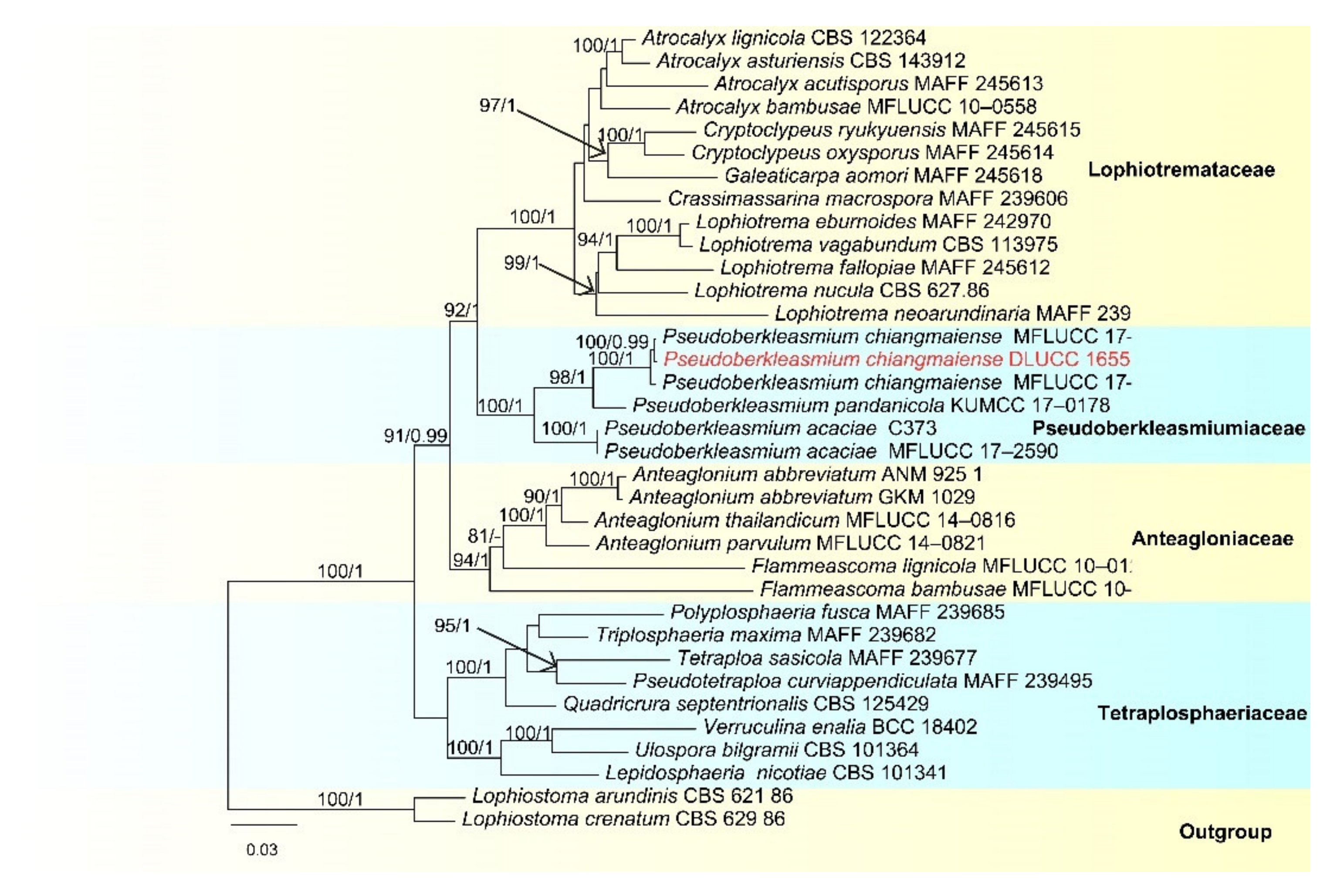
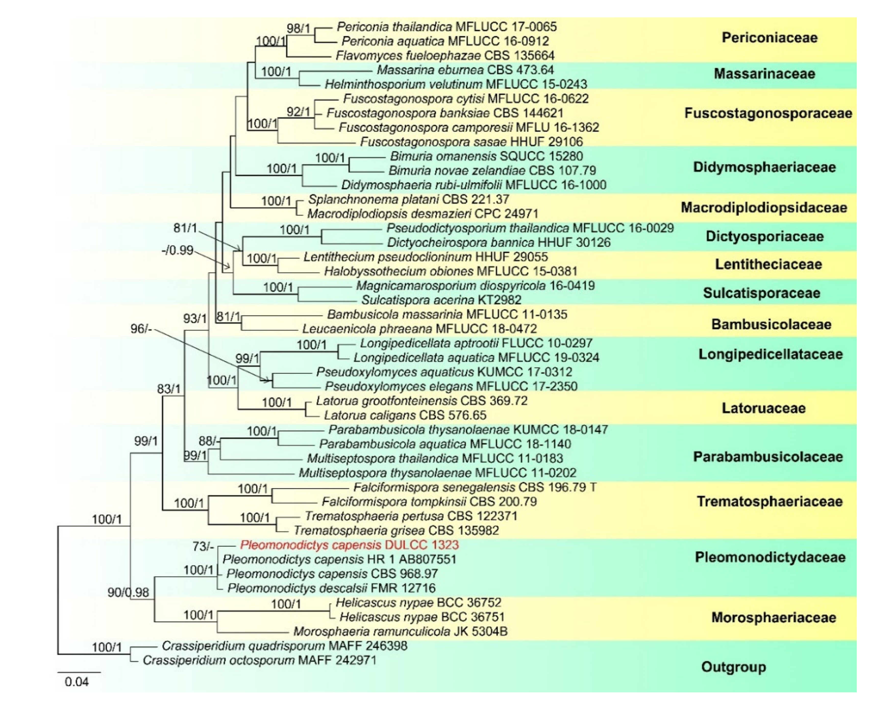
3.1.1. Dothideomycetes O.E. Erikss. and Winka, Myconet 1(1): 5 (1997)

  • Pleosporales Luttr. ex M.E. Barr 1987
  • Pseudoberkleasmiaceae Phukhams. and K.D. Hyde
  • Pseudoberkleasmium Tibpromma and K.D. Hyde
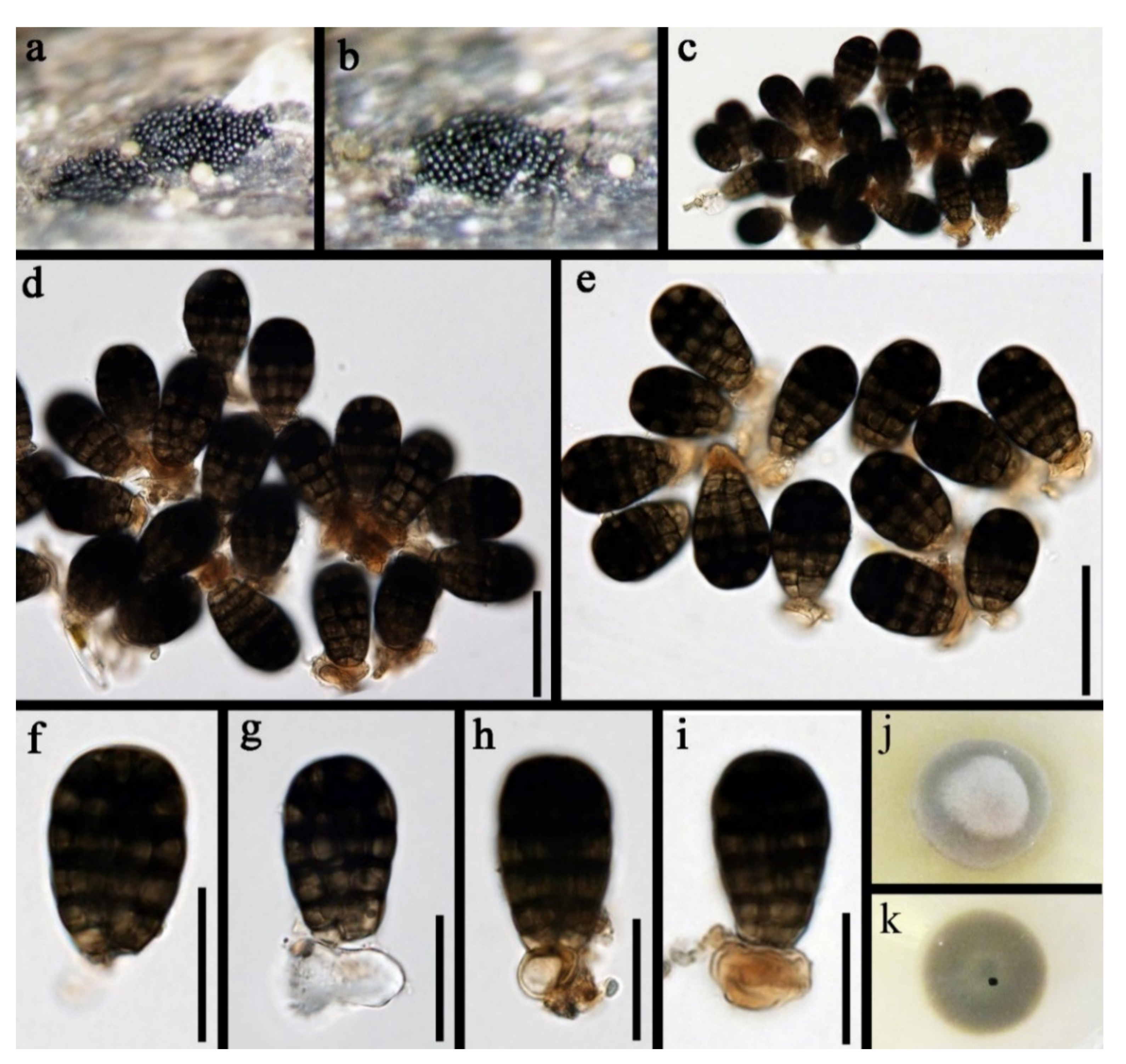
  • Pseudoberkleasmium chiangmaiense Y.Z. Lu and K.D. Hyde, in Hyde et al., Fungal Diversity 96:38 (Figure 1).
Saprobic on submerged decayed wood in freshwater habitats. Sexual morph: Undetermined. Asexual morph: Colonies on substratum superficial, sporodochia, scattered, compact, irregular, black and shining. Mycelium immersed and composed of hyaline to pale brown, branched, septate and smooth-walled hyphae. Conidiophores mononematous and micronematous, reduced to conidiogenous cells, hyaline to pale brown and smooth-walled. Conidiogenous cells monoblastic, holoblastic, determinate, terminal, globose to subglobose, hyaline when young and brown to dark brown when mature. Conidia 17–30 × 19–35 µm, (x = 33 × 18 μm, n = 30) solitary, acrogenous, muriform, obovoid to broadly ellipsoidal, flattened, dark brown to black, paler towards the base and smooth with a hyaline to brown basal cell.
Culture characteristics: Conidia germinating on PDA within 24 h. Colonies grew on PDA, reaching 20–30 mm in three weeks at 25 °C. Mycelia superficial, circular, with the entire margin flat and smooth from above, white at the center, pale grey at the edge, greyish brown from below and not producing pigmentation in culture.
Material examined: Lancang River, Yunnan Province, China, saprobic on submerged decaying wood, 12 November 2017, Z.L. Luo, S-1655 (HKAS 115794), living culture, DLUCC 1655. GenBank accession numbers: (LSU) MZ420759, (SSU) MZ420749, (ITS) MZ420744, (tef-1) MZ442693.
Notes: Pseudoberkleasmium chiangmaiense was introduced by Hyde et al. [90] from Thailand. Phylogenetic analyses showed that our new isolate (DLUCC 1655) clustered with the ex-type strain of P. chiangmaiense (MFLUCC 17–1809) with strong bootstrap support (100 ML/1.00 PP, Figure 2). Morphology of our new isolate is almost identical to the holotype of P. chiangmaiense, except the basal cell of conidia in our isolate are hyaline to brown, while in the holotype they are hyaline. Based on both phylogeny and morphology, we identified our isolate as P. chiangmaiense, which is new to China.
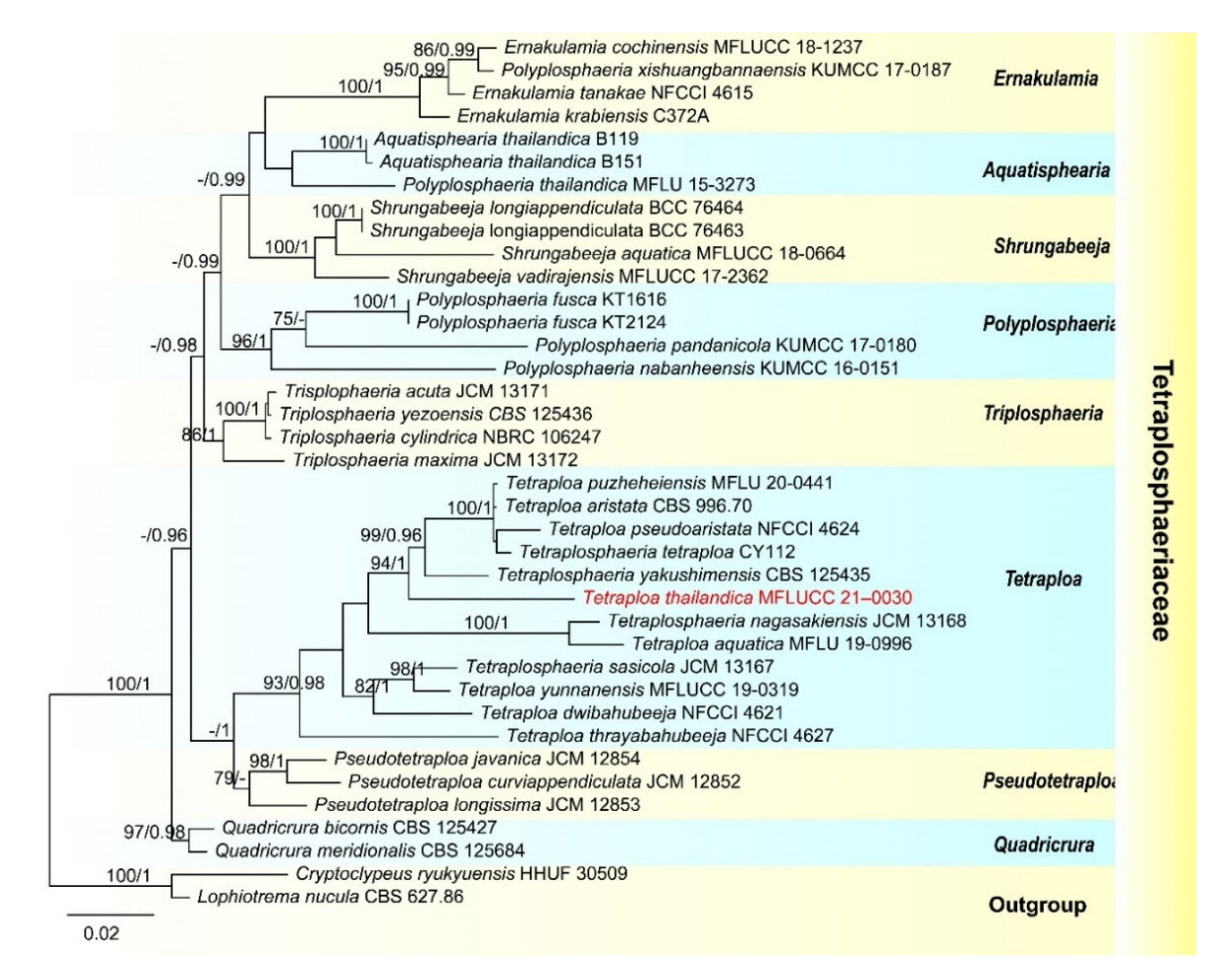
  • Tetraplosphaeriaceae Kaz. Tanaka and K. Hiray
  • Tetraploa Berk. and Broome
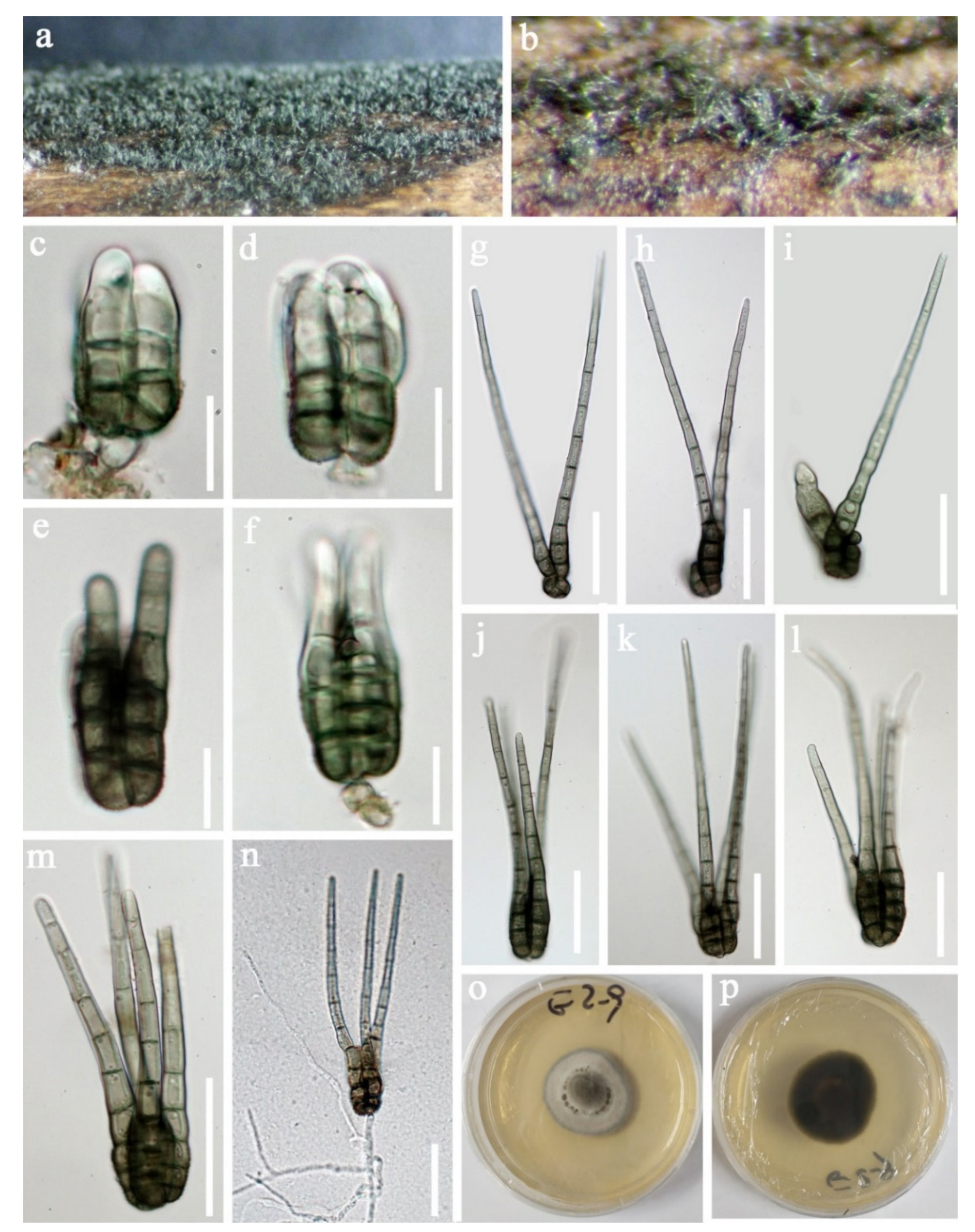
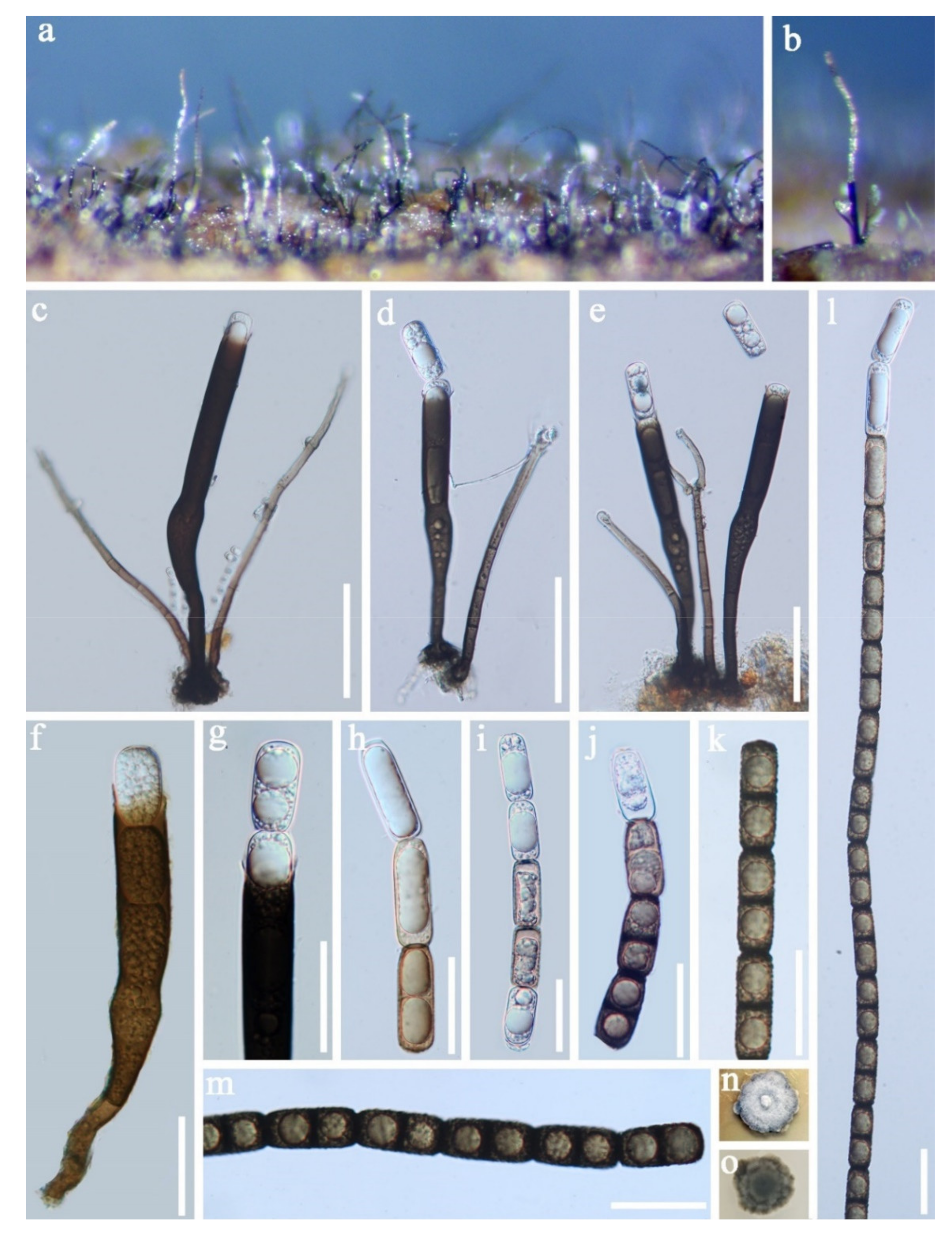
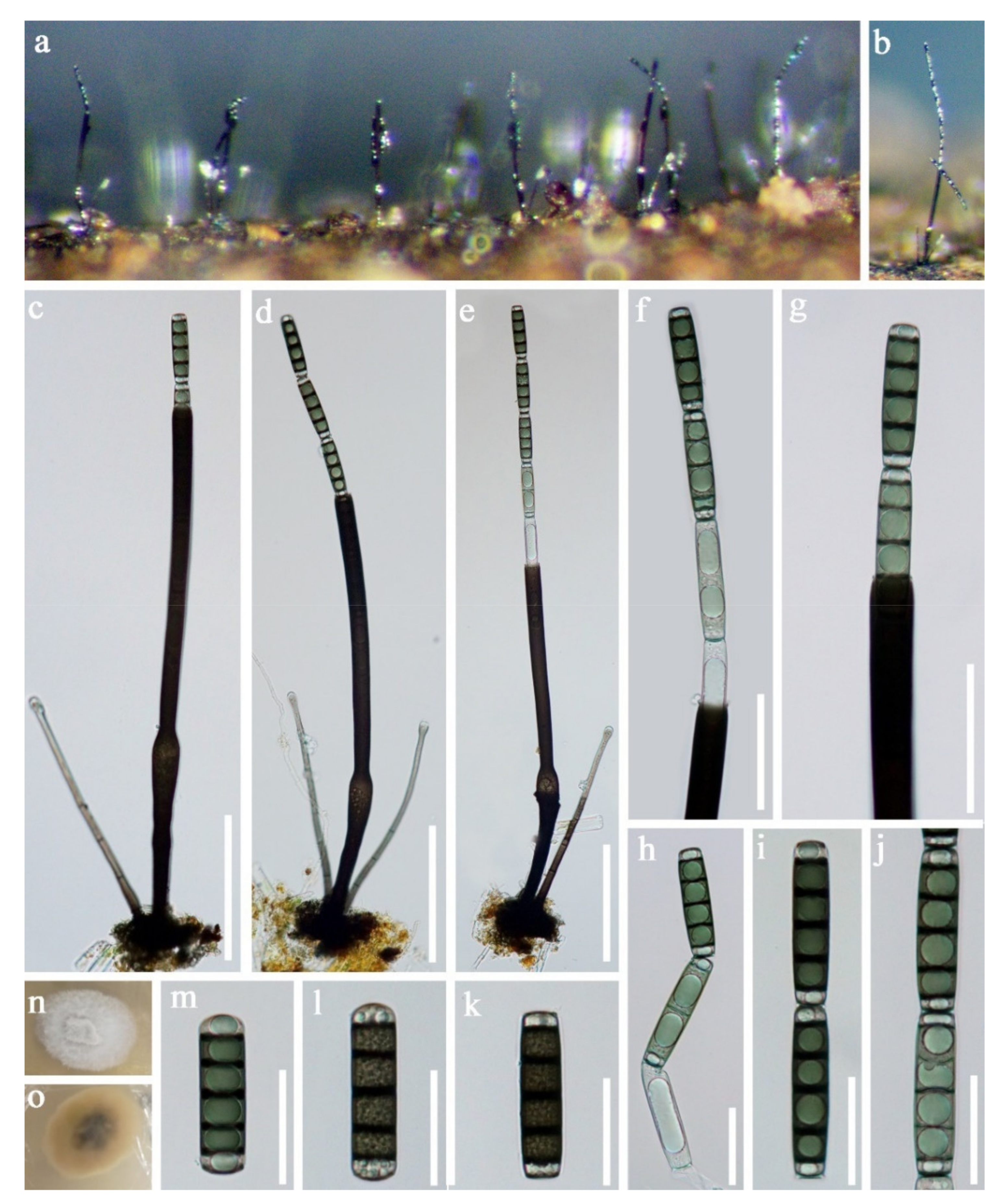
  • Tetraploa thailandica D.F. Bao, H.Y. Su, K.D. Hyde and Z.L. Luo, sp. nov. (Figure 3).
  • Index Fungorum number: IF558591; Facesoffungi number: FoF 09915
  • Holotype: MFLU 21–0030
  • Etymology—Referring to Thailand, where the fungus was collected.
Saprobic on submerged decaying wood. Sexual morph: Undetermined. Asexual morph: Colonies effuse and brown or dark greyish brown. Mycelium mostly immersed, composed of branched, septate, subhyaline and hyphae. Conidiophores indistinct. Conidiogenous cells holoblastic, monoblastic or occasionally polyblastic, integrated, determinate, terminal or intercalary and cylindrical. Conidia solitary, dry, straight, septate, verrucose and composed of a conidial body at the base with 2–4 brown to pale brown apical appendages. Conidial body 23–37 × 17–22.5 µm (x = 30 × 20 μm, n = 30) narrowly ovate or ovate, greyish-brown, pale brown to subhyaline at the apex when young, and brown to dark brown at maturity, verrucose and composed of 2–4 closely-adhered vertical columns of cell, with each column 3–5-septate. Appendages 73–136 µm long, 2–3.5 µm wide at the apex and 5.5–8 µm at the base (n = 35). Setose brown to greyish brown, 5–10-pseudoseptate and smooth-walled.
Culture characteristics: Conidia germinating on PDA within 24 h. Germ tubes were produced from the basal cell of conidia. Colonies grew on MEA, reaching 25–35 mm in three weeks at 25 °C. Mycelia was superficial and circular with the entire margin flat and smooth, greyish brown from above, dark brown to black from below and not producing pigmentation in culture.
Material examined: Sakon Nakhon, Tao Ngoi, Thailand., saprobic on submerged decaying wood, 12 November 2017, D.F. Bao, B144 (MFLU 21–0030, holotype), ex-type living culture, MFLUCC 21–0030. GenBank accession numbers: (LSU) MZ412530, (SSU) MZ413274, (ITS) MZ412518.
Notes: species of Tetraploa are quite similar and the asexual morphs are characterized by micronematous conidiophores, monoblastic conidiogenous cells and tetraploate conidia composed of four columns, which are short-cylindrical, euseptate, brown, verrucose at the base and with 2–4-setose and septate appendages at the apex [91,92,93]. Tetraploa thailandica is morphologically most similar to T. aquatica. They share some characteristicss, such as monoblastic or occasionally polyblastic conidiogenous cells and conidia composed of four closely-adhered vertical columns of cells, with 2–4 apical appendages. However, T. aquatica has a shorter but wider conidial body (23–37 × 17–22.5 vs. 22.5–27 × 20–24). Moreover, the vertical columns of T. thailandica are 3–5-septate, while those of T. aquatica are 2–3-septate [92].
In our phylogenetic analyses, Tetraploa thailandica formed a distinct lineage within the genus and was close to T. yakushimensis (Figure 4). However, T. thailandica differs from T. yakushimensis in having 3–5-celled and narrower conidia (17–22.5 vs. 20–30 µm) composed of 2–4 closely-adhered vertical columns of cell and 2–4 setose appendages. Conidia of T. yakushimensis are 4-celled, composed of four columns and four appendages [91]. Moreover, T. thailandica has more septa in the appendages than T. yakushimensis (5–10 vs. 3–8).
  • Pleomonodictydaceae Hern.-Restr., J. Mena and Gené
  • Pleomonodictys Hern.-Restr., J. Mena and Gené
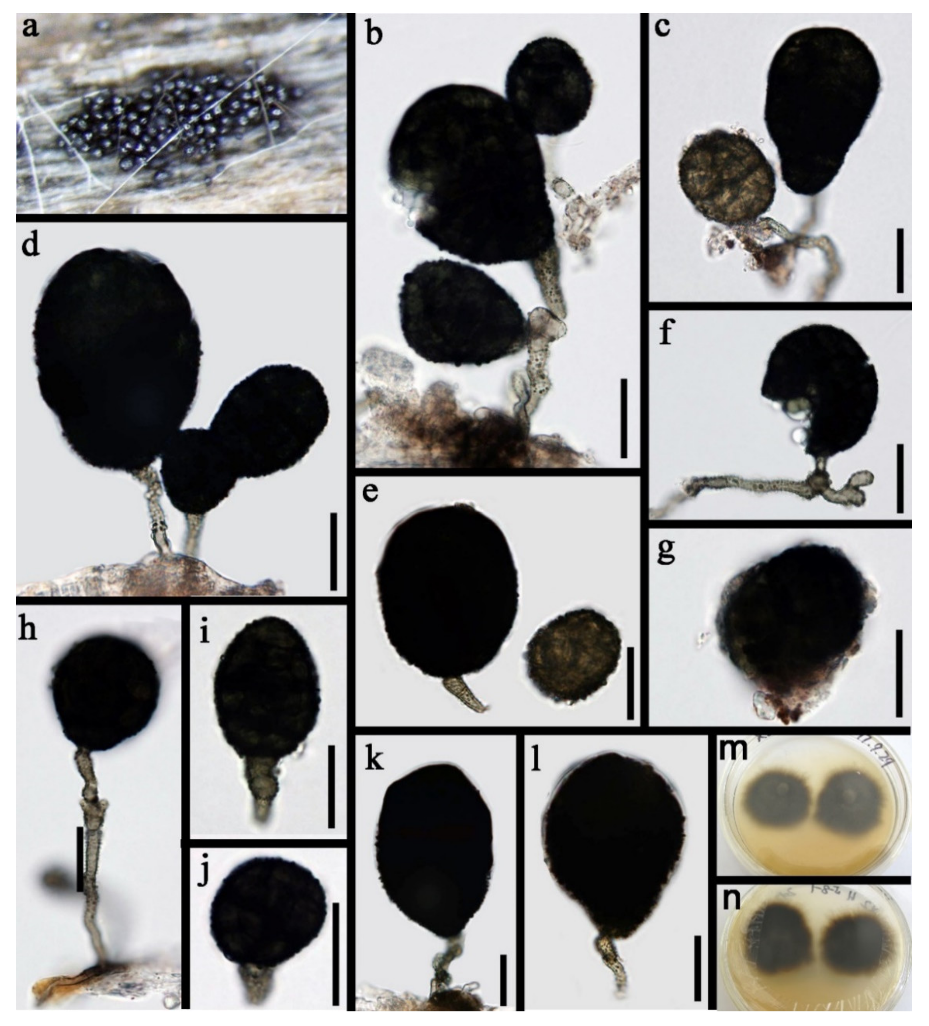
  • Pleomonodictys capensis (R.C. Sinclair, Boshoff and Eicker) Hern.-Restr., J. Mena and Gené, Studies in Mycology 86:77 (2017) (Figure 5).
Saprobic on submerged decayed wood in freshwater habitats. Sexual morph: Undetermined. Asexual morph: Colonies effuse on natural substrate, scattered, punctiform, dark brown to black and glistening. Mycelium mostly immersed and composed of branched verrucose, septate, brown to pale brown and hyphae. Conidiophores 4–6.5 μm wide (x = 5.0 μm, n = 20), mononematous, micronematous to semi-macronematous, cylindrical, straight or flexuous, septate, branched or unbranched, pale brown to brown and verrucose. Conidiogenous cells polyblastic, discrete, intercalary and brown. Conidia 28.5–60 × 23.5–44 μm (x = 44 × 33.7 μm, n = 30), pleurogenous, solitary, ellipsoid to subglobose, obovoid, pyriform to broadly clavate, muriform, dark brown to black with base rounded or truncate and thick-walled.
Culture characteristics: Conidia germinating on PDA within 24 h. Colonies grew on MEA, reaching 25–30 mm in four weeks at 25 °C, with rough surface, dense mycelia, velvety, dry, dark brown from above, dark brown to black from below; not producing pigmentation in culture.
Material examined: China, Tibet autonomous region, on submerged decaying wood, May 2017, Z.L. Luo, S-1323 (HKAS 115793), living culture, DLUCC 1323. GenBank accession numbers: (LSU) MZ420757, (ITS) MZ420742, (RPB2) MZ442696.
Notes: Pleomonodictys was established for two monodictys-like species, Pleomonodictys capensis and P. descalsii [94]. The genus is characterized by micronematous or semi-macronematous conidiophores, often reduced to conidiogenous loci on the hyphae and blastic conidia which are solitary or in short chains, variable in shape, muriform, dark brown to black. Our species fits well within the species concept of Pleomonodictys.
Morphologically, the new collection (MFLUCC 18–1499) is almost identical to the holotype of P. capensis, such as micronematous to semi-macronematous, pale brown to brown, verrucose conidiophores and muriform, obovoid, ellipsoid to subglobose, broadly clavate to pyriform, dark brown to black conidia and conidial size of our collection overlapping with the holotype (28.5–60 × 23.5–44 vs. 30–100 × 17–60 μm) [95]. Thus, we identified our collection (MFLUCC 18–1499) as P. capensis. Pleomonodictys capensis was introduced by Sinclair et al. [95], collected from terrestrial habitats in South Africa. Our collection was collected from freshwater habitat in China, a new record for China.
Our phylogenetic analyses showed that the strains of P. capensis and P. descalsii clustered together with high bootstrap support (100 ML/1.00 PP, Figure 6). Hernandez-Restrepo et al. [94] introduced P. descalsii almost based on the conidial size, which is much smaller than P. capensis. However, the conidial size of our collection is quite similar to P. descalsii (28.5–60 × 23.5–44 vs. 28–70 × 24–54 μm) and other morphological characters are indistinguishable. Thus, future morphology and molecular studies are required to confirm the relationship between these two species.

3.1.2. Sordariomycetes

  • Diaporthomycetidae
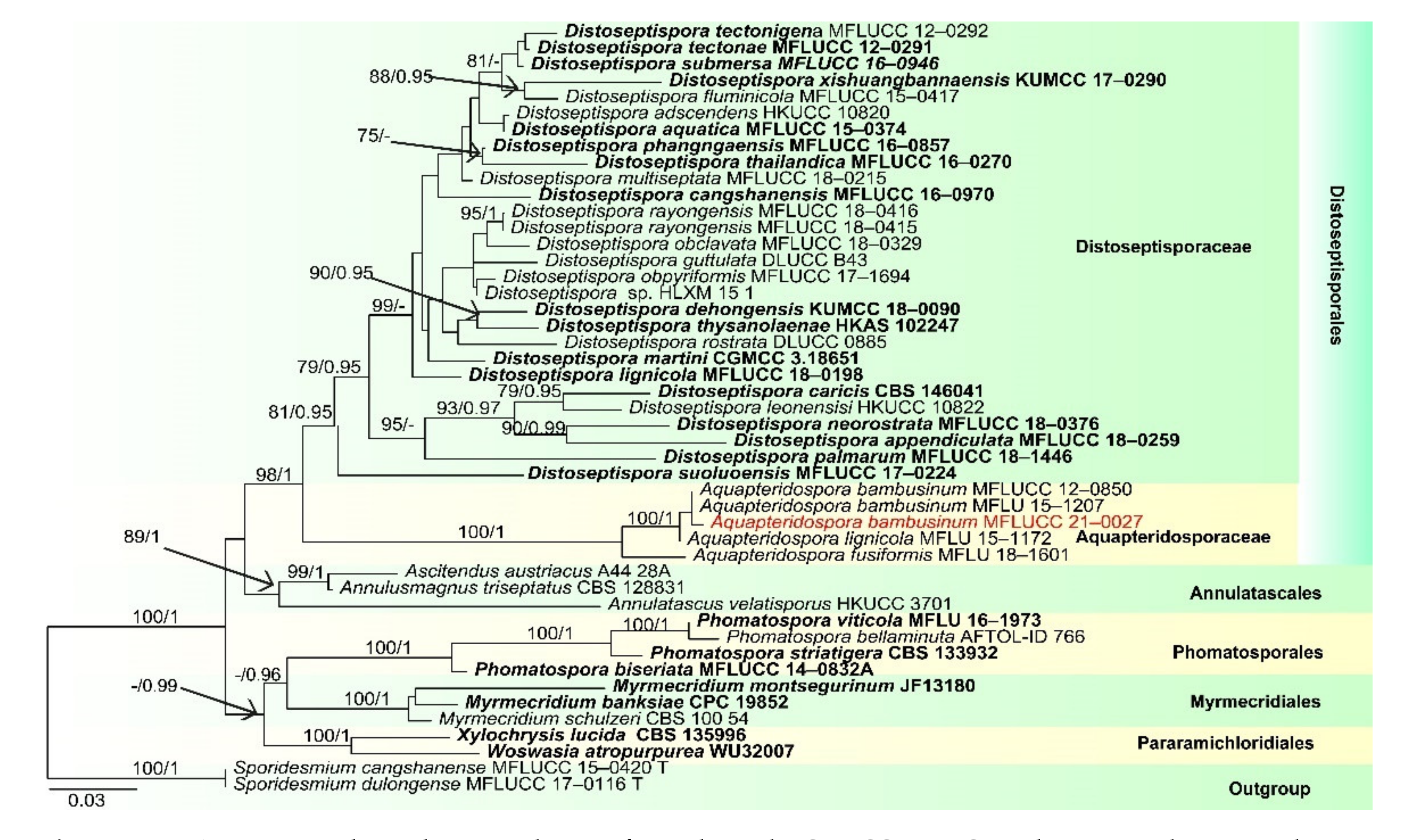
  • Distoseptisporales Z.L. Luo, K.D. Hyde and H.Y. Su
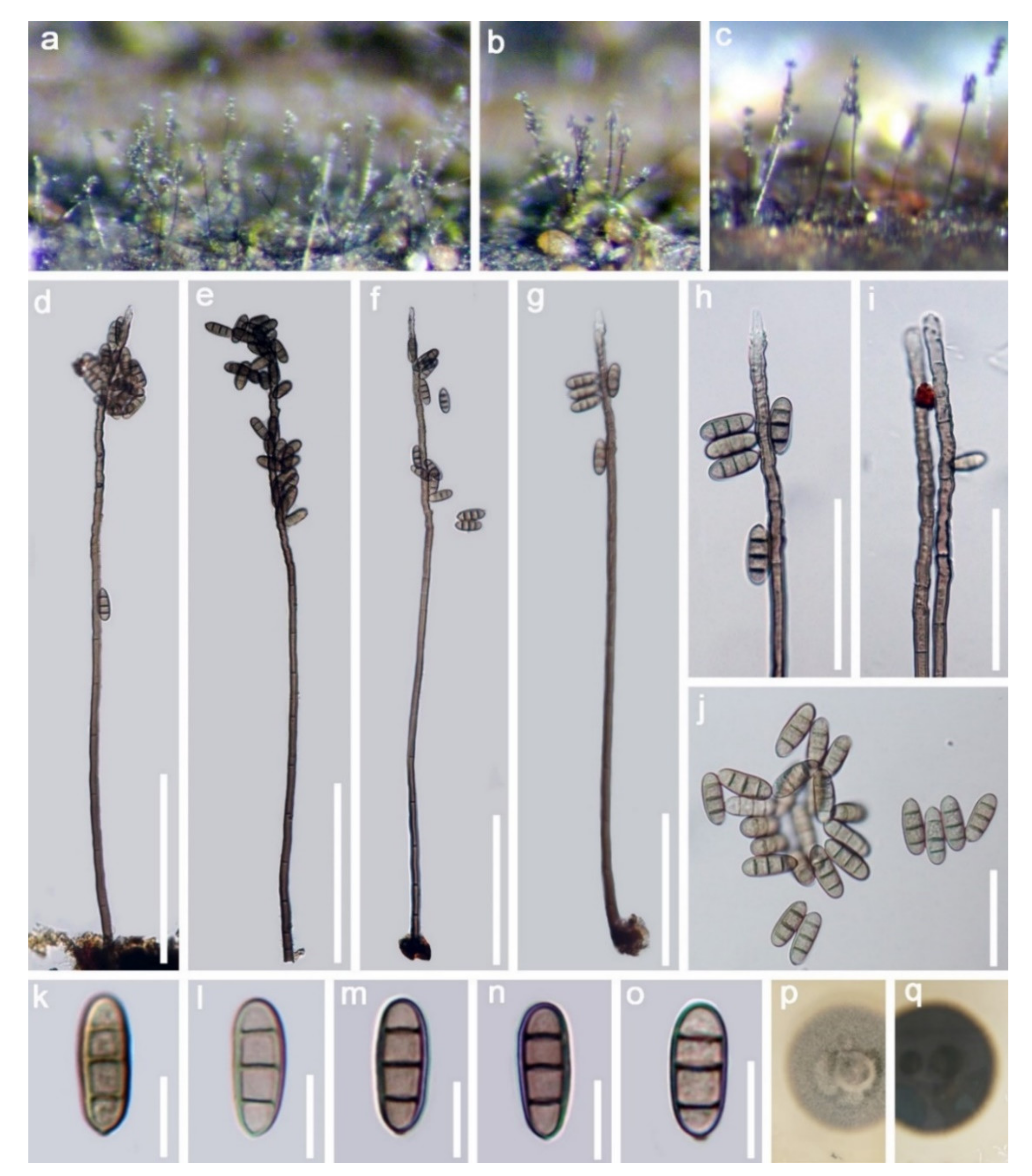
  • Aquapteridosporaceae K.D. Hyde and Hongsanan
  • Aquapteridospora J. Yang, K.D. Hyde and Maharachch
  • Aquapteridospora bambusinum (D.Q. Dai and K.D. Hyde) D.F. Bao, com. nov. (Figure 7).
  • ≡Pleurophragmium bambusinum D.Q. Dai and K.D. Hyde, in Dai et al., Fungal Diversity 82:92
  • Index Fungorum number: IF 558592
Saprobic on submerged decayed wood in freshwater habitats. Sexual morph: Undetermined. Asexual morph: Colonies on substratum, effuse, hairy and brown to dark brown. Mycelium partly immersed and composed of septate, branched, smooth and dark brown hyphae. Conidiophores 615–715 × 9–13 μm (x = 695.5 × 11 μm, n = 20), macronematous, mononematous, erect, simple, septate, straight or slightly flexuous, dark brown, paler to subhyaline towards apex and smooth. Conidiogenous cells polyblastic, sympodial, denticulate, integrated, terminal and hyaline to pale brown. Conidia 15–18 × 5.5–7 μm (x = 16.5 × 6.5 μm, n = 35) acrogenous, solitary, ellipsoid to fusiform with round ends, straight, slightly narrow towards the base, pale brown to dark brown, 3-septate, slightly constricted at the septa, thick-walled and smooth.
Culture characteristics: Conidia germinating on PDA within 24 h. Colonies grew on PDA, reaching 20–25 mm in two weeks at 25 °C. Colonies were medium sparse, circular, flat, a slightly rough surface with edge, entire margin well-defined, grey to brown from above, dark brown to black from below and not producing pigmentation in culture.
Material examined: Amphoe Thai Charoen Province, Thailand, on submerged decaying wood, acquired on 13 November 2018, D.F. Bao, B113 (MFLU 21–0027), living culture, MFLUCC 21–0027. GenBank accession numbers: (LSU) MZ412526, (SSU) MZ413270, (ITS) MZ412514, (tef-1) MZ442688.
Notes: Pleurophragmium bambusinum was described by Dai et al. [96] and was placed in Sordariomycetes incertae sedis. Their phylogenetic analyses showed that P. bambusinum clustered with Ellisembia adscendens in Sordariomycetes, genera incertae sedis. However, in our analyses, P. bambusinum was placed in Aquapteridosporaceae and clustered with Aquapteridospora species with strong support (100 ML/1.00 PP, Figure 8). P. bambusinum is characterized by mononematous, macronematous, simple conidiophores, polyblastic, denticulate, sympodial conidiogenous cells and ellipsoid, brown, 3-septate, conidia. Pleurophragmium bambusinum shares some similar characters with Aquapteridospora, such as integrated, polyblastic, terminal conidiogenous cells and acrogenous, fusiform, 3-septate conidia. Based on both phylogeny and morphology, we transferred P. bambusinum to Aquapteridospora and synonymized Aquapteridospora bambusinum instead of P. bambusinum.
In our phylogenetic analyses, our new isolate clustered with two strains of Aquapteridospora bambusinum with high bootstrap support (Figure 8). Morphology of our collection is identical to A. bambusinum. Therefore, we identified our new collection as A. bambusinum and it is phylogenetically close to A. lignicola but differs by longer conidiophores (615–715 × 9–13 vs. 70–200 × 4–7 μm) and pale brown to dark brown, ellipsoid to fusiform conidia with round ends, lacking a sheath. Conidia of A. lignicola are fusiform with obtuse ends, central cells are pale to dark brown and end cells are subhyaline, with a conspicuous sheath [27].
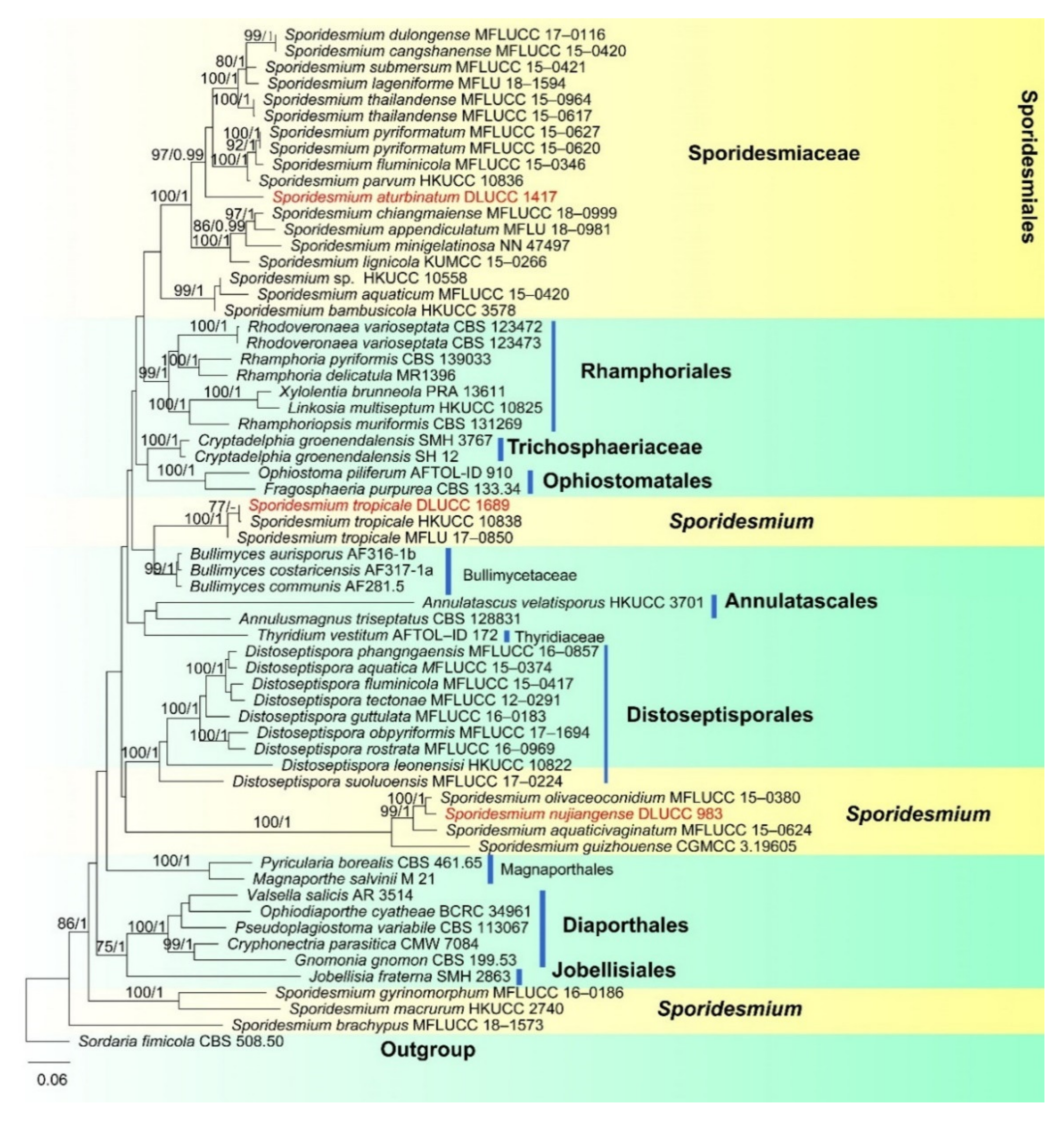
  • Sporidesmiales Crous
  • Sporidesmiaceae Fr., Summa veg
  • Sporidesmium Link, Mag. Gesell. naturf.
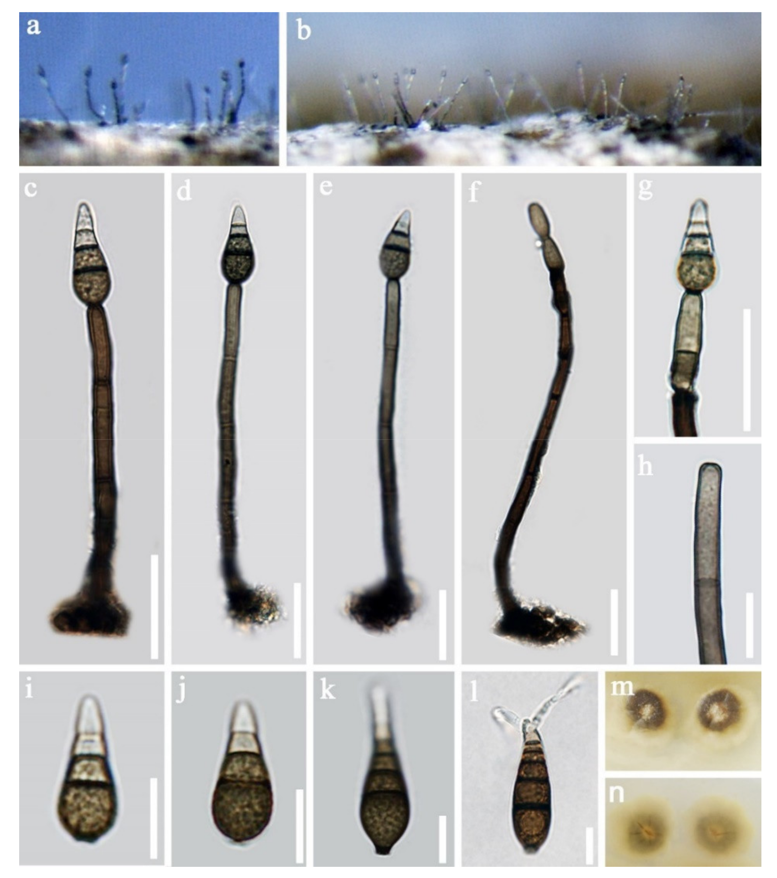
  • Sporidesmium aturbinatum (S. Hughes) M.B. Ellis, Mycol. Pap. 70: 49 (1958). (Figure 9).
Saprobic on submerged decaying wood. Sexual morph: Undetermined. Asexual morph: Colonies effuse, hairy, dark brown to black and glistening. Mycelium partly immersed in the substratum, partly superficial, composed of septate, branched and brown with smooth hyphae. Conidiophores 65–111 × 4–5 µm (x = 88.5 × 4.5 μm, n = 25), macronematous, mononematous, solitary or sometimes in a small group, cylindrical, straight or slightly flexuous, with up to three successive profilerations, septate, dark brown and paler towards the apex. Conidiogenous cells 11–28 × 4–5 µm (x = 14.5 × 4.5 μm, n = 25), integrated, terminal, holoblastic, monoblastic, cylindrical and sometimes with percurrent proliferation through the scar. Conidia 16–28 × 7–10 µm (x = 21.5 × 8.5 μm, n = 25), acrogenous, solitary, dry, pyriform, rostrate, tapering to the apex, 1.5–2.5 µm wide at apex, truncate and 2.8–3.6 µm wide at base, straight or slightly curved, 3(–4)-septate, slightly constricted at septa, two upper cells subhyaline to hyaline, lower cells dark brown and verrucose.
Culture characteristics Conidia germinating on PDA within 24 h. Colonies growing on PDA, reaching 15–20 mm in two weeks at 25 °C, surface rough, with dense mycelia, dry, rigid, from above greyish white at the center, brown at the edge, from below yellowish brown at the center, greyish brown at the edge and not producing pigmentation in culture.
Material examined: Gaoligongshan Mountain, Yunnan Province, China, on submerged decaying wood, in August 2016, H.W. Shen, S1417 (HKAS 115800), living culture, DLUCC 1417. GenBank accession numbers: (LSU) MZ420758, (ITS) MZ420743, (tef-1) MZ442692, (RPB2) MZ442697.
Notes: Sporidesmium aturbinatum was introduced by Ellis [97] and is characterized by conidiophores that are mononematous, macronematous, solitary or in groups, with up to three successive profilerations and septate, conidiogenous cells that are integrated, terminal, monoblastic and conidia that are pyriform, 2–4-septate, upper cells being subhyaline to hyaline and smooth-walled, lower cells being dark brown andverrucose. Our new collection fits well with the original description of S. aturbinatum [97]. Phylogenetic analysis showed that S. aturbinatum formed a distinct lineage within Sporidesmiaceae (Figure 10). Sporidesmium aturbinatum has been collected from England and Ireland on rotten wood of Sambucus and dead culms of Ammophila, respectively [97,98]. Our collection was from freshwater habitats in China.
Sporidesmium tropicale M.B. Ellis, Mycol. Pap. 70: 58 (1958) (Figure 11).
Saprobic on submerged decaying wood. Sexual morph: undetermined and for asexual morph, colonies on superficial substratum, scattered, hairy, effuse and brown to dark brown. Mycelium mostly immersed, composed of septate, branched, pale mid-brown to brown and with smooth-walled hyphae. Conidiophores 71–163 × 5–8 µm (x = 117 × 6.5 μm, n = 25), macronematous, mononematous, unbranched, cylindrical, erect, straight or slightly flexuous, single, 4–8-septate, dark brown, paler towards apex, smooth and thick-walled. Conidiogenous cells 13–22 × 5–6 µm (x = 17.5 × 5.5 μm, n = 25), monoblastic, holoblastic, terminal, integrated, percurrently proliferating, cylindrical and brown. Conidia 94–184 × 13–15 µm (x = 139.5 × 13.5 μm, n = 25), acrogenous, solitary, dry, pyriform, rostrate, obclavate, with a long and slender apex, straight or slightly curved, tapering to the apex, 3–5 μm wide and truncate at the base, dark brown, pale brown and 2–3 μm wide at the apex, 4–17-septate, thick-walled and with the proximal part usually verrucose.
Culture characteristics: Conidia germinating on PDA within 24 h, germ tubes produced from the basal and apical cell of conidia. Colonies growing on MEA, reaching 20–25 mm in two weeks at 25 °C, surface rough, with dense mycelia, dry, flat, rugose, from above dark brown, from below dark brown to black and not producing pigmentation in culture.
Material examined: Lancang River, Yunnan Province, China, on submerged decaying wood, acquired on July 2016, Z.L. Luo, S1689 (HKAS 115799), living culture, DLUCC 1689. (LSU) MZ420760, (ITS) MZ420745, (RPB2) MZ442698.
Notes: Sporidesmium tropicale was described by Ellis [99]. The species has a wide distribution, known from Bolivia, China, India, Ghana, Jamaica, Malaya, Nigeria, Sierra Leone, Sri Lanka, Thailand and USA [97,99,100,101]. Yang et al. [101] has provided sequence data for this species. In our phylogenetic analyses (Figure 9), our new collection clustered with two strains of S. tropicale (HKUCC 10838 and MFLU 17–0850) with strong support (100% ML and 1.00 PP). Morphology of our collection is almost the same as S. tropicale [101]. We, therefore, identified our species as S. tropicale.
Placement of Sporidesmium tropicale is still questionable. In the phylogenetic analyses of Yang et al. [101] S. tropicale clustered with Bullimyces communis, while Liu et al. [102] showed that it clustered with Cryptadelphia groenendalensis. Our phylogenetic analysis was similar to Yang et al. [101]. Sporidesmium tropicale clustered with C. groenendalensis, distant from Sporidesmiaceae (Figure 10). Further molecular studies are required to clarify the placement of this species.
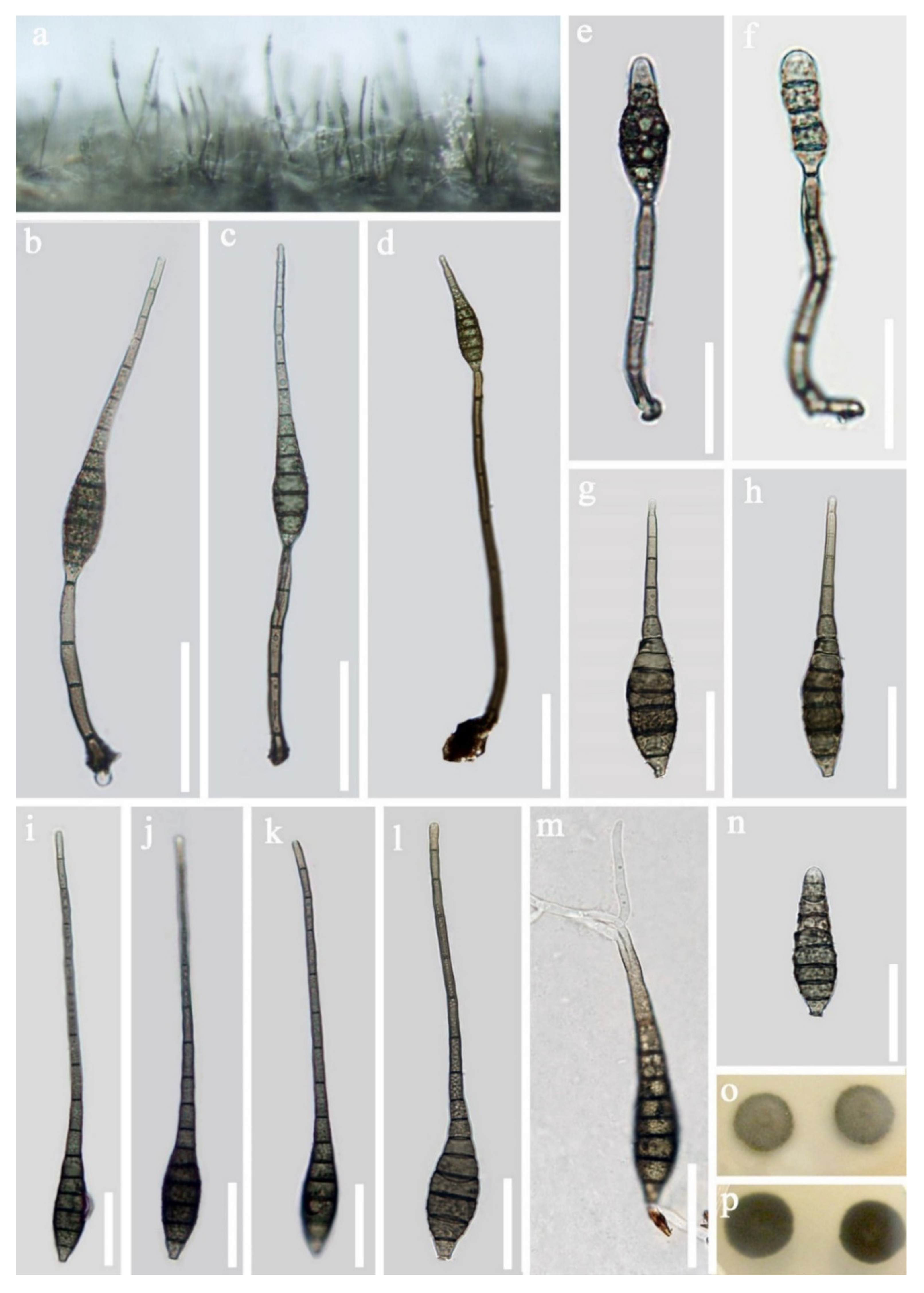
  • Sporidesmium nujiangense D.F. Bao, H.Y. Su, K.D. Hyde and Z.L. Luo, sp. nov. (Figure 12).
  • Index Fungorum number: IF558594; Faces of fungi number: FoF 09916
  • Holotype—HKAS 115795
  • Etymology—Referring to the place “Nujiang River” where this species was collected.
Saprobic on submerged decayed wood in freshwater habitats. Sexual morph: Undetermined. Asexual morph: Colonies effuse, velvety, hairy and brown to pale brown. Mycelium partly immersed, composed of septate, smooth and branched hyaline to pale brown hyphae. Conidiophores 31–51 × 4–5.5 µm (x = 40.8 × 4.8 μm, n = 27), mononematous, macronematous, erect, solitary or in small groups, cylindrical, truncate at apex, slightly swollen at the base, straight or slightly flexuous, 1–4-septate, brown to dark brown, smooth-walled. Conidiogenous cells 11.5–16.5 × 4–5 µm (x = 14 × 4.5 μm, n = 24), holoblastic, monoblastic, terminal, integrated, determinate, cylindrical and dark brown. Conidia 54–69 × 10–12.5 µm (x = 61.5 × 11 μm, n = 30), acrogenous, solitary, dry, obclavate, tapering to the apex, 2.5–3.5 µm wide at apex, truncate and 3.5–5.0 µm wide at base, brown to greyish brown, pale brown to subhyaline towards the apex, straight or slightly curved, 10–14-septate, mostly 11-septate, smooth-walled and with a mucilaginous sheath over the apex.
Culture characteristics: Conidia germinating on PDA within 24 h, with germ tubes produced from the apical cell of conidia. Colonies grew on PDA, reaching 10–15 mm in one week at 25 °C, rough surface, with dense mycelia, dry, flat, rigid, umbonate from the side view and entire edge, greyish brown from above, dark brown from below andnot producing pigmentation in culture.
Material examined: Nujiang River, Yunnan Province, China on submerged decaying wood, in July 2016, Z.L. Luo, S983 (HKAS 115795, holotype), ex-type living culture, DLUCC 983. GenBank accession numbers: (LSU) MZ420756, (SSU) MZ420748, (ITS) MZ420741, (tef-1) MZ442691, (RPB2) MZ442695.
Notes: In the phylogenetic analyses, Sporidesmium nujiangense is placed as a sister taxon to S. olivaceoconidium with strong bootstrap support (100% ML and 1.00 BYPP, Figure 10). Sporidesmium nujiangense resembles S. olivaceoconidium in having macronematous, mononematous, septate conidiophores, holoblastic, monoblastic, integrated conidiogenous cells and solitary, obclavate, brown conidia with a mucilaginous sheath at the tip. However, S. olivaceoconidium has smaller conidia (25–50 × 6–10 µm) and less septa (6–10 vs. 10–14) [103]. Comparison of ITS and RPB2 regions revealed 12 bp (3.8%) and 28 bp (2.8%) differences, respectively. Therefore, we introduce S. nujiangense as a new species [104].
Morphologically, Sporidesmium nujiangense is similar to S. appendiculatum and S. chiangmaiense in having obclavate to fusiform conidia with a mucilaginous sheath over the apex. However, conidia of both S. appendiculatum and S. chiangmaiense have a filamentous appendage at the apex, while S. nujiangense lacks this character. Moreover, our phylogenetic analysis showed that S. nujiangense is placed in a different clade to S. appendiculatum and S. chiangmaiense.
Conidia of Sporidesmium aquaticivaginatum and S. guizhouense also have a mucilaginous sheath at the conidial tip. However, S. aquaticivaginatum has longer conidiophores (60–125 × 4–6 μm) and smaller conidia with fewer septa (49.5–80.5 × 10.5–14 μm, 6–10-septate) [103]. S. guizhouense has larger conidia (46–86 × 7–11.4 µm) and more conidial septa (9–16-septate) [102].
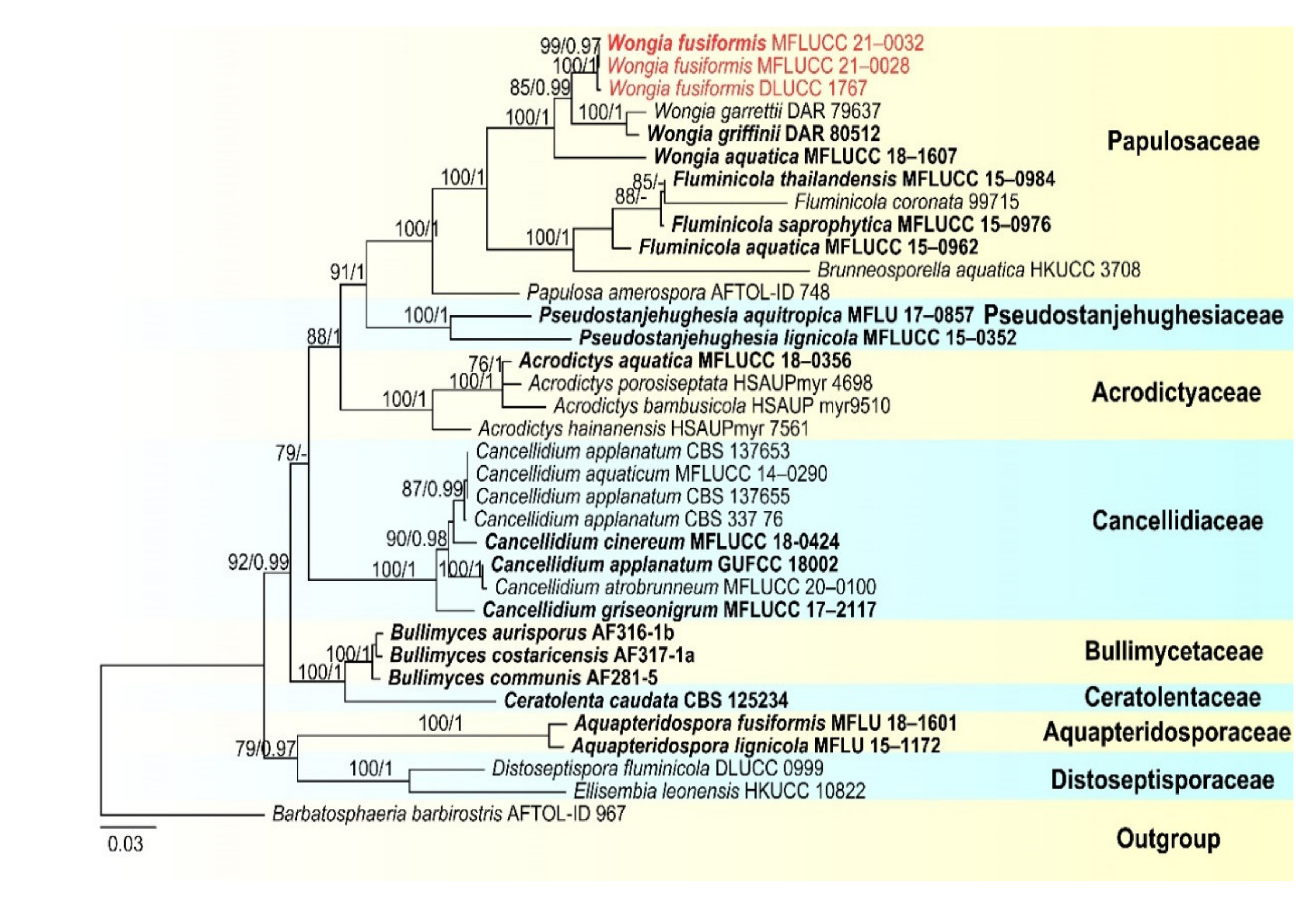
  • Papulosaceae Winka and O.E. Erikss., Mycoscience 41 (2):102 (2000)
  • Wongia Khemmuk, Geering and R.G. Shivas
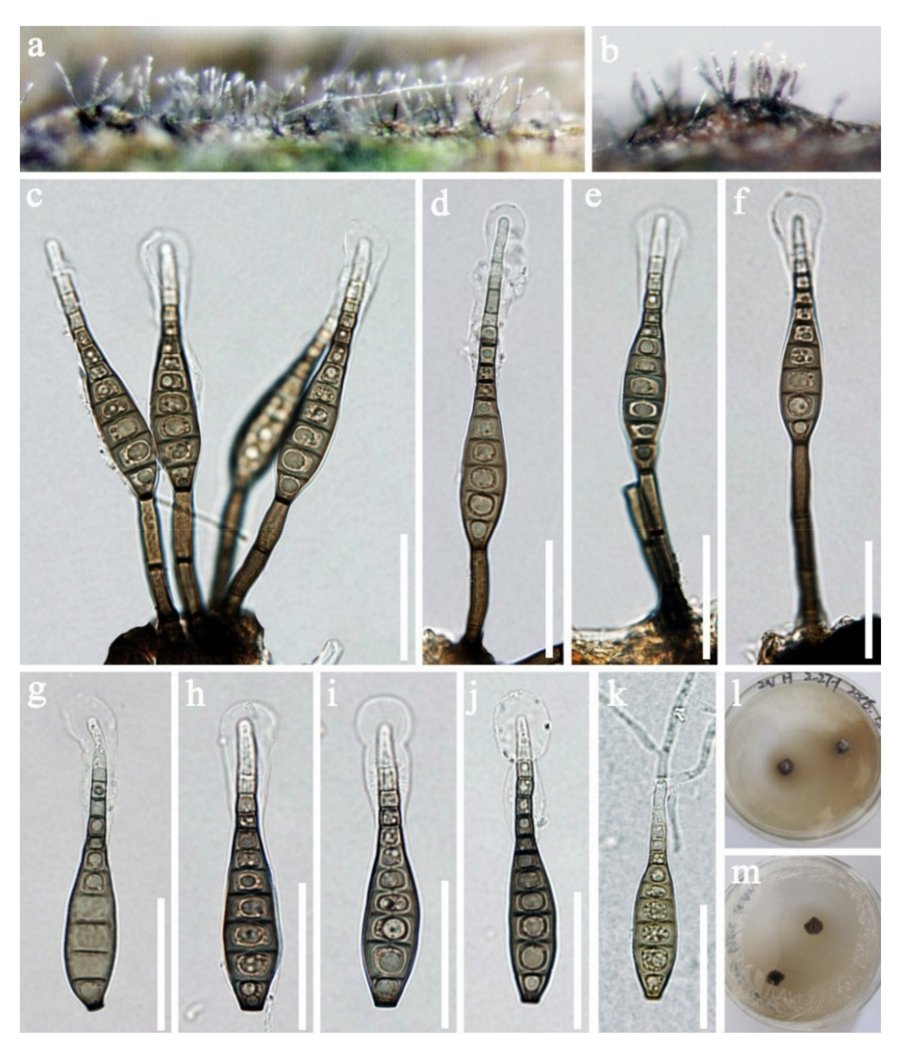
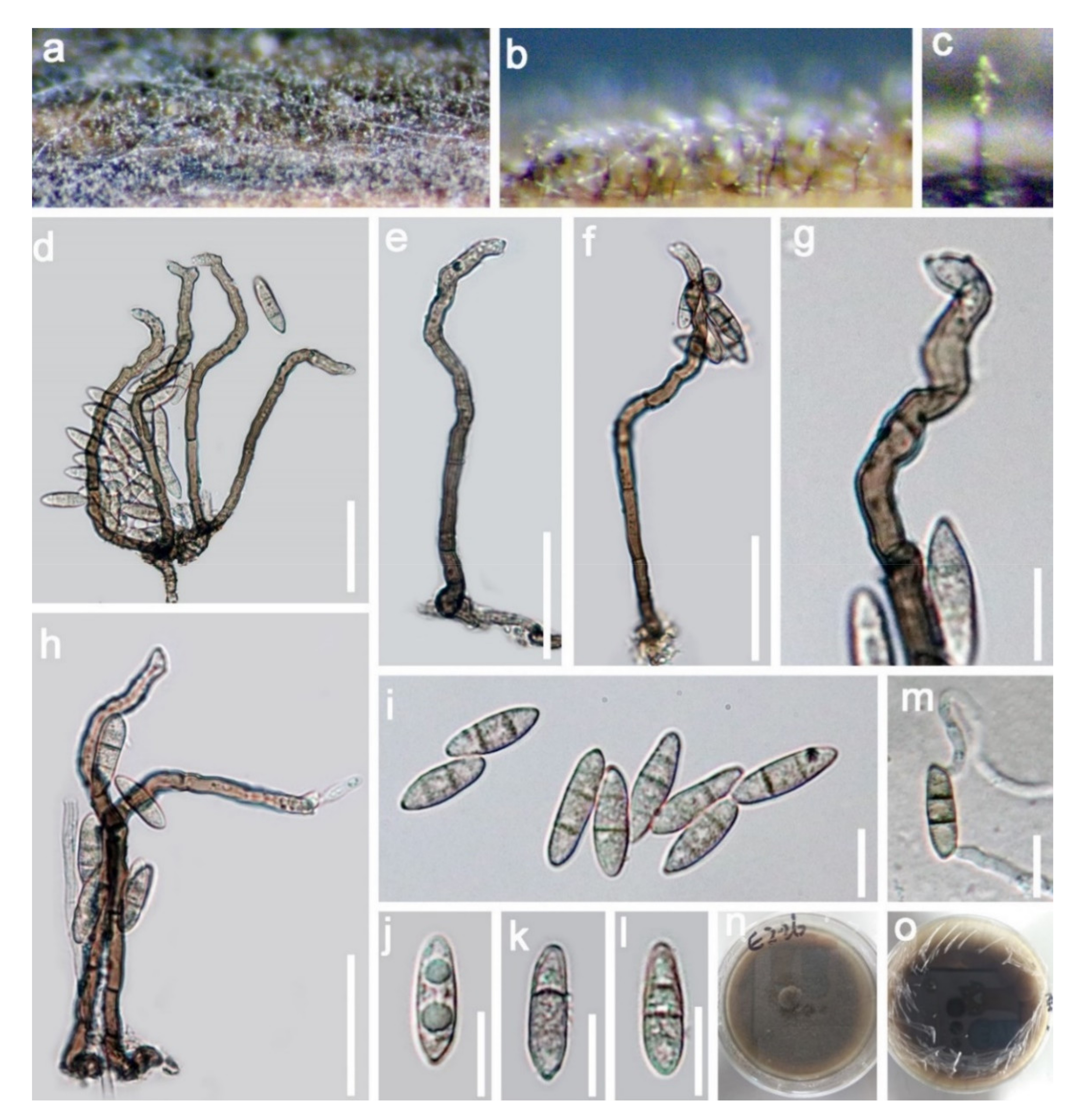
  • Wongia fusiformis D.F. Bao, H.Y. Su, K.D. Hyde and Z.L. Luo, sp. nov. (Figure 13).
  • Index Fungorum number: IF558595; Facesoffungi number: FoF 09917
  • Holotype—MFLU 21–0028
  • Etymology—Referring to the fusiform conidia of this fungus
Saprobic on submerged decayed wood in freshwater habitats. Sexual morph: Undetermined. Asexual morph: Colonies on superficial substratum, hairy, scattered, granulate and black. Mycelium partly superficial and partly immersed, composed of branched, septate, smooth and with brown to pale brown hyphae. Conidiophores 70–105 × 4–5 µm, (x = 87 × 4 μm, n = 25) macronematous, mononematous, simple, solitary, septate, erect, straight or slightly flexuous, dark brown, paler towards apex and smooth-walled. Conidiogenous cells polyblastic, denticulate, terminal, sympodial and subhyaline to pale brown. Conidia 13–18 × 4–5 µm, (x = 15 × 5 μm, n = 30) acropleurogenous, solitary, fusiform, clavate, (1)–2-septate, pale brown to brown, tapering and pointed at both ends, with a guttule in each cell when young and smooth-walled.
Culture characteristics: Conidia germinating on PDA within 24 h and germ tubes produced from the basal and apical cell of conidia. Colonies grew on PDA, reaching 45–50 mm in four weeks at 25 °C, rough surface, with dense mycelia, velvety, dry, umbonate in the middle from the side view, edge undulate, brown to dark brown from above, dark brown from below and not producing pigmentation in culture.
Material examined: Sakon Nakhon, Tao Ngoi, Thailand, on submerged decaying wood, 12 November 2017, D.F. Bao, B117 (MFLU 21–0029, holotype), ex-type living culture, MFLUCC 21–0032. GenBank accession numbers: (LSU) MZ412527, (SSU) MZ413271, (ITS) MZ412515, (tef-1) MZ442689. Tao Ngoi, Sakon Nakhon, Thailand, on submerged decaying wood, 12 November 2017, D.F. Bao, B140 (MFLU 21–0028), living culture, MFLUCC 21–0028. (LSU) MZ412529, (SSU) MZ413273, (ITS) MZ412517, (tef-1) MZ442690. Lancang River, Yunnan Province, China, on submerged decaying wood, 12 November 2017, Z.L. Luo, S-1797 (HKAS 115798), living culture, DLUCC 1767. GenBank accession numbers: (LSU) MZ420761, (SSU) MZ420750, (ITS) MZ420746.
Notes: in the current phylogenetic analyses, our three new isolates clustered together, sister to the clade of Wongia garrettii and W. griffinii with strong bootstrap support (100 ML/1.00 PP, Figure 14). Wongia garrettii and W. griffinii were introduced with a sexual morph [105], but W. fusiformis is known only from the asexual morph. Thus, we are unable to compare their morphology but, they are phylogenetically distinct (Figure 14). Wongia fusiformis resembles W. aquatica in having macronematous, mononematous, solitary, unbranched conidiophores, polyblastic, sympodial, denticulate conidiogenous cells and acropleurogenous, solitary and fusiform conidia [28]. However, W. fusiformis differs from W. aquatica by the color (pale brown to brown vs. dark brown at the two central cells and paler at end cells), shape (tapering and pointed at both ends vs. rounded and narrow at apex and truncate at base) and septation (1–2-septate vs. 3-septate) of conidia. Phylogenetic analyses also showed that they formed different clades in Wongia; therefore, we identified our isolate as a new species.
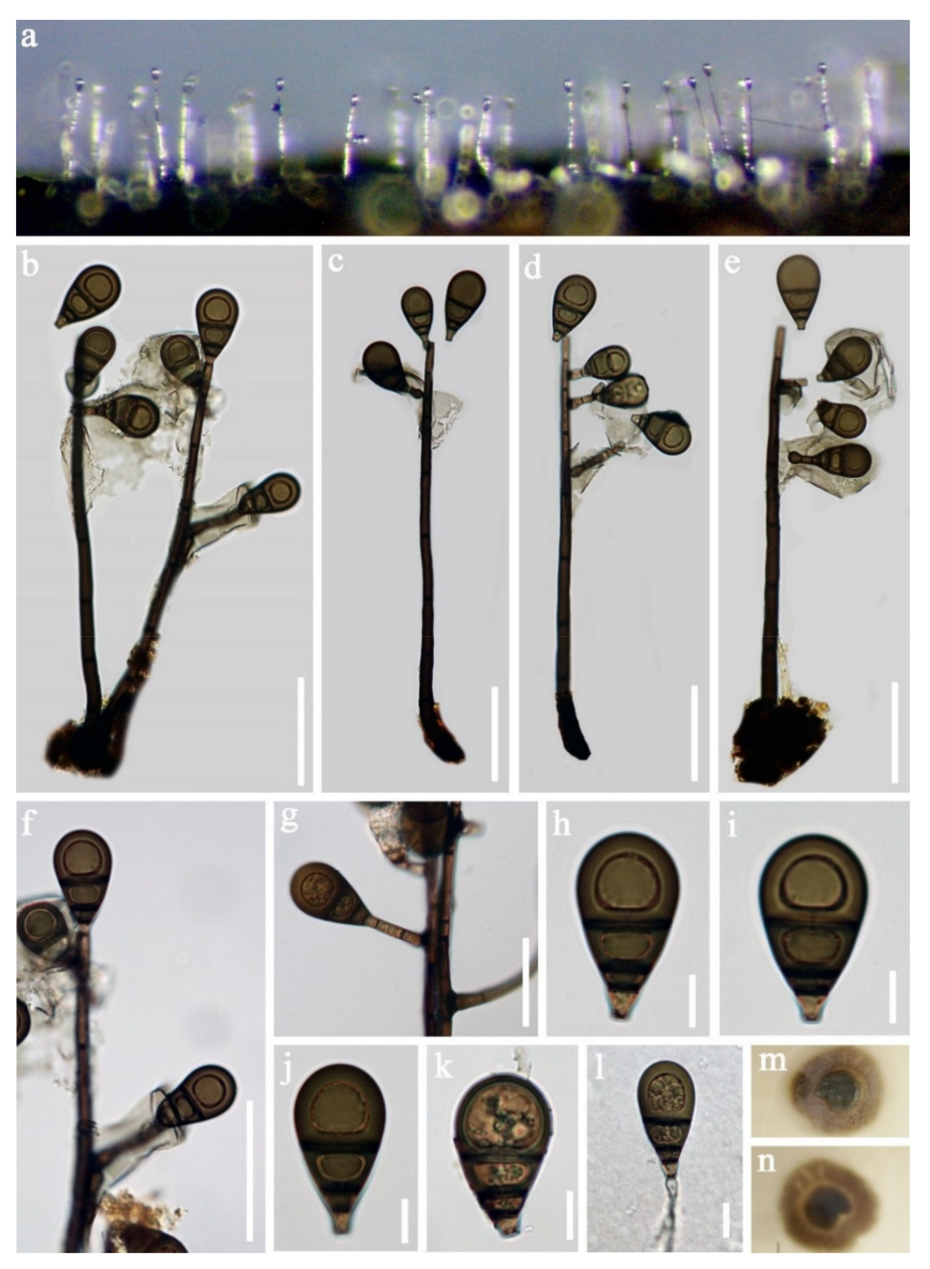
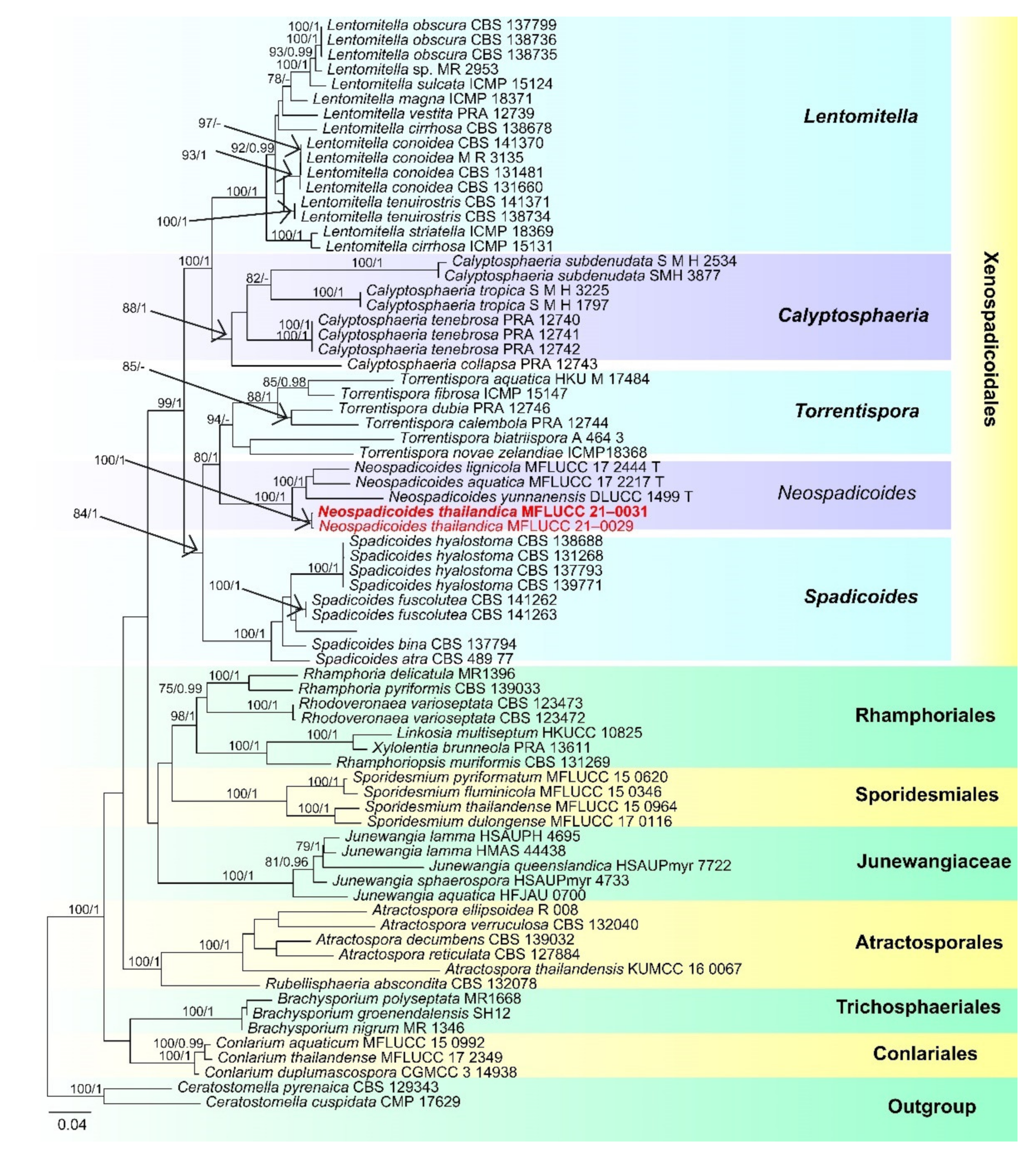
  • Xenospadicoidales Hern.-Restr., J. Mena and Gené
  • Xenospadicoidaceae Hern.-Restr., J. Mena and Gené
  • Neospadicoides thailandica D.F. Bao, H.Y. Su, K.D. Hyde and Z.L. Luo, sp. nov. (Figure 15).
  • Index Fungorum number: IF 558596; Faces of fungi number: FoF 09918
  • Holotype: MFLU 21–0032
  • Etymology: Referring to the species was collected from Thailand.
Saprobic on submerged decayed wood in freshwater habitats. Sexual morph: Undetermined. Asexual morph: Colonies on superficial substratum, scattered, granulate and dark brown to black. Mycelium partly superficial and partly immersed, composed of branched, septate, brown to dark brown and branched hyphae. Conidiophores 165–235 × 5–7 μm (x = 200 × 6 μm, n = 15), macronematous, mononematous, erect, solitary, cylindrical, straight or flexuous, dark brown to black, smooth, with thick, dark brown to black septa, with 2–4-branches in the middle to upper part, branches 15–35 × 4–6 μm (x =24 × 5 μm, n = 15), 1–3-septate, medium to dark brown, with a hyaline to brown sheath over the branches. Conidiogenous cells holoblastic, monoblastic, integrated, terminal and dark brown. Conidia 29–35 µm long, 15–20 µm wide at apex, 5–8 µm wide at base (n = 30), acrogenous or acropleurogenous, solitary, obovoid, 3-septate, brown to dark brown, rounded at the apex, truncate at the base, thick- and smooth-walled and sometimes with a subhyaline to brown sheath.
Culture characteristics: Conidia germinated on PDA within 24 h and germ tubes produced from the basal cell of conidia. Colonies growing on PDA, reached 20–25 mm in two weeks at 25 °C, rough surface, dense, circular, slightly raised to umbonate with the entire edge, floccose, brown from above, dark brown from below and not producing pigmentation in culture.
Material examined: Sakon Nakhon, Tao Ngoi, Thailand, on submerged decaying wood, 12 November 2017, D.F. Bao, B159 (MFLU 21–0032, holotype), ex-type living culture, MFLUCC 21–0031. GenBank accession numbers: (LSU) MZ412532, (SSU) MZ413275, (ITS) MZ412520. Thailand, Tao Ngoi, Sakon Nakhon, on submerged decaying wood, 12 November 2017, D.F. Bao, B168 (MFLU 21–0031), living culture, MFLUCC 21–0029. GenBank accession numbers: (LSU) MZ412533, (SSU) MZ413276, (ITS) MZ412521.
Notes: Neospadicoides was introduced by Luo et al. [28] with three new species: N. lignicola, N. aquatica and N. yunnanensis. The genus is characterized by macronematous, mononematous, septate conidiophores, holoblastic, terminal, integrated conidiogenous cells and acrogenous or acropleurogenous, fusiform, obovoid and septate conidia. Neospadicoides thailandica fits well with the generic concept. However, N. thailandica is unique in the genus by its branched conidiophores, sometimes with a hyaline to brown sheath over the branches and conidia. Other species in the genus lack a sheath and the conidiophores are unbranched. Phylogenetic analyses showed that our collections formed a distinct and stable lineage within Neospadicoides (Figure 16). Therefore, we introduce our collections as a new species. All species in the genus were reported from freshwater habitats in China [28], while N. thailandica was collected from Thailand.
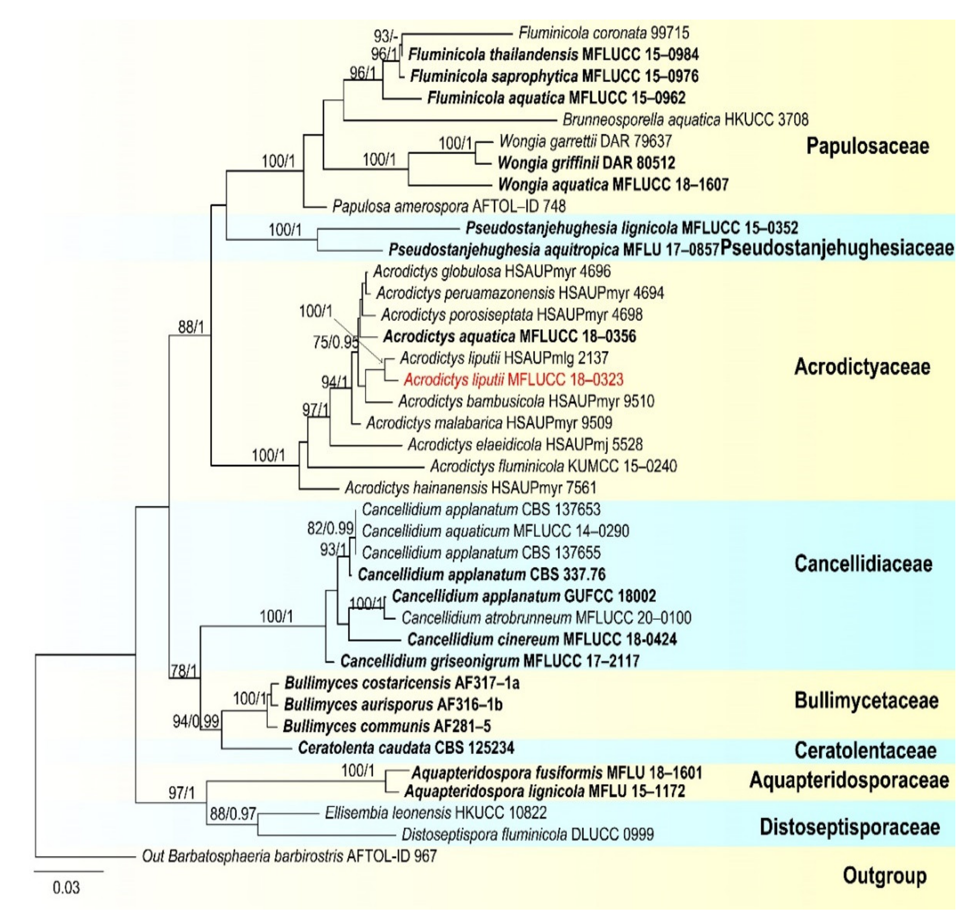
  • Acrodictyaceae J.W. Xia and X.G. Zhang
  • Acrodictys M.B. Ellis
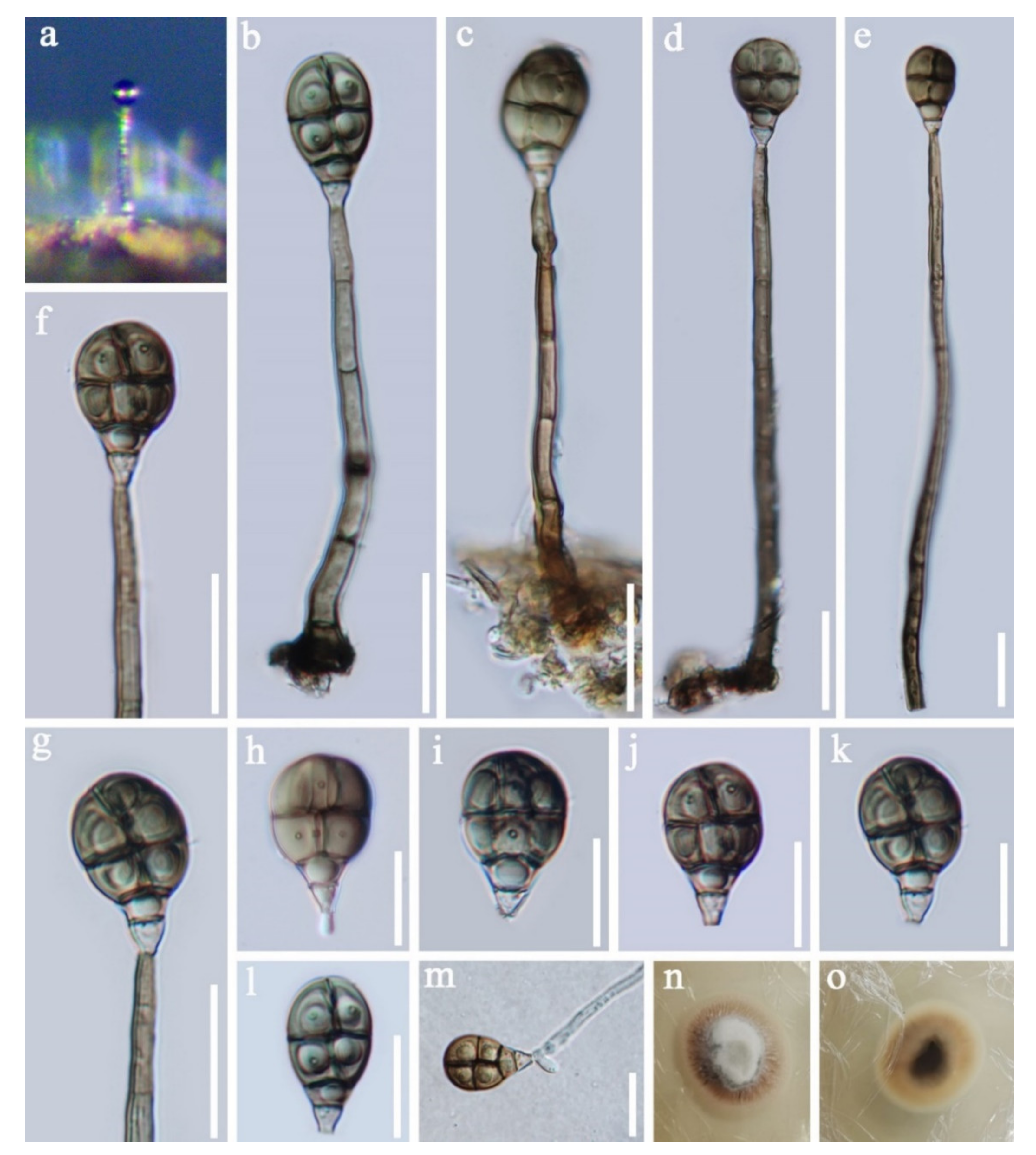
  • Acrodictys liputii L. Cai, K.Q. Zhang, McKenzie, W.H. Ho and K.D. Hyde, Nova Hedwigia 75 (3–4): 526, 2002. (Figure 17).
Saprobic on submerged decaying wood. Sexual morph: Undetermined. Asexual morph: Colonies on superficial substratum, effuse, scattered, hairy and brown to dark brown. Mycelium partly superficial and partly immersed, composed of branched, septate, brown to dark brown and with smooth hyphae. Conidiophores 97–180 × 4.5–6 µm (x = 138.5 × 5.5 μm, n = 20), macronematous, mononematous, simple, cylindrical, erect, 4–8-septate, straight or slightly flexuous, pale brown to brown and smooth-walled. Conidiogenous cells holoblastic, monoblastic, terminal, integrated, cylindrical and truncate, brown. Conidia 11.5–15 × 3.4–5 µm (x = 13 × 4 μm, n = 30), acrogenous, dry, solitary, muriform, subglobose, obovoid to pyriform, truncated at base, rounded at apex, pale brown to subhyaline at the basal cell, brown to greyish brown at other parts, with 3–4- transverse septa and 2- longitudinal septa, constricted at the septa, with conspicuous pores in the septa and smooth.
Culture characteristics: Conidia germinating on PDA within 24 h, germ tubes produced from the basal cell of conidia. Colonies grew on PDA, reaching 20–25 mm in two weeks at 25 °C, surface rough, circular, with dense mycelia, velvety, slightly raised to umbonate, with edge entire, floccose, white at center from above, yellow at the edge, dark brown at center from below, pale yellow to greyish yellow at the edge and not producing pigmentation in culture.
Material examined: Phra Khanong Nuea, Bangkok Province, Thailand, on submerged decaying wood, 1 October 2017, Z.L. Luo, B-50 (MFLU 21-0034), ex-type living culture MFLUCC 18–0323. GenBank accession numbers: (LSU) MZ412524, (SSU) MZ413269, (ITS) MZ412512.
Notes: In the phylogenetic analyses, our isolate clustered with Acrodictys liputii (HSAUPmlg 2137) with strong bootstrap support (100% ML/1.00 PP, Figure 18). Morphology of our isolate and the holotype of A. liputii are indistinguishable. Acrodictys liputii was introduced by Cai et al. [106] and collected on submerged bamboo culm from the Philippines. Xia et al. [107] provided sequence data for this species and first reported this species from China. Our collection is the first record from Thailand.
  • Chaetosphaeriaceae Réblová, M.E. Barr and Samuels, Sydowia 51(1): 56 (1999)
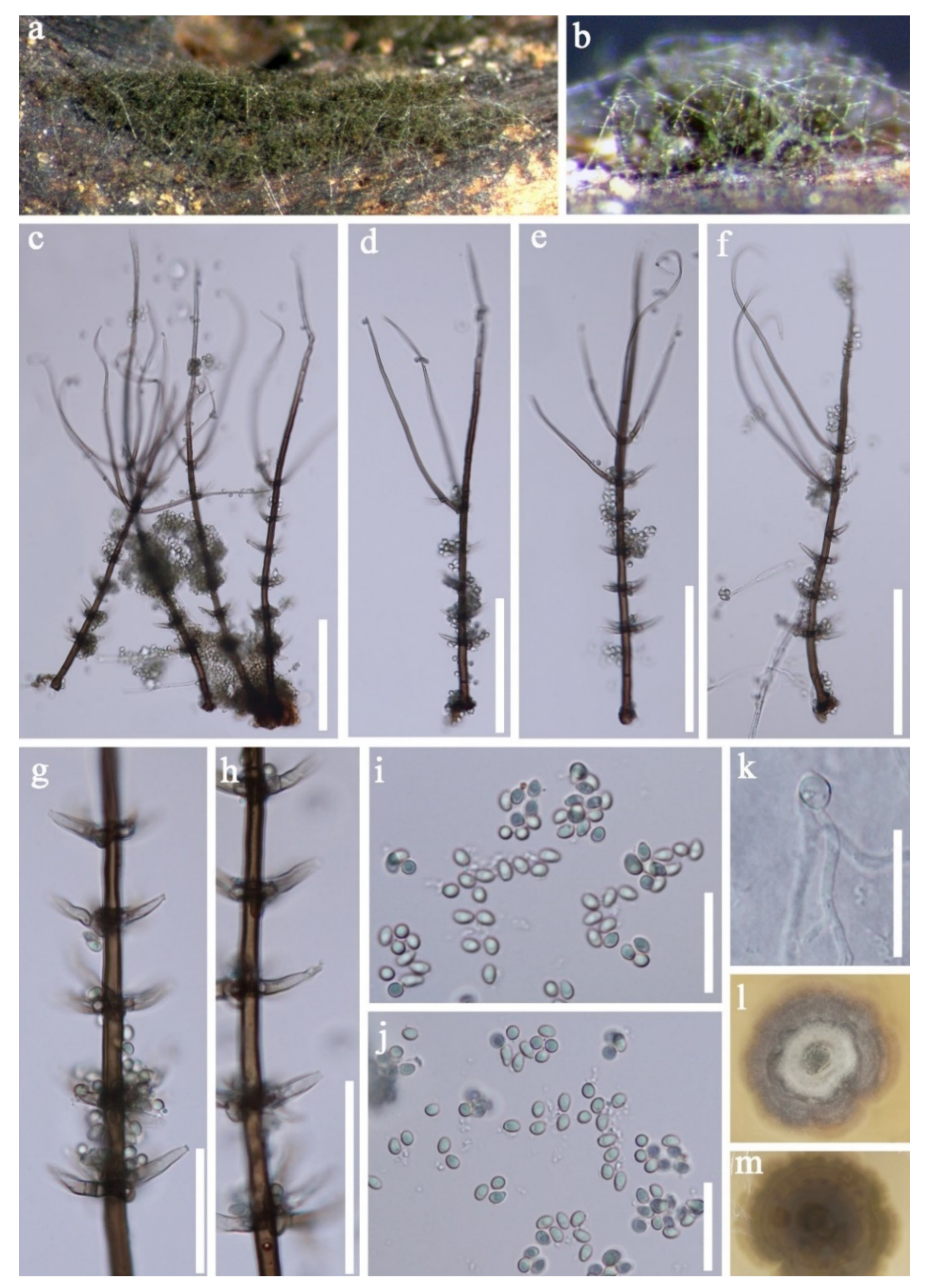
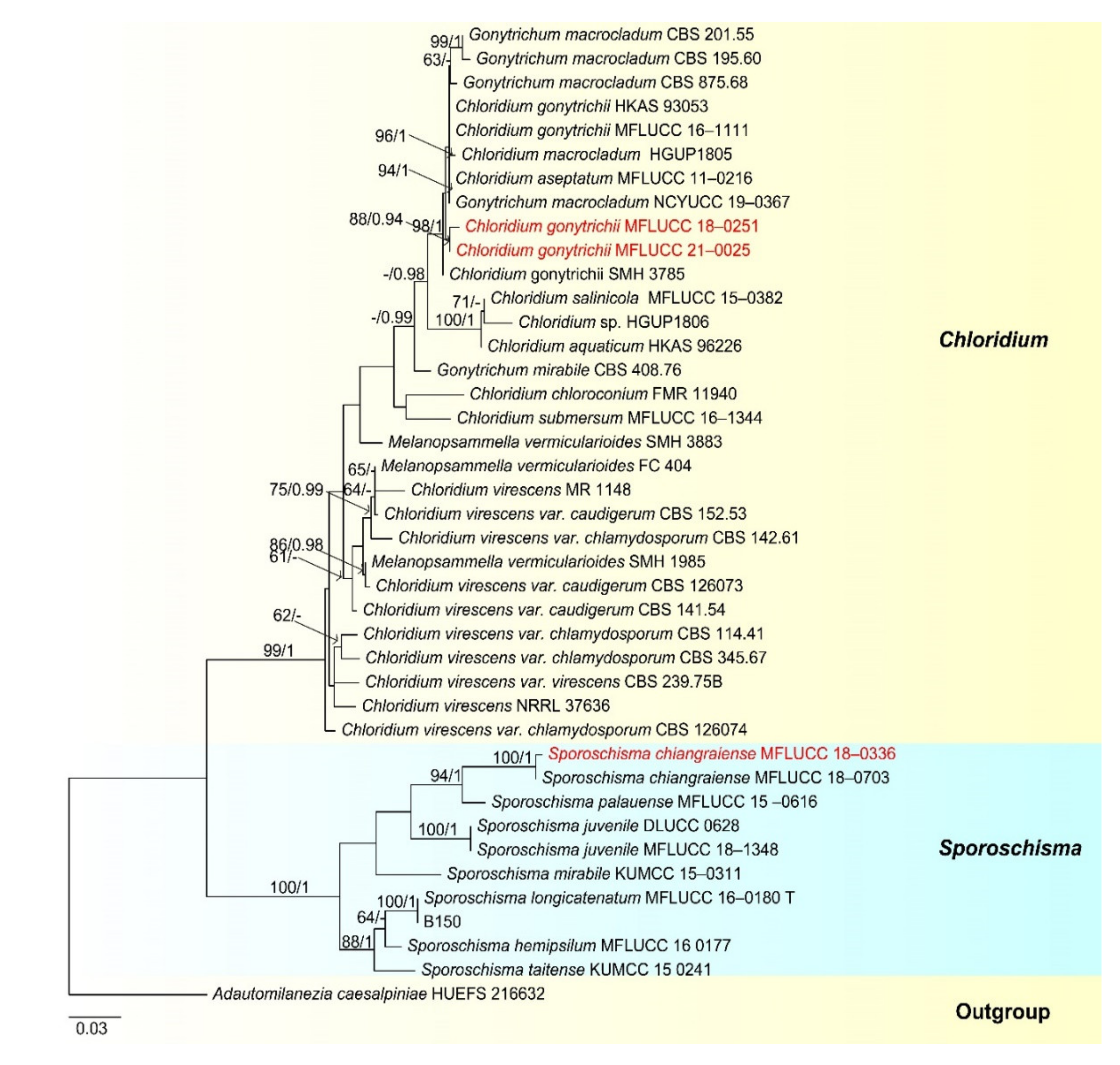
  • Chloridium Link, Mag. Gesell. naturf. Freunde, Berlin 3(1–2): 13 (1809)
  • Chloridium gonytrichii (F.A. Fernández and Huhndorf) Réblová & Seifert, in Réblová et al., IMA Fungus 7(1): 134 (2016) (Figure 19).
  • Gonytrichum macrocladum (Sacc.) S. Hughes, Trans. Br. Mycol. Soc. 34(4): 565 (1952) (1951)
  • Chloridium aseptatum M.J. Wei and H. Zhang, in Wei, Zhang, Dong, Boonmee & Zhang, Phytotaxa 362(2): 191 (2018)
  • Chloridium macrocladum (Sacc.) Karun, Maharachch., C.H.Kuo and K.D.Hyde, in Yuan et al., Fungal Diversity [74] (2020)
Saprobic on decaying wood submerged in freshwater habitats. Sexual morph: Undetermined. Asexual morph: Colonies effuse, hairy and dark green to dark brown. Mycelium superficial. Conidiophores 190–336 × 4.5–6.5 µm, (x = 262.5 × 5.5 μm, n = 15), macronematous, mononematous, single, unbranched, septate, gradually becoming narrower towards the apex, with 3–4 long branches at the upper part, with 2–5 whorls of phialides in the midsection to lower section and a single phialide at the apex, dark brown and paler towards the apex. Conidiogenous cells 9–16.5 × 3–4 µm, (x = 12.5 × 3.5 μm, n = 25), cylindrical to lageniform, phialides, producing conidia from multiple entero-blastic conidiogenous loci and phialides borne on collar hyphae around the conidiophore. Conidia 3.5–4.5 × 2.5–3.0 µm, (x = 4 × 2.5 μm, n = 35), globose to subglobose, aseptate and hyaline to subhyaline.
Culture characteristics: Conidia germinated on PDA within 24 h. Colonies grew on PDA, reaching 20–25 mm in two weeks at 25 °C, with rough surface and dense mycelia, dry, rigid, umbonate from the side view and entire edge, greyish brown to pale brown at the margin from above, white to grey at the center, dark grey to brown from below and not producing pigmentation in culture.
Material examined: Sakon Nakhon, Tao Ngoi, Thailand, saprobic on submerged decaying wood, on 12 November 2018, D.F. Bao, B130 (MFLU 21–0026), living culture, MFLUCC 21–0026. GenBank accession numbers: (LSU) MZ412528, (SSU) MZ413272, (ITS) MZ412516. Khwaeng Phra Khanong Nuea, Khet Watthana Krung Thep Maha Nakhon, Thailand, saprobic on decaying wood submerged in a freshwater stream was acquired on 1 October 2017, Z.L. Luo, B-47 (MLU 21–0035, holotype), living culture MFLUCC 18–0251. GenBank accession numbers: (LSU) MZ412523, (ITS) MZ412511.
Notes: our new collection is identified as Chloridium gonytrichii based on both phylogeny and morphology. Chloridium gonytrichii is characterized by single, unbranched conidiophores with 5–8 whorls of phialides in the midsection, a single phialide at the apex, phialide and cylindrical to lageniform conidiogenous cells ellipsoid conidia [28,108]. Our collection is almost the same as the holotype of C. gonytrichii [108]. Therefore, we identified our new collection as C. gonytrichii.
Chloridium gonytrichii was described by Fernández et al. [108] with its sexual and asexual morphs. It was originally placed in Melanopsammella. Crous et al. [109] provided sequence data for this species and phylogenetic analysis showed that C. gonytrichii clustered with Melanopsammella vermicularioides. Réblová et al. [110] transferred it to Chloridium, synonymized C. gonytrichii under Melanopsammella gonytrichii. Phylogenetic analyses of Luo et al. [28] and Lin et al. [111] showed that gonytrichii formed a stable and well-supported lineage within Chloridium. In our analyses, C. aseptatum (MFLUCC 11–0216), four strains of C. macrocladum (CBS 201.55, CBS 195.60, CBS 875.68 and NCYUCC 19–0367) and five strains of C. gonytrichii (MFLUCC 16–1111, HKAS:93053, SMH 3785, MFLUCC 18–0251, MFLUCC 21–0025) clustered together in a distinct clade within Chloridium with strong bootstrap support (98 ML/1.00 PP). Both morphology and phylogeny of C. macrocladum is indistinguishable to C. gonytrichii (Figure 20) [28,108,112], thus we propose C. macrocladum as a synonym of C. gonytrichii.
Chloridium aseptatum was introduced by Wei et al. [113], based on morphology and ITS sequence data, Yuan et al. [112] synonymized C. macrocladum instead C. aseptatum, our phylogenetic analysis also obtained the same result, C. aseptatum (MFLUCC 11–0216) clustered with C. gonytrichii (Figure 20). Hence, we are in agreement with Yuan et al. [112], and synonymized both C. macrocladum and C. aseptatum under C. gonytrichii, based on phylogeny and morphology.
  • Sporoschisma Berk. & Broome
  • Sporoschisma chiangraiense N.G. Liu and K.D. Hyde, Fungal Diversity 96: 160 (2019) (Figure 21).
Saprobic on decaying wood submerged in freshwater habitats. Sexual morph: Undetermined. Asexual morph: Colonies were effuse, hairy and black, with long chains of conidia. Mycelium immersed, composed of brown to dark brown and unbranched hyphae. Setae 99−162 × 4–6 µm, capitate, scattered or in groups mixed with conidiophores, straight or slightly flexuous, apex swollen, surrounded by hyaline mucilage, median brown, subhyaline to pale brown near the apex and septate. Conidiophores 120–170 µm long, 12–16 µm wide at venter, 5–8 µm wide below venter, 13–15 µm wide above, macronematous, mononematous, solitary or in a small group of 2–3, erect, unbranched, straight or slightly flexuous, arising from dark brown to black bulbous base, dark brown to black, paler at the torn apex, composed of a cylindrical stipe and a swollen venter with a long cylindrical neck, sometimes proliferating percurrently and smooth. Conidiogenous cells monophialidic, integrated, percurrent, lageniform, brown and frayed at the apex. Conidia 27–32 × 11–13 µm (x = 29 × 12 µm, n = 30), catenate, cylindrical, hyaline and smooth-walled when young, becoming olivaceous brown to dark brown and verrucose at maturity, with a big guttule in each cell when mature, 1-septate, conspicuously darkened and slightly constricted at the septa and darkened at both ends.
Culture characteristics: Conidia germinated on PDA within 24 h and colonies grew on MEA, reaching 15–25 mm in two weeks at 25 °C, withrough surface and dense mycelia, velvety, dry, umbonate in the middle from the side view, edge undulate, white to pale grey from above, dark grey to brown at the margin from below andnot producing pigmentation in culture.
Material examined: Phra Khanong Nuea, Bangkok Province, Thailand, on submerged decaying wood, 1 October 2017, Z.L. Luo, B81 (MFLU 21–0036, holotype), ex-type living culture MFLUCC 18–0336. GenBank accession numbers: (LSU) MZ412525, (ITS) MZ412513.
Notes: Sporoschisma chiangraiense was introduced by Hyde et al. [90], it is unique in Sporoschisma by having 1-septate, verrucose conidia, while conidia of other species are multi-septate with smooth-walled. Phylogenetic analyses showed that our new isolate MFLUCC 18–0336 clustered with the ex-type strain of S. chiangraiense (MFLUCC 18–0703) with high bootstrap support (Figure 19). Morphology of our isolate and holotype of S. chiangraiense are almost the same, except the wall of conidia; conidia of our isolate are verrucose at maturity, while this character was not observed in holotype. Based on both phylogenetic analyses and morphology characters, we identified our new isolate as S. chiangraiense.
Sporoschisma longicatenatum Jing Yang, Jian K. Liu and K.D. Hyde, in Yang et al., Phytotaxa 289(2): 152 (2016) (Figure 22).
Saprobic on decaying wood submerged in freshwater habitats. Sexual morph: Undetermined. Asexual morph: Colonies effuse, hairy, black and with long chains of conidia. Mycelium immersed, composed of brown to dark brown, branched and with smooth hyphae. Setae 143−177 × 5–7 μm, capitate, simple or in groups, cylindrical, swollen at apex, pale to medium brown, paler towards apex, straight or slightly flexuous, 3−5-septate and smooth. Conidiophores macronematous, mononematous, solitary with 0−4 setae, erect, straight or slightly flexuous, smooth, dark brown to black, arising from dark brown to black bulbous base, composed of a cylindrical stipe and a swollen venter with a long cylindrical neck, 315–365 μm long, 11–13 μm wide below venter and 12–14 μm wide above, 15–22 μm wide at venter. Conidiogenous cells monophialidic, terminal, integrated, determinate, lageniform, dark brown to black and frayed at the apex. Conidia 43–48 × 9–12 μm (x = 45 × 11 μm, n = 35), catenate, cylindrical to doliiform, rounded at both ends, mostly 5-euseptate, conspicuously darkened at the septa, hyaline when young, dark olive green to brown at maturity, end cells were hyaline to pale brown and shorter than the inner cells, with guttules in each cell.
Culture characteristics: Conidia germinated on PDA within 24 h and colonies grew on PDA, reaching 20–25 mm in two weeks at 25 °C, with rough surface and dense mycelia, dry, velvety, raised from the side view and entire edge, white to pale grey from above; greyish white from below and not producing pigmentation in culture.
Material examined: Sakon Nakhon, Tao Ngoi, Thailand, on submerged decaying wood, 12 November 2017, D.F. Bao, B150 (MFLU 21–0033), living culture, MFLUCC 21–0033. GenBank accession numbers: (LSU) MZ412531, (ITS) MZ412519.
Notes: Morphology of our new collection is indistinguishable with Sporoschisma longicatenatum. Phylogenetic analyses showed that our new collection clustered with the ex-type strain of S. longicatenatum with high bootstrap (Figure 20), we therefore identified our collection as S. longicatenatum. Sporoschisma longicatenatum was introduced by Yang et al. [28] from freshwater habitat in Thailand and this species were exclusively reported from freshwater habitats and only known from Thailand.
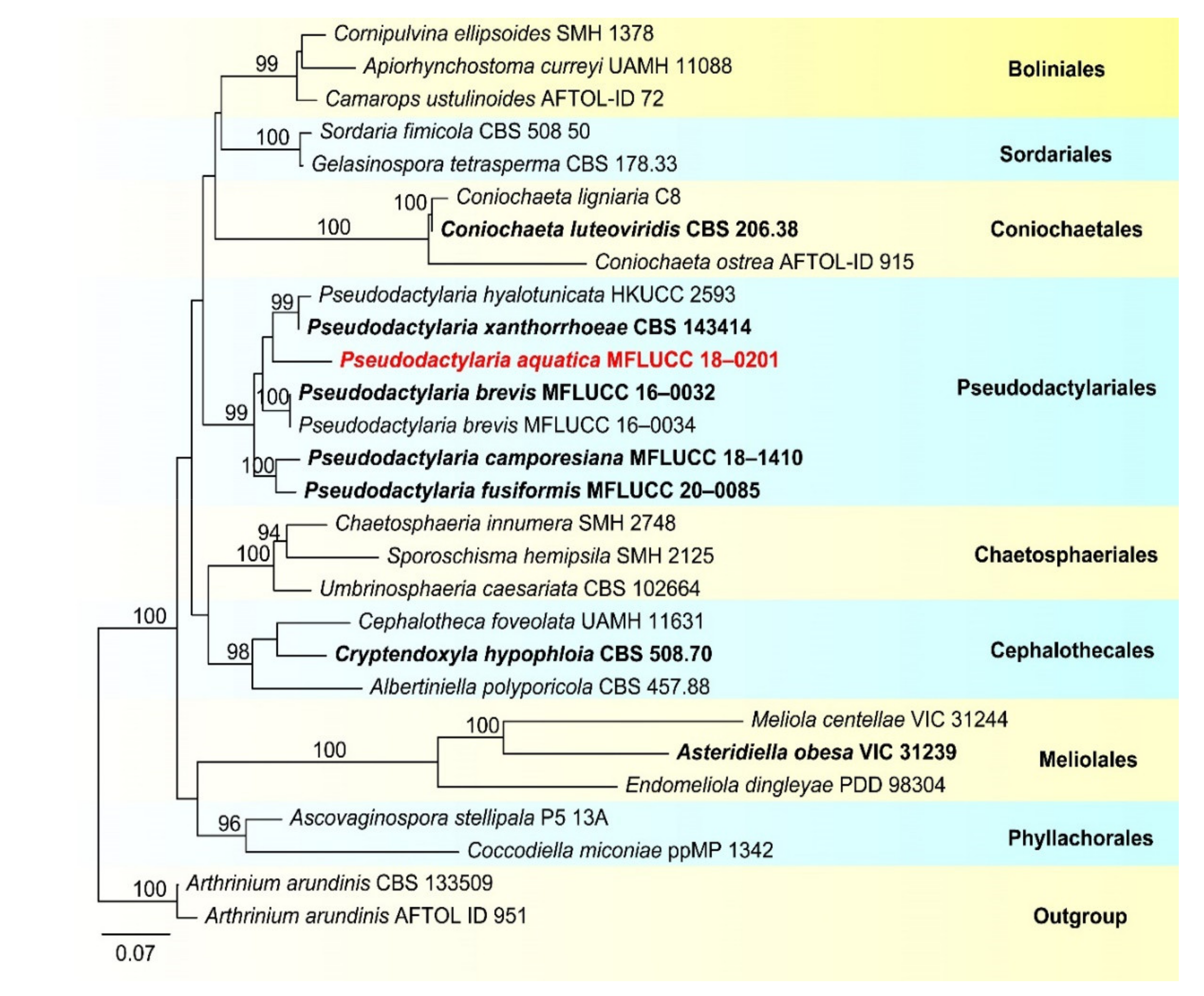
  • Pseudodactylariales Crous,
  • Pseudodactylariaceae Crous
  • Pseudodactylaria Crous
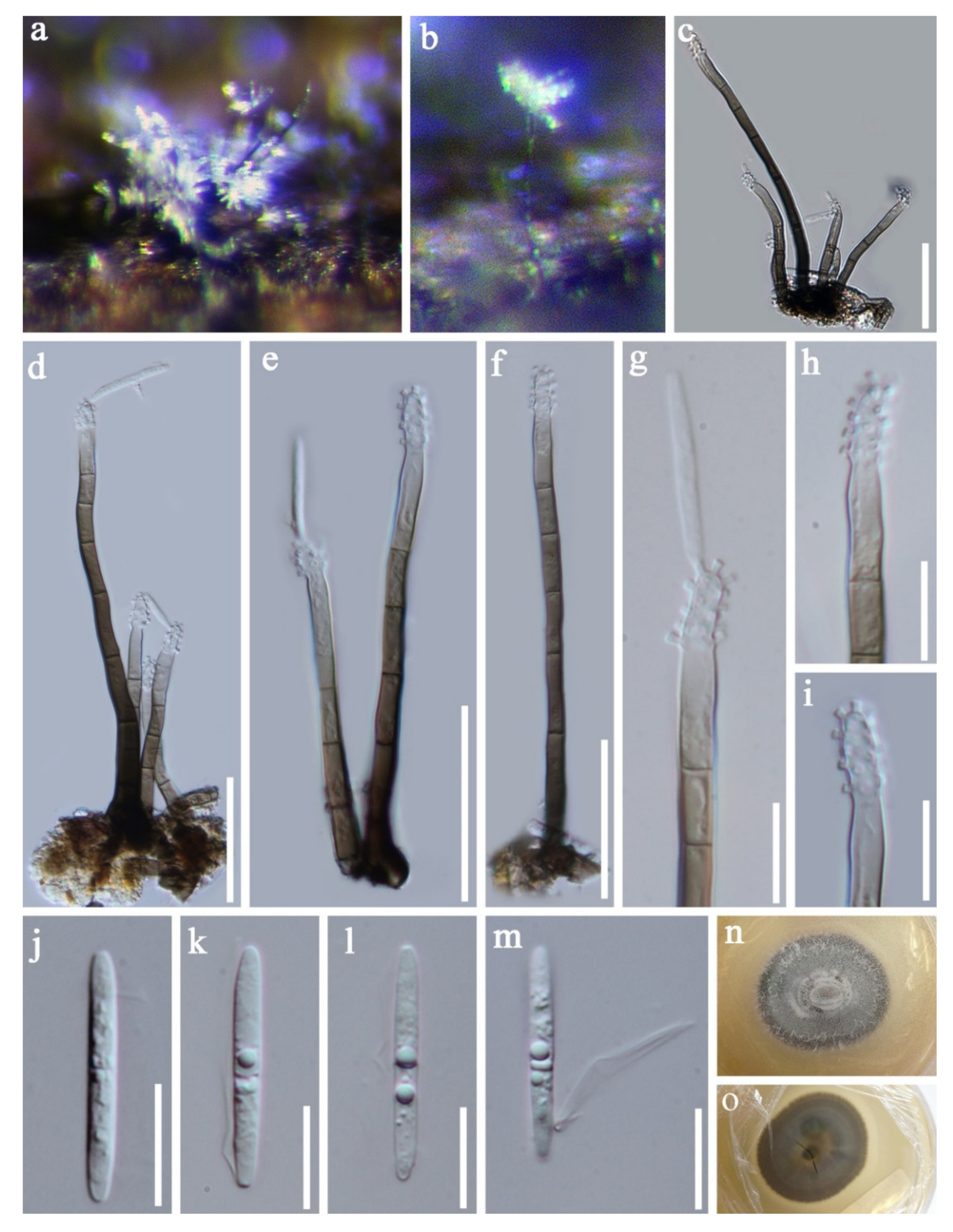
  • Pseudodactylaria aquatica D.F. Bao, H.Y. Su, K.D. Hyde and Z.L. Luo, sp. nov. (Figure 23).
  • Index Fungorum number: IF 558597; Faces of fungi number: FoF 09919
  • Holotype—MFLU 21–0037
  • Etymology—Referring the fungus was collected form aquatic habitat.
Saprobic on decaying wood submerged in freshwater habitats. Sexual morph: Undetermined. Asexual morph: Colonies effuse, shining, greyish brown, velvety and in groups. Mycelium partly immersed, composed of septate, brown, branched and with smooth hyphae. Conidiophores 40–100 × 3.5–4.5 µm, (x = 70 × 4.0 μm, n = 30), macronematous, mononematous, erect, unbranched, 3–6-septate, straight or slightly flexuous, brown to dark brown, paler towards the apex, often in a small group of 3–5 and smooth-walled. Conidiogenous cells 11.5–22 × 3.0–4.5 µm, (x = 11.5 × 3.0 μm, n = 30), polyblastic, denticulate, integrated, cylindrical and hyaline. Conidia 20–23 × 2.5–3.5 µm, (x = 21.5 × 3.0 μm, n = 35), acrogenous, solitary, cylindrical, narrowly fusiform, straight, rounded at the both bends, 0-1-septate, guttules, hyaline and smooth-walled, with a hyaline appendage at the base.
Culture characteristics: Conidia germinated on PDA within 24 h and colonies grew on MEA, reaching 25–30 mm in three weeks at 25 °C, with rough surface and dense mycelia, dry, raised from the side view and entire edge, dark grey at the margin from above, pale grey at the middle, dark brown at the margin from below, greyish brown at the middle and not producing pigmentation in culture.
Material examined: Khok Pho District, Thailand, on submerged decaying wood, 28 August 2017, C.G. Lin, 1F–8–1. B6 (MFLU 21–0037, holotype), ex-type living culture, MFLUCC 18–0201. GenBank accession numbers: (LSU) MZ412522, (ITS) MZ412510.
Notes: Species of Pseudodactylaria are characterized by single, unbranched, septate, hyaline conidiophores, polyblastic, denticulate conidiogenous cells and solitary, fusoid-ellipsoid, hyaline conidia [69,114,115]. Pseudodactylaria aquatica fits well with the generic concept of Pseudodactylaria, such as, single, unbranched, septate conidiophores, polyblastic, denticulate conidiogenous cells and solitary, fusoid-ellipsoid and hyaline conidia. However, our species can be distinguished from other Pseudodactylaria species in having brown to dark brown conidiophores, which are in groups of 3–5, and cylindrical, narrowly fusiform conidia with a hyaline appendage at the base. While other Pseudodactylaria species have single conidiophores and conidia are lacking an appendage. Phylogenetic analysis showed that P. aquatica formed a distinct lineage within the genus (Figure 24) [69,114,116].
  • Pseudodactylaria species were reported as saprobes from freshwater or terrestrial habitats in Australia [116], China [55,69] and Thailand [114]. Our new species was collected from freshwater habitats in Thailand.
  • Xylariales Nannf., Nova Acta R. Soc
  • Xylariaceae Tul. and C. Tul
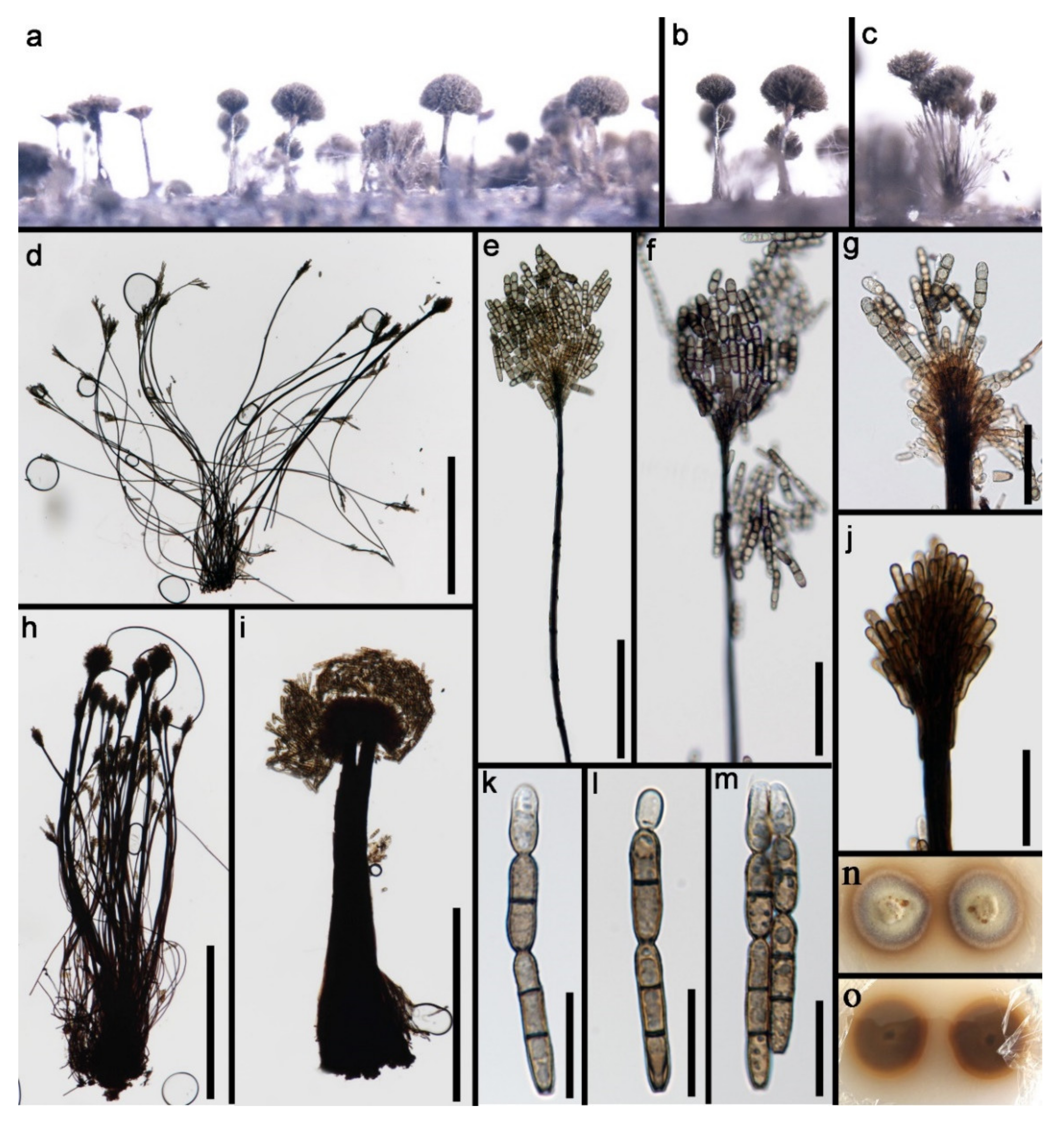
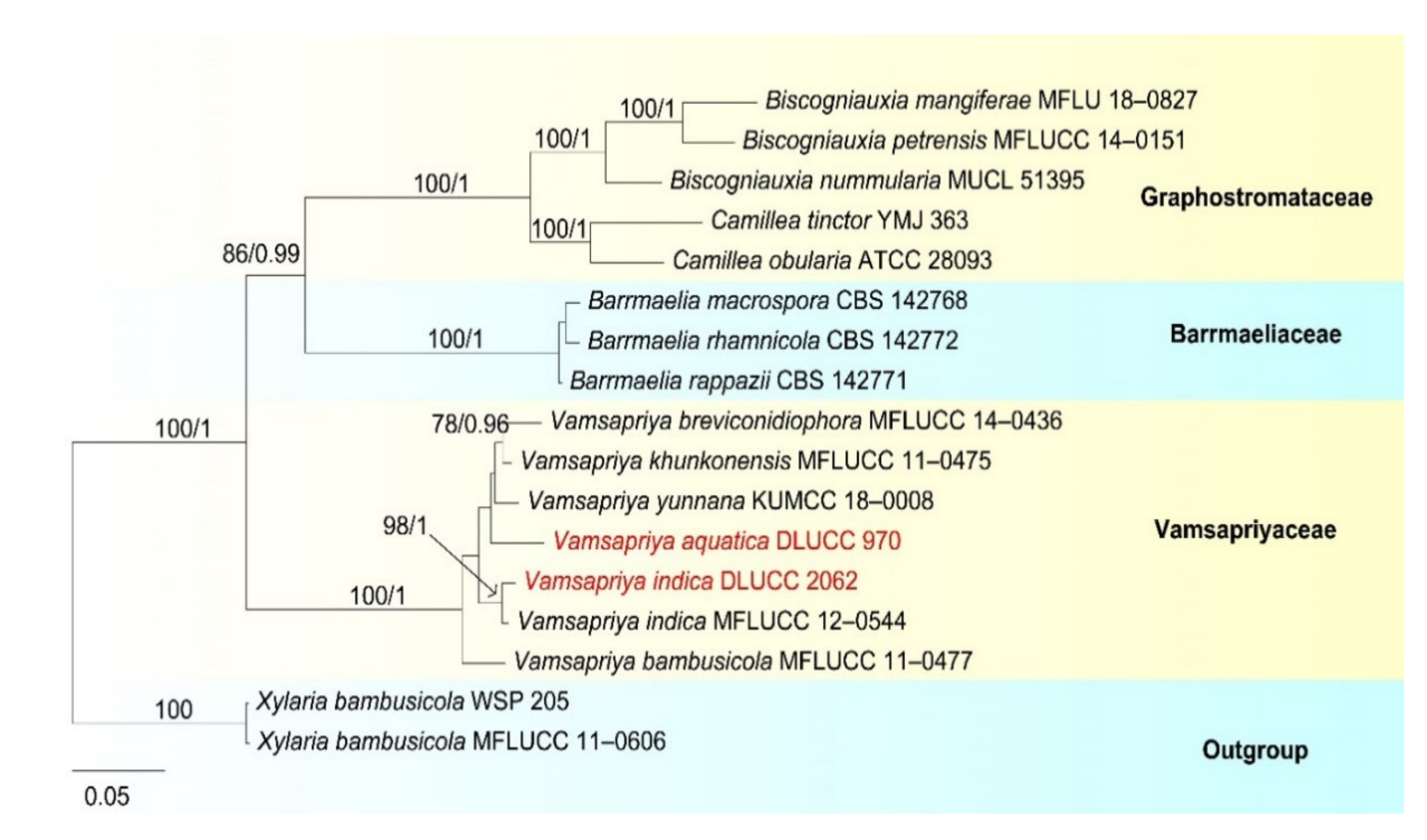
  • Vamsapriya Gawas and Bhat, Mycotaxon 94: 150 (2006)
  • Vamsapriya aquatica D.F. Bao, H.Y. Su, K.D. Hyde and Z.L. Luo, sp. nov. (Figure 25).
  • Index Fungorum number: IF 558598; Faces of fungi number: FoF 09920
  • Holotype—HKAS 115791
  • Etymology—Referring the fungus was collected form aquatic habitat.
Saprobic on decaying wood submerged in freshwater habitats. Sexual morph: Undetermined. Asexual morph: Colonies effuse on natural substrate and dark brown to black. Mycelium immersed, composed of branched, septate and brown hyphae. Conidiophores 450–947 μm long, 97–177 μm wide at the base, 60–100 μm wide in the middle, 40–120 μm wide at the apical fertile region, macronematous, synnematous, branched, septate, brown to dark brown and smooth. Synnemata erect, rigid, velvety, dark brown, smooth and composed of compact, parallel, adpressed conidiophores. Conidiogenous cells 4.5–6.5 × 1.8–2.2 µm, (x = 5.3 × 2.0 μm, n = 30), monotretic, enteroblastic, terminal, discrete, ellipsoidal, brown to dark brown and smooth. Conidia 16.5–33 × 5–6 µm, (x = 24.5 × 5.5 μm, n = 35), catenate, initially subhyaline to pale brown, brown to dark brown when mature, minutely verrucose, cylindrical to obclavate, rounded at the apex, truncate at base, straight or slightly curved, mostly 2-septate, up to 4-septate at maturity and slightly constricted at the septa, smooth.
Culture characteristics: Conidia germinated on PDA within 24 h and colonies grew on MEA, reaching 25–30 mm in three weeks at 25 °C, dense, floccose, dry, raised from the side view and entire edge, greyish yellow at the middle from above, pale yellow at the margin, dark yellow at the middle from below, pale yellow at the margin and not producing pigmentation in culture.
Material examined: Nujiang River, Yunnan Province, China on submerged bamboo culms, acquired in May 2016, Z.L. Luo, S-970 (HKAS 115791, holotype), ex-type living culture, DLUCC 970. GenBank accession numbers: (ITS) MZ420740,
Notes: Vamsapriya aquatica is morphologically quite similar to V. indica. V. aquatica shares some similar characters with V. indica, such as, macronematous, synnematous, septate, branched, brown to dark brown, conidiophores, monotretic, enteroblastic, terminal Conidiogenous cells and catenate, cylindrical and septate conidia. However, V. aquatica can be distinguished from V. indica by the shorter conidiophores (450–947 vs. 700–1100 μm), smaller conidia (16.5–33 × 5–6 vs. 35–290 × 4–6.5 μm) and less conidial septum (1–3-septate, mostly 2-septate vs. 1–3-septate when young, more than 20-septate at maturity) [117]. In addition, we compared the nucleotides of ITS region between V. aquatica and V. indica; there were 39 bp (6.5%) differences, which is strongly support our species to be a new species.
Phylogenetic analysis showed that Vamsapriya aquatica close to V. yunnana (Figure 26). However, V. aquatica differs from V. yunnana by the conidial shape (cylindrical to obclavate truncate at base, rounded at the apex vs. fusiform, broad at middle, obtuse at base, rostrate at tip) and size (16.5–33 × 5–6 vs. 50–70 × 9–11 μm) [118]. Thus, we introduce the new collection as a new species.
  • Vamsapriya indica Gawas & Bhat, Mycotaxon 94: 150 (Figure 27).
Saprobic on decaying wood submerged in freshwater habitats. Sexual morph: Undetermined. Asexual morph: Colonies effuse on natural substrate and dark brown to black. Mycelium mostly immersed in the substratum, composed of subhyaline, septate, branched and smooth hyphae. Conidiophores 1145–1475 μm long, 105–235 μm wide at the base, 50–80 μm wide in the middle, 70–155 μm wide at the apical fertile region, macronematous, synnematous, cylindrical, straight, or slightly flexuous, septate, smooth, dark brown and unbranched. Synnemata erect, rigid, dark brown and composed of compact parallel conidiophores. Conidiogenous cells 5–9 × 3–5 μm (= 20 × 5 μm, n = 30), monotretic, integrated or discrete, terminal, clavate, slightly curved toward the exterior. Conidia 15–30 × 4–6.5 μm (= 20 × 5 μm, n = 30) acrogenous, catenate, dry, cylindrical to obclavate, straight, 1-4-septate, constricted at the septa, brown dark brown, smooth to slightly verrucose and developing acropetal chains.
Culture characteristics: Conidia germinated on PDA within 24 h and colonies grew on MEA, reaching 30–35 mm in three weeks at 25 °C, dense, floccose, dry, raised from the side view with entire edge, pale grey at the middle from above, white to greyish white at the margin, dark yellow at the middle from below, pale yellow at the margin and not producing pigmentation in culture.
Material examined: Yunnan Province, China, and were saprobic on bamboo culms submerged in Lancang River, acquired in May 2017, Z.L. Luo, S-2062 (HKAS 115803), living culture, DLUCC 2062. GenBank accession numbers: (LSU) MZ420762, (SSU) MZ420751, (ITS) MZ420747, (tef-1) MZ442694, (RPB2) MZ442699.
Notes: The new collection is identified as Vamsapriya indica based on morphology and phylogeny. In the phylogenetic analysis, our new isolate clustered with the ex-type of V. indica (MFLUCC 12–0544) with strong bootstrap support (95 ML/1.00 PP, Figure 25). Morphology of our collection is almost identical to the holotype of V. indica, except for the conidial size; the conidia of holotype are much longer than our collection (DLUCC 2062).
Vamsapriya indica was introduced by Dai et al. [117] and was collected on bamboo culms in Thailand and India. While our collection was collected from freshwater habitats in China, this is the first report of this species from China.

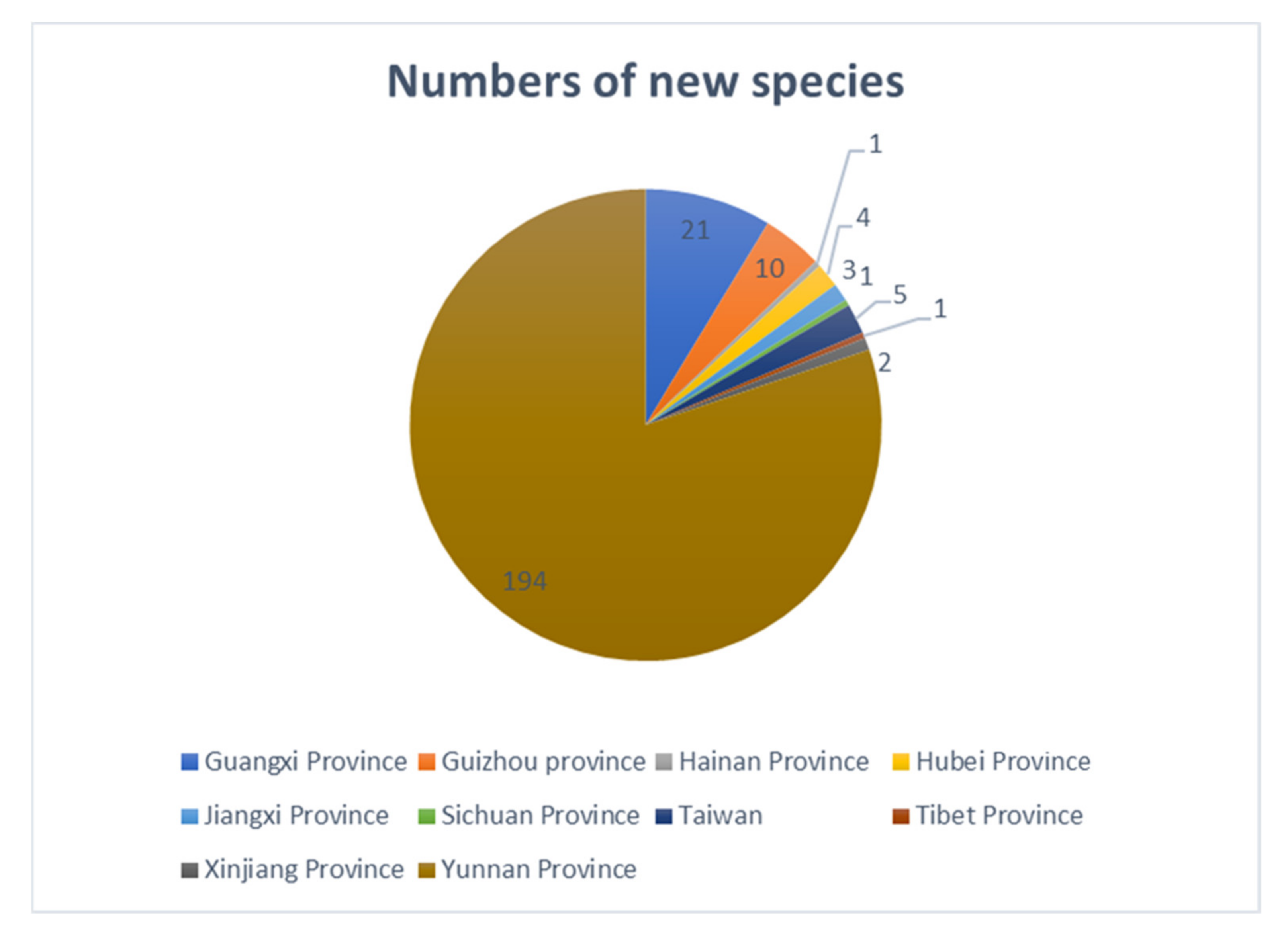
3.2. Biodiversity of Freshwater Fungi in China

Hu et al. [63] documented the biodiversity of aquatic fungi in China and reported 782 freshwater fungi from 24 provinces/districts of China. In recent years, many new species have been introduced from China and over the last five years, studies of freshwater fungi in China have been rapidly increasing. In this paper, a checklist of freshwater fungi in China from 2015–2020 is provided (Table S1). A total 243 freshwater fungi have been described in China during last five years, of which 170 species are new species (Figure 28). Thus, until 2020, about 1025 freshwater fungi have been reported from China.
Freshwater fungi reported in China are highly diverse in Ascomycota, a few are Zygomycetes [63]. All the 243 freshwater fungi reported in China belong to Ascomycota as follows: 241 species belong to 56 families and 25 orders, of which 25 families and 5 orders belong to Dothideomycetes, one family and one order belong to Eurotiomycetes, 30 families and 19 orders belong to Sordariomycetes, two species have been referred to as ascomycota incertae sedis (Table S1). The most common orders in Dothideomycetes are Pleosporales, followed by Tubeufiales, while the common orders in Sordariomycetes are Chaetosphaeriales, followed by Pleurotheciales and Hypocreales (Table S1).
During the last five years, studies of freshwater fungi in China have mainly been focused on southern China (Yunnan, Guizhou, Guangxi, Hainan, Jiangxi, Sichuan and Tibet province, Figure 29), while other regions are poorly reported. In southern China, most species were reported from Yunnan province (194 species), flowed by Guangxi (21 species), Guizhou (10 species), Hainan (1species), Jiangxi (3 species), Sichuan (1 species) and Tibet provinces (1 species). Few species were reported from other regions of China, e.g., Hubei, Jiangxi, Xinjiang provinces and Taiwan (Figure 29), and this points out the regional limitations in research on freshwater fungi in China. Freshwater fungi in many regions of China are still to be studied and the current trend suggests that a large proportion of new species awaits discovery in China.
In the present study, we report sixteen freshwater hyphomycetes from Thailand and China. Six new species, six new country records (four new records for China and two new records for Thailand), one new combination and three new collections are introduced. Of the sixteen freshwater hyphomycetes, thirteen of them are Sordariomycetes and three are Dothideomycetes. These taxa are from twelve genera, namely. Aquapteridospora, Acrodictys, Chloridium, Neospadicoides, Pseudoberkleasmium, Pseudodactylaria, Pleomonodictys, Sporidesmium, Sporoschisma, Tetraploa, Vamsapriya and Wongia, of which two genera, Aquapteridospora and Neospadicoides were exclusively from freshwater habitats and were known only from China and Thailand [28,29]. Acrodictys, Chloridium, Pseudoberkleasmium, Pseudodactylaria, Sporidesmium, Sporoschisma and Tetraploa are commonly found freshwater habitats. Pleomonodictys and Vamsapriya were first reported from freshwater habitats and China in this study [94,95,117,118,119,120,121].

4. Discussion

Freshwater hyphomycetes are a highly diverse group, with a worldwide distribution. They can adapt to different habitats and environments. They have unique adaptions to adapt and survive in the freshwater habitats with the distinct conidia morphologies [122]. The conidia of freshwater hyphomycetes have diverse shapes and sizes. For example, most Ingoldian fungi have tetraradiate, branched or filiform conidia and these can help attach to surfaces and colonize substrates [6,123,124]. When a tetraradiate spore attaches to the surface of a substrate, it attaches at three points, with the spore acting as a tripod, representing a very stable attachment form [123]. Some species have conidia with prominent sheaths or appendages and these characters strengthen adhesion to surfaces of substrate, which also helps to prevent conidial detachment by strong water currents [124,125]. Freshwater hyphomycetes are dominant fungi on submerged leaves and woody substrates. The main advantage of these fungi on submerged leaves and woody substrates is that they have the ability to maintain activity at low temperatures and degrade submerged organic matter under various climatic conditions [126,127,128].
Traditional identification of freshwater hyphomycetes was mainly based on morphological characters but with the advent of DNA sequence analyses, the identification and classification of freshwater hyphomycetes have greatly improved with better insights into species complexes and linking sexual–asexual morphs as well resolving problems associated with dual nomenclature. Freshwater hyphomycetes are distributed in different phyla, classes and orders. Despite some species having similar morphologies, phylogenetic analyses showed that they are polyphyletic and associated with different orders, families, and classes, e.g., Acrodictys-like taxa and Sporidesmium-like taxa [101,124]. Sporidesmium is a typical example to show the polyphyletic nature of freshwater hyphomycetes, the genus was redescribed by Ellis [97] with a broad generic concept, characterized by solitary or gregarious conidiophores, monoblastic, determinate or percurrent conidiogenous cells, cylindrical, fusiform, obclavate, obpyriform and sometimes rostrate conidia [71,97,99,129]. Later studies have segregated Sporidesmium in several different genera, namely Ellisembia, Imimyces, Linkosia, Penzigomyces, Polydesmus, Repetophragma, Sporidesmiella and Stanjehughesia based on morphological characters [130,131]. Recent studies found that Sporidesmium is polyphyletic and related to different orders and families in Dothidieomycetes and Sordariomycetes [71,101,102,132]. Our study herein reports similar results.
In this study, three Sporidesmium species are introduced. Phylogenetic analyses showed that the three species grouped in three different clades within Diaporthomycetidae S. aturbinatum were within Sporidesmiaceae with strong bootstrap support (Figure 13), while S. tropicale and S. nujiangense were phylogenetically distant from Sporidesmiaceae in two other clades. S. tropicale clustered as a sister taxon to Bullimycetaceae with low support, which is consistent with the analyses of Yang et al. [101]. The placement of this species is still questionable and further phylogenetic analyses are needed to resolve its placement. The novel species, Sporidesmium nujiangense clustered with S. olivaceoconidium, S. aquaticivaginatum and S. guizhouense in a stable and well-supported clade sister to Distoseptisporales. Yang et al. [101] showed that S. olivaceoconidium and S. aquaticivaginatum are basal to Sporidesmiaceae with low support, while, in the analyses of Liu et al. [102], S. olivaceoconidium and S. aquaticivaginatum and S. guizhouense formed a stable clade close to Jobellisiales. Morphology of these taxa are not that different from other taxa in the Sporidesmiaceae; however, they are phylogenetically distinct.
Freshwater fungi in Thailand have been studied for several decades [18,47,48,50,53,133]. Calabon et al. [134] summarized the studies of freshwater fungi in Thailand during 2015–2020 with a total 129 new species. Fungal numbers have been estimated between 2.2 and 3.8 million; however, only around 2.6 and 4.5% species of fungi and fungus-like taxa have been accepted [135]. Over the past five years, 298 new species have been reported from China and Thailand, which indicates a high diversity of freshwater fungi, and many new species are still not yet discovered. Recently, the new generation sequencing (NGS) method has been used to better assess fungal diversity, and this greatly improved our understanding on the diversity, ecology and distribution of fungi [136,137,138,139,140]. Studies of freshwater fungi are mainly focused on lignicolous freshwater fungi but fungi on other hosts are poorly reported. The diversity of freshwater fungi is much higher than previously thought. Using NGS method to investigate freshwater fungal diversity may help for a better understanding of their ecology and distribution.

Supplementary Materials

The following are available online at https://www.mdpi.com/article/10.3390/jof7080669/s1, Table S1: Checklist of freshwater fungi in China from 2015–2020.

Author Contributions

Conceptualization: D.-F.B., K.D.H.; methodology: D.-F.B., Z.-L.L.; formal analysis and investigation: D.-F.B., Z.-L.L., R.J.; resources: K.D.H., H.-Y.S.; writing—original draft preparation, D.-F.B.; writing—review and editing, K.D.H., R.J., E.H.C.M., Z.-L.L., S.N.; supervision, K.D.H., H.-Y.S., S.N.; funding acquisition, K.D.H., H.-Y.S. All authors have read and agreed to the published version of the manuscript.

Funding

This work was mainly funded by the National Natural Science Foundation of China (Project ID: 32060005, 31970021), Fungal diversity conservation and utilization innovation team (ZKLX2019213) and the Thailand Research Fund for the grant entitled “Impact of climate change on fungal diversity and biogeography in the Greater Mekong Subregion” (Grant No. RDG6130001).

Institutional Review Board Statement

Not applicable.

Informed Consent Statement

Not applicable.

Data Availability Statement

All sequences generated in this study were submitted to GenBank.

Acknowledgments

D.-F.B. would like to thank Shaun Pennycook from Landcare Research, Auckland, New Zealand, for advising on the taxon names, C.G.L is thanked for helping to collect the samples. K.D.H. thanks Chiang Mai University for the award of Visiting Professor. R.J. thanks University of Mauritius for research support.

Conflicts of Interest

The authors declare no conflict of interest.

References

  1. Shearer, C.A.; Descals, E.; Kohlmeyer, B.; Kohlmeyer, J.; Marvanová, L.; Padgett, D.; Porter, D.; Raja, H.A.; Schmit, J.P.; Thorton, H.A.; et al. Fungal biodiversity in aquatic habitats. Biodivers. Conserv. 2007, 16, 49–67. [Google Scholar] [CrossRef]
  2. Wong, K.M.K.; Goh, T.K.; Hodgkiss, I.J.; Hyde, K.D.; Ranghoo, V.M.; Tsui, C.K.M.; Ho, W.H.; Wong, W.S.W.; Yuen, T.K. Role of fungi in freshwater ecosystems. Biodivers. Conserv. 1998, 7, 1187–1206. [Google Scholar] [CrossRef]
  3. Hyde, K.D.; Fryar, S.; Tian, Q.; Bahkali, A.H.; Xu, J.C. Lignicolous freshwater fungi along a north–south latitudinal gradient in the Asian/Australian region; can we predict the impact of global warming on biodiversity and function? Fungal Ecol. 2016, 19, 190–200. [Google Scholar] [CrossRef]
  4. Raja, H.A.; Shearer, C.A.; Tsui, C.K.M. Freshwater Fungi. eLS 2018, 1–13. [Google Scholar] [CrossRef]
  5. Goh, T.K.; Hyde, K.D. Biodiversity of freshwater fungi. J. Ind. Microbiol. 1996, 17, 328–334. [Google Scholar] [CrossRef]
  6. Jones, E.B.G.; Hyde, K.D.; Pang, K.L. Freshwater Fungi and Fungal-Like Organisms; De Gruyter: Berlin, Germany, 2014. [Google Scholar]
  7. Da Silva, G.V.R.; Castañeda-Ruiz, R.F.; Malosso, E. Comparison of aquatic hyphomycetes communities between lotic and lentic environments in the Atlantic rain forest of Pernambuco, Northeast Brazil. Fungal Biol. 2019, 123, 660–668. [Google Scholar] [CrossRef]
  8. Ingold, C.T. Aquatic hyphomycetes of decaying alder leaves. Trans. Brit. Mycol. Soc. 1942, 25, 339–417. [Google Scholar] [CrossRef]
  9. Ingold, C.T. Aquatic ascomycetes: Ceriospora caudaesuis n. sp. and Ophiobolus tyhae. Trans. Brit. Mycol. Soc. 1951, 34, 210–215. [Google Scholar] [CrossRef]
  10. Ingold, C.T.; Chapman, B. Aquatic ascomycetes: Loramyces juncicola Weston and L. macrospora n. sp. Trans. Brit. Mycol. Soc. 1952, 35, 268–272. [Google Scholar] [CrossRef]
  11. Hyde, K.D.; Goh, T.K. Fungi on submerged wood in Lake Barrine, north Queensland, Australia. Mycol. Res. 1998, 102, 739–749. [Google Scholar] [CrossRef]
  12. Cai, L.; Tsui, C.; Zhang, K.; Hyde, K.D. Aquatic fungi from Lake Fuxian, Yunnan, China. Fungal Divers. 2002, 9, 57–70. [Google Scholar]
  13. Voglmayr, H.; Delgado-Rodriguez, G. Dendroclathra caeruleofusca gen. nov. et sp. nov., an aeroaquatic hyphomycete from Cuba. Can. J. Bot. 2001, 79, 995–1000. [Google Scholar] [CrossRef]
  14. Sridhar, K.R.; Kaveriappa, K.M. Colonization of leaves by water-borne hyphomycetes in a tropical stream. Mycol. Res. 1989, 92, 392–396. [Google Scholar] [CrossRef]
  15. Minoura, K.; Muroi, T. Some freshwater ascomycetes from Japan. Trans. Mycol. Soc. 1978, 19, 129–134. [Google Scholar]
  16. Nawawi, A. Some interesting hyphomycetes from water. Mycotaxon 1985, 24, 217–226. [Google Scholar]
  17. Kuthubutheen, A.J.; Nawawi, A. Dictyochaeta guadalcanalensis comb. nov. and several new records of the genus in Malaysia. Mycol. Res. 1991, 95, 1220–1223. [Google Scholar] [CrossRef]
  18. Sivichai, S.; Hywel-Jones, N. Biflagellospora (aero-aquatic hyphomycetes) from submerged wood in Thailand. Mycol. Res. 1999, 103, 908–914. [Google Scholar] [CrossRef]
  19. Sivichai, S.; Jones, E.B.G.; Hywel-Jones, N. Fungal colonisation of wood in a freshwater stream at Tad Ta Phu, Khao Yai National Park, Thailand. In: Fungal Succession (eds. KD Hyde, EB GarethJones). Fungal Divers. 2002, 10, 113–129. [Google Scholar]
  20. Shearer, C.A.; Raja, H.A. Freshwater Ascomycetes Database. Available online: http://fungi.life.illinois.Edu/ (accessed on 11 May 2013).
  21. Vijaykrishna, D.; Jeewon, R.; Hyde, K.D. Fusoidispora aquatica: New freshwater ascomycetes from Hong Kong based on morphology and molecules. Sydowia 2005, 57, 267–280. [Google Scholar]
  22. Hirayama, K.; Tanaka, K.; Raja, H.A.; Miller, A.N.; Shearer, C.A. A molecular phylogenetic assessment of Massarina ingoldiana sensu lato. Mycologia 2010, 102, 729–746. [Google Scholar] [CrossRef] [Green Version]
  23. Ferrer, A.; Miller, A.N.; Shearer, C.A. Minutisphaera and Natipusilla: Two new genera of freshwater Dothideomycetes. Mycologia 2011, 103, 411–423. [Google Scholar] [CrossRef] [Green Version]
  24. Barbosa, F.R.; Gusmao, L.F.P.; Raja, H.A.; Shearer, C.A. New species and new records of freshwater ascomycetes from Brazil and Costa Rica. Mycologia 2013, 105, 335–343. [Google Scholar] [CrossRef] [PubMed]
  25. Raja, H.A.; Oberlies, N.H.; Figueroa, M.; Tanaka, K.; Hirayama, K.; Hashimoto, A.; Miller, A.N.; Zelski, S.E. Freshwater ascomycetes: Minutisphaera (Dothideomycetes) revisited, including one new species from Japan. Mycologia 2013, 105, 959–976. [Google Scholar] [CrossRef] [Green Version]
  26. Raja, H.A.; El-Elimat, T.; Oberlies, N.H.; Shearer, C.A.; Miller, A.N.; Tanaka, K.; Hashimoto, A.; Fournier, J. Minutisphaerales (Dothideomycetes, Ascomycota): A new order of freshwater ascomycetes including a new family, Minutisphaeraceae, and two new species from North Carolina, USA. Mycologia 2015, 107, 845–862. [Google Scholar] [CrossRef] [Green Version]
  27. Yang, J.; Maharachchikumbura, S.S.N.; Hyde, K.D.; Bhat, D.J.; McKenzie, E.H.C.; Bahkali, A.H.; Jones, E.B.G.; Liu, Z.Y. Aquapteridospora lignicola gen. et sp. nov., a new hyphomycetous taxon (Sordariomycetes) from wood submerged in a freshwater stream. Cryptogam. Mycol. 2015, 36, 469–479. [Google Scholar] [CrossRef]
  28. Yang, J.; Liu, J.K.; Hyde, K.D.; Bhat, D.J.; Jones, E.B.G.; Liu, Z.Y. New species of Sporoschisma (Chaetosphaeriaceae) from aquatic habitats in Thailand. Phytotaxa 2016, 289, 147–157. [Google Scholar] [CrossRef] [Green Version]
  29. Luo, Z.L.; Hyde, K.D.; Liu, J.K.; Maharachchikumbura, S.S.N.; Jeewon, R.; Bao, D.F.; Bhat, D.J.; Lin, C.G.; Li, W.L.; Yang, J.; et al. Freshwater Sordariomycetes. Fungal Divers. 2019, 99, 451–660. [Google Scholar] [CrossRef] [Green Version]
  30. Dong, W.; Wang, B.; Hyde, K.D.; McKenzie, E.H.C.; Bhat, D.J.; Raja, H.A.; Tanaka, K.; Abdel-Wahab, M.A.; Abdel-Aziz, F.A.; Doilom, M.; et al. Freshwater Dothideomycetes. Fungal Divers. 2020, 105, 319–575. [Google Scholar] [CrossRef]
  31. Chapela, I.H.; Petrini, O.; Hagann, L. Monolignol glucosides as specific recognition messengers in fungus-plant symbioses. Physiol. Mol. Plant. Pathol. 1991, 39, 289–298. [Google Scholar] [CrossRef]
  32. Feau, N.; Vialle, A.; Allaire, M.; Tanguay, P.; Joly, D.L.; Frey, P.; Callan, B.E.; Hamelin, R.C. Fungal pathogen (mis-) identifications: A case study with DNA barcodes on Melampsora rusts of aspen and white poplar. Mycol. Res. 2009, 113, 713–724. [Google Scholar] [CrossRef] [Green Version]
  33. Mortimer, P.E.; Jeewon, R.; Xu, J.K.; Lumyong, S.; Wanasinghe, D.N. Morpho-Phylo taxonomy of novel Dothideomycetous fungi Associated with dead woody twigs in Yunnan Province, China. Front. Microbiol. 2021, 12, 654683. [Google Scholar] [CrossRef]
  34. Guarro, J.; Gene, J.; Stchigel, A.M. Developments in Fungal Taxonomy. Clin. Microbiol. Rev. 1999, 12, 454–500. [Google Scholar] [CrossRef] [Green Version]
  35. Hyde, K.D.; Bao, D.F.; Hongsanan, S.; Chethana, K.W.T.; Yang, J.; Suwanarach, N. Evolution of freshwater Diaporthomycetidae (Sordariomycetes) provides evidence for five new orders and six new families. Fungal Divers. 2021, 107, 71–105. [Google Scholar] [CrossRef]
  36. Calabon, M.; Hyde, K.D.; Jones, E.B.G.; Chandrasiri, S.; Dong, W.; Fryar, S.C.; Yang, J.; Luo, Z.L.; Lu, Y.Z.; Bao, D.F. www. freshwaterfungi. org, an online platform for the taxonomic classification of freshwater fungi. Asian J. Mycol. 2020, 3, 419–445. [Google Scholar] [CrossRef]
  37. Wijayawardene, N.N.; Hyde, K.D.; Rajeshkumar, K.C.; Hawksworth, D.L.; Madrid, H.; Kirk, P.M.; Braun, U.; Singh, R.V.; Crous, W.; Kukwa, M.; et al. Notes for genera: Ascomycota. Fungal Divers. 2017, 86, 1–594. [Google Scholar] [CrossRef] [Green Version]
  38. Liu, X.Y.; Udayanga, D.; Luo, Z.L.; Chen, L.J.; Zhou, D.Q.; Su, H.Y.; Hyde, K.D. Backbone tree for Chaetothyriales with four new species of Minimelanolocus from aquatic habitats. Fungal Biol. 2015, 119, 1046–1062. [Google Scholar] [CrossRef]
  39. Wang, G.N.; Yu, X.D.; Dong, W.; Bhat, D.J.; Boonmee, S.; Zhang, D.; Zhang, H. Freshwater hyphomycetes in Eurotiomycetes: A new species of Minimelanolocus and a new collection of Thysanorea papuana (Herpotrichiellaceae). Mycol. Prog. 2019, 18, 511–522. [Google Scholar] [CrossRef]
  40. Dong, W.; Hyde, K.D.; Doilom, M.; Yu, X.D.; Bhat, D.J.; Jeewon, R.; Boonmee, S.; Wang, G.N.; Nalumpang, S.; Zhang, H. Pseudobactrodesmium (Dactylosporaceae, Eurotiomycetes, Fungi) a novel lignicolous genus. Front. Microbiol. 2020, 11, 456. [Google Scholar] [CrossRef] [Green Version]
  41. Sivichai, S.; Jones, E.B.G.; Hywel-Jones, N.L. Lignicolous freshwater Ascomycota from Thailand: Hymenoscyphus varicosporoides and its Tricladium anamorph. Mycologia 2003, 95, 340–346. [Google Scholar] [CrossRef]
  42. Hyde, K.D.; McKenzie, E.H.C.; KoKo, T.W. Towards incorporating anamorphic fungi in a natural classification-checklist and notes for 2010. Mycosphere 2011, 2, 1–98. [Google Scholar]
  43. Sati, S.C.; Pathak, R. Anamorph (asexual stage) teleomorph (sexual stage) connections in aquatic hyphomycetes. Int. J. Plant Biol. 2016, 8, 128–135. [Google Scholar]
  44. Tanney, J.; Miller, A.N. Asexual-sexual morph connection in the type species of Berkleasmium. IMA Fungus 2017, 8, 99–105. [Google Scholar] [CrossRef] [Green Version]
  45. Luo, Z.L.; Hyde, K.D.; Bhat, D.J.; Jeewon, R.; Maharachchikumbura, S.S.N.; Bao, D.F.; Li, W.L.; Su, X.J.; Yang, X.Y.; Su, H.Y. Morphological and molecular taxonomy of novel species Pleurotheciaceae from freshwater habitats in Yunnan, China. Mycol. Prog. 2018, 17, 511–530. [Google Scholar] [CrossRef]
  46. Tubaki, K.; Watanabe, K.; Manoch, L. Aquatic hyphomycetes from Thailand. Trans. Brit. Mycol. Soc. 1983, 24, 451–457. [Google Scholar]
  47. Sivichai, S.; Jones, N.H.; Jones, E.B.G. Lignicolous freshwater Ascomycota from Thailand: 1. Ascotaiwania sawada and its anamorph state Monotosporella. Mycoscience 1998, 39, 307–311. [Google Scholar] [CrossRef]
  48. Sivichai, S.; Jones, N.H.; Somrithipol, S. Lignicolous freshwater Ascomycota from Thailand: Melanochaeta and Sporoschisma anamorphs. Mycol. Res. 2000, 104, 478–485. [Google Scholar] [CrossRef] [Green Version]
  49. Sivichai, S.; Jones, E.B.G. Teleomorphic-anamorphic connections of freshwater fungi. In: Tsui CKM, Hyde KD, eds. Freshwater Mycology. Fungal Divers. 2003, 10, 259–272. [Google Scholar]
  50. Jones, E.B.G.; Wong, S.; Sivichai, S.; Au, D.T.W.; Hywel-Jones, N.L. Lignicolous freshwater Ascomycota from Thailand: Micropeltopsis quinquecladiopsis sp. nov. Mycol. Res. 1999, 103, 729–735. [Google Scholar] [CrossRef]
  51. Xu, L.; Bao, D.F.; Luo, Z.L.; Su, X.J.; Shen, H.W.; Su, H.Y. Lignicolous freshwater ascomycota from Thailand: Phylogenetic and morphological characterization of two new freshwater fungi: Tingoldiago hydei sp. nov. and T. clavata sp. nov. from eastern Thailand. MycoKeys 2020, 65, 119–138. [Google Scholar] [CrossRef] [Green Version]
  52. Sivichai, S.; Boonyene, N. Freshwater fungi. In Thai Fungal Diversity; Jones, E.B.G., Tanticharoen, M., Hyde, K.D., Eds.; BIOTEC: Pathum Thani, Thailand, 2004; pp. 95–106. [Google Scholar]
  53. Zhang, H.; Jones, E.B.G.; Zhou, D.; Bahkali, A.H.; Hyde, K.D. Checklist of freshwater fungi in Thailand. Cryptogam. Mycol. 2011, 32, 199–217. [Google Scholar] [CrossRef]
  54. He, S.C. A new record genus and species of aquatic cup fungi from China. Acta Mycol. Sinica. 1988, 7, 120–121. [Google Scholar]
  55. Tsui, K.M.; Goh, T.K.; Hyde, K.D. A new species of Dactylaria from Hong Kong. Sydowia 1997, 49, 182–186. [Google Scholar]
  56. Tsui, C.K.M.; Hyde, K.D.; Hodgkiss, I.J. Biodiversity of fungi on submerged wood in Hong Kong streams. Aquat. Microb. Ecol. 2000, 21, 289–298. [Google Scholar] [CrossRef] [Green Version]
  57. Tsui, C.K.M.; Goh, T.K.; Hyde, K.D.; Hodgkiss, I.J. New records or species of Dictyochaeta, Endophragmiella and Ramichloridium from submerged wood in Hong Kong freshwater streams. Cryptogam. Mycol. 2001, 22, 139–145. [Google Scholar] [CrossRef]
  58. Tsui, C.K.M.; Goh, T.K.; Hyde, K.D.; Hodgkiss, I.J. New species or records of Cacumisporium, Helicosporium, Monotosporella and Bahusutrabeeja on submerged wood in Hong Kong streams. Mycologia 2001, 93, 389–397. [Google Scholar] [CrossRef]
  59. Tsui, C.K.M.; Hodgkiss, I.J.; Hyde, K.D. Three new species of Aquaticola (Ascomycetes) from tropical freshwater habitats. Nova Hedwigia 2003, 77, 161–168. [Google Scholar] [CrossRef]
  60. Goh, T.K.; Tsui, K.M.; Hyde, K.D. Elegantimyces sporidesmiopsis gen. et sp. nov. on submerged wood from Hong Kong. Mycol. Res. 1998, 102, 239–242. [Google Scholar] [CrossRef]
  61. Ho, W.H.; Hyde, K.D.; Hodgkiss, I.J. Digitodesmium recurvum, a new species of chirosporous hyphomycete from Hong Kong. Mycologia 1999, 91, 900–904. [Google Scholar] [CrossRef] [Green Version]
  62. Luo, J.; Yin, L.; Cai, L.; Zhang, K.; Hyde, K.D. Freshwater fungi in Lake Dianchi, a heavily polluted lake in Yunnan, China. Fungal Divers. 2004, 16, 93–112. [Google Scholar]
  63. Hu, D.M.; Liu, F.; Cai, L. Biodiversity of aquatic fungi in China. Mycol. Int. J. Fungal Biol. 2013, 4, 125–168. [Google Scholar] [CrossRef]
  64. Li, W.L.; Luo, Z.L.; Liu, J.K.; Bhat, D.J.; Bao, D.F.; Su, H.Y.; Hyde, K.D. Lignicolous freshwater fungi from China I: Aquadictyospora lignicola gen. et sp. nov. and new record of Pseudodictyosporium wauense from northwestern Yunnan Province. Mycosphere 2017, 8, 1587–1597. [Google Scholar] [CrossRef]
  65. Luo, Z.L.; Hyde, K.D.; Liu, J.K.; Bhat, D.J.; Bao, D.F.; Li, W.L.; Su, H.Y. Lignicolous freshwater fungi from China II: Novel Distoseptispora (Distoseptisporaceae) species from northwestern Yunnan Province and a suggested unified method for studying lignicolous freshwater fungi. Mycosphere 2018, 9, 444–461. [Google Scholar] [CrossRef]
  66. Bao, D.F.; Luo, Z.L.; Liu, J.K.; Bhat, D.J.; Sarunya, N.; Li, W.L.; Su, H.Y.; Hyde, K.D. Lignicolous freshwater fungi in China III: New species and record of Kirschsteiniothelia from northwestern Yunnan Province. Mycosphere 2018, 9, 755–768. [Google Scholar] [CrossRef]
  67. Liu, N.G.; Hyde, K.D.; Bhat, D.J.; Jumpathong, J.; Liu, J.K. Morphological and phylogenetic studies of Pleopunctum gen. nov. (Phaeoseptaceae, Pleosporales) from China. Mycosphere 2019, 10, 757–775. [Google Scholar] [CrossRef]
  68. Bao, D.F.; Su, H.Y.; Maharachchikumbura, S.S.N.; Liu, J.K.; Nalumpang, S.; Luo, Z.L.; Hyde, K.D. Lignicolous freshwater fungi from China and Thailand: Multi-gene phylogeny reveals new species and new records in Lophiostomataceae. Mycosphere 2019, 10, 1080–1099. [Google Scholar] [CrossRef]
  69. Lu, Y.Z.; Zhang, J.Y.; Lin, C.G.; Luo, Z.L.; Liu, J.K. Pseudodactylaria fusiformis sp. nov. from freshwater habitat in China. Phytotaxa 2020, 446, 95–102. [Google Scholar] [CrossRef]
  70. Su, H.Y.; Udayanga, D.; Luo, Z.L.; Manamgoda, D.S.; Zhao, Y.C.; Yang, J.; Liu, X.Y.; McKenzie, E.H.C.; Zhou, D.Q.; Hyde, K.D. Hyphomycetes from aquatic habitats in Southern China: Species of Curvularia (Pleosporaceae) and Phragmocephala (Melannomataceae). Phytotaxa 2015, 226, 201–4216. [Google Scholar] [CrossRef]
  71. Su, H.Y.; Hyde, K.D.; Maharachchikumbura, S.S.N.; Ariyawansa, H.A.; Luo, Z.L.; Promputtha, I.; Tian, Q.; Lin, C.G.; Shang, Q.J.; Zhao, Y.C.; et al. The families Distoseptisporaceae fam. nov., Kirschsteiniotheliaceae, Sporormiaceae and Torulaceae, with new species from freshwater in Yunnan Province, China. Fungal Divers. 2016, 80, 375–409. [Google Scholar] [CrossRef]
  72. Song, H.Y.; Zhong, P.A.; Liao, J.L.; Wang, Z.H.; Hu, D.M.; Huang, Y.J. Junewangia aquatica (Junewangiaceae), a new species from freshwater habitats in China. Phytotaxa 2018, 336, 272–278. [Google Scholar] [CrossRef]
  73. Song, H.Y.; Huo, G.H.; Hu, D.M. Dictyosporella hydei sp. nov., an asexual species from freshwater habitats in China. Phytotaxa 2018, 358, 181–188. [Google Scholar] [CrossRef]
  74. Senanayake, I.C.; Rathnayaka, A.R.; Marasinghe, D.S.; Calabon, M.S.; Gentekaki, E.; Lee, H.B.; Hurdeal, V.G.; Pem, D.; Dissanayake, L.S.; Wijesinghe, S.N.; et al. Morphological approaches in studying fungi: Collection, examination, isolation, sporulation and preservation. Mycosphere 2020, 11, 2678–2754. [Google Scholar] [CrossRef]
  75. Jayasiri, S.C.; Hyde, K.D.; Ariyawansa, H.A.; Bhat, D.J.; Buyck, B.; Cai, L.; Dai, Y.C.; Abd-Elsalam, K.A.; Ertz, D.; Hidayat, I.; et al. The faces of fungi database: Fungal names linked with morphology, phylogeny and human impacts. Fungal Divers. 2015, 74, 3–18. [Google Scholar] [CrossRef]
  76. Vilgalys, R.; Hester, M. Rapid genetic identification and mapping of enzymatically amplified ribosomal DNA from several Cryptococcus species. J. Bacteriol. Res. 1990, 172, 4238–4246. [Google Scholar] [CrossRef] [Green Version]
  77. White, T.J.; Bruns, T.; Lee, S.; Taylor, J. Amplification and direct sequencing of fungal ribosomal RNA genes for phylogenetics. In PCR Protocols: A Guide to Methods and Applications; Innis, G.M., Shinsky, D., White, T., Eds.; Academic: New York, NY, USA, 1990; pp. 315–322. [Google Scholar]
  78. Liu, Y.J.; Whelen, S.; Hall, B.D. Phylogenetic relationships among ascomycetes: Evidence from an RNA polymerase II subunit. Mol. Biol. Evol. 1999, 16, 1799–1808. [Google Scholar] [CrossRef]
  79. Katoh, K.; Standley, D.M. MAFFT multiple sequence alignment software version 7: Improvements in performance and usability. Mol. Biol. Evol. 2013, 30, 772–780. [Google Scholar] [CrossRef] [Green Version]
  80. Glez-Peña, D.; Gómez-Blanco, D.; Reboiro-Jato, M.; Fdez-Riverola, F. ALTER: Programoriented conversion of DNA and protein alignments. Nucleic Acids Res. 2010, 38, 14–18. [Google Scholar] [CrossRef]
  81. Stamatakis, A. RAxML-VI-HPC: Maximum likelihood-based phylogenetic analyses with thousands of taxa and mixed models. Bioinformatics 2006, 22, 2688–2690. [Google Scholar] [CrossRef]
  82. Stamatakis, A.; Hoover, P.; Rougemont, J. A rapid bootstrap algorithm for the RAxML web-servers. Syst. Biol. 2008, 75, 758–771. [Google Scholar] [CrossRef]
  83. Miller, M.A.; Pfeiffer, W.; Schwartz, T. Creating the CIPRES Science Gateway for inference of large phylogenetic trees. In Proceedings of the 2010 Gateway Computing Environments Workshop (GCE), New Orleans, LA, USA, 14 November 2010; pp. 1–8. [Google Scholar]
  84. Ronquist, F.; Teslenko, M.; van der Mark, P.; Ayres, D.L.; Darling, A.; Höhna, S.; Larget, B.; Liu, L.; Suchard, M.A.; Huelsenbeck, J.P. MrBayes 3.2: Efficient Bayesian phylogenetic inference and model choice across a large model space. Syst. Biol. 2012, 61, 539–542. [Google Scholar] [CrossRef] [Green Version]
  85. Nylander, J.A.A. MrModeltest v2 Program Distributed by the Author; Evolutionary Biology Centre, Uppsala University: Uppsala, Sweden, 2004. [Google Scholar]
  86. Rannala, B.; Yang, Z. Probability distribution of molecular evolutionary trees: A new method of phylogenetic inference. J. Mol. Evol. 1996, 43, 304–311. [Google Scholar] [CrossRef]
  87. Zhaxybayeva, O.; Gogarten, J.P. Bootstrap, Bayesian probability and maximum likelihood mapping: Exploring new tools for comparative genome analyses. BMC Genom. 2002, 3, 4. [Google Scholar] [CrossRef] [PubMed] [Green Version]
  88. Huelsenbeck, J.P.; Ronquist, F. MRBAYES: Bayesian inference of phylogenetic trees. Bioinformatics 2001, 17, 754–755. [Google Scholar] [CrossRef] [PubMed] [Green Version]
  89. Rambaut, A. FigTree v1.4: Tree Figure Drawing Tool. Available online: http://tree.bio.ed.ac.uk/software/figtree/ (accessed on 12 August 2021).
  90. Hyde, K.D.; Tennakoon, D.S.; Jeewon, R.; Bhat, D.J.; Maharachchikumbura, S.S.N.; Rossi, W.; Leonardi, M.; Lee, H.M.; Mun, H.Y.; Houbraken, J.; et al. Fungal diversity notes 1036–1150: Taxonomic and phylogenetic contributions on genera and species of fungal taxa. Fungal Divers. 2019, 96, 1–242. [Google Scholar] [CrossRef]
  91. Tanaka, K.; Hirayama, K.; Yonezawa, H.; Hatakeyama, S.; Harada, Y.; Sano, T.; Shirouzu, T.; Hosoya, T. Molecular taxonomy of bambusicolous fungi: Tetraplosphaeriaceae, a new pleosporalean family with Tetraploa-like anamorphs. Stud. Mycol. 2009, 64, 175–209. [Google Scholar] [CrossRef] [Green Version]
  92. Li, W.L.; Bao, D.F.; Bhat, D.J.; Su, H.Y. Tetraploa aquatica (Tetraplosphaeriaceae), a new freshwater fungal species from Yunnan Province, China. Phytotaxa 2020, 459, 181–189. [Google Scholar] [CrossRef]
  93. Hongsanan, S.; Hyde, K.D.; Phookamsak, R.; Wanasinghe, D.N.; McKenzie, E.H.C.; Sarma, V.V.; Boonmee, S.; Lücking, R.; Bhat, D.J.; Liu, N.G. Refined families of Dothideomycetes: Dothideomycetidae and Pleosporomycetidae. Mycosphere 2020, 11, 1553–2107. [Google Scholar] [CrossRef]
  94. Hernandez-Restrepo, M.J.; Gené, R.; Castañeda-Ruiz, J.; Mena-Portales, J.; Crous, P.W.; Guarro, J. Phylogeny of saprobic microfungi from Southern Europe. Stud. Mycol. 2017, 86, 53–97. [Google Scholar] [CrossRef]
  95. Sinclair, R.C.; Boshoff, S.; Eicker, A. A new species of Monodictys from South Africa. Mycotaxon 1996, 59, 359–366. [Google Scholar]
  96. Dai, D.Q.; Phookamsak, R.; Wijayawardene, N.N.; Li, W.J.; Bhat, D.J.; Xu, J.C.; Taylor, J.E.; Hyde, K.D.; Chukeatirote, E. Bambusicolous fungi. Fungal Divers. 2017, 82, 1–105. [Google Scholar] [CrossRef]
  97. Ellis, M.B. Clasterosporium and some allied Dematiaceae-Phragmosporae. I. Mycol. Pap. 1958, 70, 1–89. [Google Scholar]
  98. Ellis, M.B. More Dematiaceous Hyphomycetes; Commonwealth Mycological Institute; Kew Publisher: London, UK, 1976; p. 507. [Google Scholar]
  99. Ellis, M.B. Dematiaceous Hyphomycetes; Kew; Commonwealth Mycological Institute; Kew Publisher: London, UK, 1971; p. 608. [Google Scholar]
  100. Wu, W.P.; Zhuang, W.Y. Sporidesmium, Endophragmiella and related genera from China. Fungal Divers. 2005, 15, 1–531. [Google Scholar]
  101. Yang, J.; Maharachchikumbura, S.S.N.; Liu, J.K.; Hyde, K.D.; Jones, E.B.G.; Al-Sadi, A.M.; Liu, Z.Y. Pseudostanjehughesia aquitropica gen. et sp. nov. and Sporidesmium sensu lato species from freshwater habitats. Mycol. Prog. 2018, 17, 591–616. [Google Scholar] [CrossRef]
  102. Liu, L.L.; Yang, J.; Liu, N.G.; Chen, Y.Y.; Gui, X.X.; Liu, Z.Y. Sporidesmium guizhouense sp. nov. (Sordariomycetes incertae sedis), a new species from a freshwater habitat in Guizhou Province, China. Phytotaxa 2019, 422, 144–156. [Google Scholar] [CrossRef]
  103. Hyde, K.D.; Hongsanan, S.; Jeewon, R.; Bhat, D.J.; McKenzie, E.H.C.; Jones, E.B.G.; Phookamsak, R.; Ariyawansa, H.A.; Boonmee, S.; Zhao, Q.; et al. Fungal diversity notes 367–490: Taxonomic and phylogenetic contributions to fungal taxa. Fungal Divers. 2016, 80, 1–270. [Google Scholar] [CrossRef]
  104. Jeewon, R.; Hyde, K.D. Establishing species boundaries and new taxa among fungi: Recommendations to resolve taxonomic ambiguities. Mycosphere 2016, 7, 1669–1677. [Google Scholar] [CrossRef]
  105. Khemmuk, W.; Geering, A.D.; Shivas, R.G. Wongia gen. nov.(Papulosaceae, Sordariomycetes), a new generic name for two root-infecting fungi from Australia. IMA Fungus 2016, 7, 247–252. [Google Scholar] [CrossRef]
  106. Cai, L.; Zhang, K.; McKenzie, E.H.C.; Ho, W.H.; Hyde, K.D. Acrodictys liputii sp. nov. and Digitodesmium bambusicola sp. nov. from bamboo submerged in Liput River in the Philippines. Nova Hedwigia. 2002, 75, 525–532. [Google Scholar] [CrossRef]
  107. Xia, J.W.; Ma, Y.R.; Li, Z.; Zhang, X.G. Acrodictys-like wood decay fungi from southern China, with two new families Acrodictyaceae and Junewangiaceae. Sci. Rep. 2017, 7, 1–21. [Google Scholar] [CrossRef]
  108. Fernández, F.A.; Huhndorf, S.M. New species of Chaetosphaeria, Melanopsammella and Tainosphaeria gen. nov. from the Americas. Fungal Divers. 2005, 18, 15–57. [Google Scholar]
  109. Crous, P.W.; Verkley, G.J.M.; Christensen, M.; Castaneda-Ruiz, R.F.; Groenewald, J.Z. How important are conidial appendages? Persoonia 2012, 28, 126–137. [Google Scholar] [CrossRef] [PubMed]
  110. Réblová, M.; Miller, A.N.; Rossman, A.Y.; Seifert, K.A.; Crous, P.W.; Hawksworth, D.L.; Abdel-Wahab, M.A.; Cannon, P.F.; Daranagama, D.A.; De Beer, Z.W.; et al. Recommendations for competing sexual-asexually typified generic names in Sordariomycetes (except Diaporthales, Hypocreales, and Magnaporthales). IMA Fungus 2016, 7, 131–153. [Google Scholar] [CrossRef]
  111. Lin, C.G.; McKenzie, E.H.C.; Liu, J.K.; Jones, E.B.G.; Hyde, K.D. Hyaline-spored chaetosphaeriaceous hyphomycetes from Thailand and China, with a review of the family Chaetosphaeriaceae. Mycosphere 2019, 10, 655–700. [Google Scholar] [CrossRef]
  112. Yuan, H.S.; Lu, X.; Dai, Y.C.; Hyde, K.D.; Kan, Y.H.; Kušan, Y.; He, S.H.; Liu, N.G.; Sarma, V.V.; Zhao, C.L.; et al. Fungal diversity notes 1277–1386: Taxonomic and phylogenetic contributions to fungal taxa. Fungal Divers. 2020, 104, 1–266. [Google Scholar] [CrossRef]
  113. Wei, M.J.; Zhang, H.; Dong, W.; Boonmee, S.; Zhang, D. Introducing Dictyochaeta aquatica sp. nov. and two new species of Chloridium (Chaetosphaeriaceae, Sordariomycetes) from aquatic habitats. Phytotaxa 2018, 362, 187–199. [Google Scholar] [CrossRef]
  114. Lin, C.G.; McKenzie, E.H.C.; Bhat, D.J.; Liu, J.K.; Hyde, K.D.; Lumyong, S. Pseudodactylaria brevis sp. nov. from Thailand confirms the status of Pseudodactylariaceae. Phytotaxa 2018, 369, 241–250. [Google Scholar] [CrossRef]
  115. Hyde, K.D.; Norphanphoun, C.; Maharachchikumbura, S.S.N.; Bhat, D.J.; Jones, E.B.G.; Bundhun, D.; Chen, Y.J.; Bao, D.F.; Boonmee, S.; Calabon, M.S.; et al. Refined families of Sordariomycetes. Mycosphere 2020, 11, 305–1059. [Google Scholar] [CrossRef]
  116. Crous, P.W.; Wingfield, M.J.; Burgess, T.I.; Carnegie, A.J.; Hardy, G.; Smith, D. Fungal Planet description sheets: 625–715. Persoonia 2017, 39, 270–467. [Google Scholar] [CrossRef]
  117. Dai, D.Q.; Bahkali, A.H.; Li, Q.R.; Bhat, D.J.; Wijayawardene, N.N.; Li, W.J.; Chukeatirote, E.; Zhao, R.L.; Xu, J.C.; Hyde, K.D. Vamsapriya (Xylariaceae) re-described, with two new species and molecular sequence data. Cryptogam. Mycol. 2014, 35, 339–357. [Google Scholar] [CrossRef]
  118. Jiang, H.B.; Phookamsak, R.; Bhat, D.J.; Khan, S.; Bahkali, A.H.; Elgorban, A.M.; Hyde, K.D. Vamsapriya yunnana, a new species of Vamsapriya (Xylariaceae, Xylariales) associated with bamboo from Yunnan, China. Phytotaxa 2018, 356, 61–70. [Google Scholar] [CrossRef]
  119. Gawas, P.; Bhat, D.J. Vamsapriya indica gen. et sp. nov., a bambusicolous, synnematous fungus from India. Mycotaxon 2005, 94, 149–154. [Google Scholar]
  120. Ling, Y.; Li, H.H.; Xia, J.W.; Zhang, X.G.; Zhuang, L. Vamsapriya jinniuensis sp. nov., and a first record of Garnaudia elegans from southern China. Mycotaxon 2018, 133, 367–372. [Google Scholar] [CrossRef]
  121. Castañeda-Ruiz, R.F.; Zhang, X.G.; Li, D.W.; Gusmão, L.F.P.; Pérez-Martínez, S.; Sosa, D. Notes on Vamsapriya and V. camagueyensis comb. nov. Mycotaxon 2017, 132, 553–557. [Google Scholar] [CrossRef]
  122. Jones, E.B.G. Form and function of fungal spore appendages. Mycoscience 2006, 47, 167–183. [Google Scholar] [CrossRef]
  123. Chan, S.; Goh, T.; Hyde, K.D. Ingoldian fungi in Hong Kong. Fungal Divers. 2000, 5, 89–107. [Google Scholar]
  124. Sridhar, K. Freshwater hyphomycetes. Available online: https://www.researchgate.net/publication/267452702_Freshwater_hyphomycetes (accessed on 12 August 2021).
  125. Read, S.J.; Moss, S.T.; Jones, E.B.G. Attachment and germination of conidia. In The Ecology of Aquatic Hyphomycetes; Baerlocher, F., Ed.; Springer: Berlin, Germany, 1992. [Google Scholar]
  126. Baerlocher, F.; Kendrick, B. Dynamics of the fungal population on leaves in a stream. J. Ecol. 1974, 62, 761–791. [Google Scholar] [CrossRef]
  127. Godfrey, B.E.S. Growth of two terrestrial microfungi on submerged alder leaves. Trans. Brit. Mycol. Soc. 1983, 79, 418–421. [Google Scholar] [CrossRef]
  128. Graça, M.A.S.; Ferreira, C.F. The ability of selected aquatic hyphomycetes and terrestrial fungi to decompose leaves in freshwater. Sydowia 1995, 47, 167–179. [Google Scholar]
  129. Dubey, M.K.; Upadhyay, R.S.; Kamil, D.; Gupta, R.C. Sporidesmium binserum sp. nov. from Binser forest of Almora, Himalaya, India. Indian Phytopathol. 2018, 71, 457–462. [Google Scholar] [CrossRef]
  130. Subramanian, C. A reassessment of Sporidesmium (Hyphomycetes) and some related taxa. Proc. Indian Natn. Sci. Acad. 1992, 58, 179–190. [Google Scholar]
  131. Hernandez-Gutierrez, A.; Sutton, B.C. Imimyces and Linkosia, two new genera segregated from Sporidesmium sensu lato, and redescription of Polydesmus. Mycol. Res. 1997, 101, 201–209. [Google Scholar] [CrossRef]
  132. Shenoy, B.D.; Jeewon, R.; Wu, W.P.; Bhat, D.J.; Hyde, K.D. Ribosomal and RPB2 DNA sequence analyses suggest that Sporidesmium and morphologically similar genera are polyphyletic. Mycol. Res. 2006, 110, 916–928. [Google Scholar] [CrossRef]
  133. Sri-indrasutdhi, V.; Boonyuen, N.; Suetrong, S.; Chuaseeharonnachai, C.; Sivichai, S.; Jones, E.B.G. Wood-inhabiting freshwater fungi from Thailand: Ascothailandia grenadoidia gen. et sp. nov., Canalisporium grenadoidia sp. nov. with a key to Canalisporium species (Sordariomycetes, Ascomycota). Mycoscience 2010, 51, 411–420. [Google Scholar] [CrossRef]
  134. Calabon, M.S.; Jones, E.B.G.; Boonmee, S.; Doilom, M.; Lumyong, S.; Hyde, K.D. Five Novel freshwater ascomycetes indicate high undiscovered diversity in lotic habitats in Thailand. J. Fungi. 2021, 7, 117. [Google Scholar] [CrossRef]
  135. Hyde, K.D.; Jeewon, R.; Chen, Y.J.; Bhunjun, C.S.; Calabon, M.S.; Jiang, H.B.; Lin, C.G.; Norphanphoun, C.; Sysouphanthong, P.; Pem, D. The numbers of fungi: Is the descriptive curve flattening? Fungal Divers. 2020, 103, 219–271. [Google Scholar] [CrossRef]
  136. Nagy, L.G.; Petkovits, T.; Kovács, G.M.; Voigt, K.; Vágvölgyi, C.; Papp, T. Where is the unseen fungal diversity hidden? A study of Mortierella reveals a large contribution of reference collections to the identification of fungal environmental sequences. New Phytol. 2011, 191, 789–794. [Google Scholar] [CrossRef]
  137. Sota, T.; Kagata, H.; Ando, Y.; Utsumi, S.; Osono, T. Metagenomic Approach Yields Insights into Fungal Diversity and Functioning. Species Divers. Community Struct. 2013, 1–23. [Google Scholar] [CrossRef]
  138. Fouquier, J.; Schwartz, T.; Kelley, S.T. Rapid assemblage of diverse environmental fungal communities on public restroom floors. Indoor. Air. 2016, 26, 869–879. [Google Scholar] [CrossRef]
  139. Aline, B.M.V.; Paula, L.C.F.; Laura, R.L.; Fernanda, B.; Anna, C.M.S.; Flavio, M.G.A.; Sara, C.O.; Ângelo, A.D.; Carlos, A.R.; Guilherme, O.; et al. Using next-generation sequencing (NGS) to uncover diversity of wood-decaying fungi in neotropical atlantic forests. Phytotaxa 2017, 295, 1–21. [Google Scholar] [CrossRef] [Green Version]
  140. Dissanayake, A.J.; Purahong, W.; Wubet, T.; Hyde, K.D.; Zhang, W.; Xu, H.; Zhang, G.J.; Fu, C.H.; Liu, M.; Xin, Q.K.; et al. Direct comparison of culture-dependent and culture-independent molecular approaches reveal the diversity of fungal endophytic communities in stems of grapevine (Vitis vinifera). Fungal Divers. 2018, 90, 85–107. [Google Scholar] [CrossRef]
Figure 1. Pseudoberkleasmium chiangmaiense (HKAS 115794). (a,b) Colony on submerged decaying wood; (ci) Conidia with basal cell; (j,k) Colonies on PDA from above and below. Scale bars: (ce) 30 µm; (fi) 20 µm.
Jof 07 00669 g001
Figure 2. RAxML tree based on analysis of combined LSU, SSU, ITS, TEF1-a and RPB2 dataset. The combined dataset comprises 65 strains with 4833 characters including gaps (LSU: 1300 bp, SSU: 1036 bp, ITS: 528 bp, TEF1-a: 957 bp, RPB2: 1012 bp). The tree is rooted with Lophiostoma crenatum (CBS 629.86) and L. arundinis (CBS 621.86). Tree topology of the maximum likelihood analysis and Bayesian analysis are similar. The RAxML analysis of the combined dataset yielded a best scoring tree (Figure 2) with a final ML likelihood value of −24484.351178. The matrix had 1464 distinct alignment patterns, with 26.42% undetermined characters or gaps. Estimated base frequencies were as follows: A = 0.248305, C = 0.251085, G = 0.269428, T = 0.231183; substitution rates AC = 1.513073, AG = 3.989931, AT = 1.420330, CG = 1.195982, CT = 9.915363, GT = 1.000000; gamma distribution shape parameter α = 0.160321. Bootstrap values for ML ≥ 75% and Bayesian posterior probabilities (PP) ≥ 0.95 are labelled on the nodes. The ex-type strains are in bold and black. The newly obtained sequence is indicated in red.
Jof 07 00669 g002
Figure 3. Tetraploa thailandica (MFLU 21–0030, holotype). (a,b) Colonies on submerged wood; (cm) Conidia; (n) Germinating conidium; (o,p) Colonies on MEA from above and below. Scale bars: (cf) 15 µm; (gn) 40 µm.
Jof 07 00669 g003
Figure 4. RAxML tree based on analysis of combined ITS, LSU and SSU dataset. The combined analyses include 38 strains with 2673 characters including gaps (LSU: 1064 bp, SSU: 1013 bp and ITS: 596). The tree is rooted with Cryptoclypeus ryukyuensis (HHUF 30509) and Lophiotrema nucula (CBS 627.86). Tree topology of the maximum likelihood analysis and Bayesian analysis are similar. The RAxML analysis of the combined dataset yielded a best scoring tree (Figure 4) with a final ML likelihood value of −10303.690850. The matrix had 614 distinct alignment patterns, with 21.18% undetermined characters or gaps. Estimated base frequencies were as follows: A = 0.249102, C = 0.235801, G = 0.278062, T = 0.237035; substitution rates AC = 3.429408, AG = 3.446748, AT = 1.814192, CG = 1.255022, CT = 8.630115, GT = 1.000000; gamma distribution shape parameter α = 0.117960. Bootstrap values for ML ≥ 75% and Bayesian posterior probabilities (PP) ≥ 0.95 are labelled on the nodes. The newly obtained sequence is indicated in red.
Jof 07 00669 g004
Figure 5. Pleomonodictys capensis (HKAS 115793). (a) Colony on submerged decaying wood; (bf,hl) Conidiophores with attached conidia; (g) Conidia. (m,n) Colonies on MEA from above and below. Scale bars: (bl) 20 µm.
Jof 07 00669 g005
Figure 6. RAxML tree based on analysis of combined LSU, ITS, SSU, TEF and RPB2 dataset. T The combined analyses include 48 strains with 4622 characters including gaps (LSU: 933 bp, ITS: 633 bp SSU: 1040 bp, TEF: 930 bp and RPB2: 1086 bp). The tree is rooted with Crassiperidium quadrisporum (MAFF 246398) and C. octosporum (MAFF 242971). Tree topology of the maximum likelihood analysis and Bayesian analysis are similar. he RAxML analysis of the combined dataset yielded a best scoring tree (Figure 6) with a final ML likelihood value of −31661.985748. The matrix had 1955 distinct alignment patterns, with 41.80% undetermined characters or gaps. Estimated base frequencies were as follows: A = 0.244236, C = 0.244678, G = 0.272346, T = 0.238739; substitution rates AC = 1.552502, AG = 3.228919, AT = 1.382042, CG = 1.340908, CT = 6.470873, GT = 1.000000; gamma distribution shape parameter α = 0.204896. Bootstrap values for ML ≥ 75% and Bayesian posterior probabilities (PP) ≥ 0.95 are labelled on the nodes. The newly obtained sequence is indicated in red.
Jof 07 00669 g006
Figure 7. Aquapteridospora bambusinum (MFLU 21–0027). (ac) Colonies on submerged decaying wood; (dg) Conidiophores with conidia; (h,i) Conidiogenous cells with attached conidia; (jo) Conidia; (p,q) Culture on PDA from above and below. Scale bars: (dg) 100 µm; (h,i) 50 µm; (j) 30 µm; (ko) 10 µm.
Jof 07 00669 g007
Figure 8. RAxML tree based on analysis of combined LSU, SSU, ITS and TEF1-a dataset. The combined analyses include 47 strains with 3464 characters including gaps (LSU: 943 bp, SSU: 1036 bp, ITS: 528 bp and TEF1-a: 957 bp). The tree is rooted to Sporidesmium cangshanense (MFLUCC 15–0420) and S. dulongense (MFLUCC 17–0116). Tree topology of the maximum likelihood analysis Bayesian analysis are similar. The RAxML analysis of the combined dataset yielded a best scoring tree (Figure 8) with a final ML likelihood value of −17099.345044. The matrix had 1090 distinct alignment patterns, with 48.94% undetermined characters or gaps. Estimated base frequencies were as follows: A = 0.241037, C = 0.253983, G = 0.288692, T = 0.216288; substitution rates AC = 1.213787, AG = 2.314464, AT = 1.495483, CG = 1.069500, CT = 6.672100, GT = 1.000000; gamma distribution shape parameter α = 0.250596. Bootstrap values for ML ≥ 75% and Bayesian posterior probabilities (PP) ≥ 0.95 are labelled on the nodes. The ex-type strains are in bold and black. The newly obtained sequence is indicated in red.
Jof 07 00669 g008
Figure 9. Sporidesmium aturbinatum (HKAS 115800). (a,b) Colonies on submerged decaying wood; (ce) Conidiophores with conidia; (f) Conidiophores; (g) Conidiogenous cells with attached conidium; (h) Conidiogenous cells; (ik) conidia; (l) Germinating conidia. (m,n) Culture on PDA from above and below. Scale bars: (cg) 20 µm; (hl) 10 µm.
Jof 07 00669 g009
Figure 10. RAxML tree based on analysis of combined LSU, SSU, ITS TEF1-a and RPB2 dataset. The combined analyses include 62 strains with 4832 characters including gaps (LSU: 967 bp, SSU: 1235 bp, ITS: 624 bp TEF1-a: 935 bp and RPB2: 1071 bp). The tree is rooted to Sordaria fimicola (CBS 508 50). Tree topology of the maximum likelihood analysis and Bayesian analysis are similar. The RAxML analysis of the combined dataset yielded a best scoring tree with a final ML likelihood value of −44,590.479128. The matrix had 2439 distinct alignment patterns, with 50.13% undetermined characters or gaps. Estimated base frequencies were as follows: A = 0.244673, C = 0.255940, G = 0.282757, T = 0.216629; substitution rates AC = 1.228225, AG = 2.563347, AT = 1.222728, CG = 1.236937, CT = 5.801299, GT = 1.000000; gamma distribution shape parameter α = 0.277316. Bootstrap values for ML ≥ 75% and Bayesian posterior probabilities (PP) ≥ 0.95 are labelled on the nodes. The newly obtained sequences are indicated in red.
Jof 07 00669 g010
Figure 11. Sporidesmium tropicale (HKAS 115799). (a) Colonies on submerged decaying wood; (bf) Conidiophores with conidia; (gl,n) Conidia; (m) Germinating conidium; (o,p) Cultures on MEA from above and below. Scale bars: (bd) 50 µm; (en) 30 µm.
Jof 07 00669 g011
Figure 12. Sporidesmium nujiangense (HKAS 115795, holotype). (a,b) Colonies on submerged decaying wood; (cf) Conidiophores with conidia; (gj) Conidia; (k) Germinating conidium; (l,m) Culture on PDA from above and below. Scale bars: (ck) 30 µm.
Jof 07 00669 g012
Figure 13. Wongia fusiformis (MFLU 21–0028, holotype). (ac) Colonies on submerged decaying wood; (df,h) Conidiophores and conidia; (g) Conidiogenous cells; (il) conidia; (m) Germinating conidium; (n,o) Culture on PDA from above and below. Scale bars: (df,h) 30 µm; (g,im) 10 µm.
Jof 07 00669 g013
Figure 14. RAxML tree based on analysis of combined LSU, SSU, ITS, TEF1-a and RPB2 dataset. The combined analyses include 35 strains with 4270 characters including gaps (LSU: 1022 bp, SSU: 937 bp, ITS: 482 bp, TEF1-a: 838 bp and RPB2: 991 bp). The tree is rooted to Barbatosphaeria barbirostris (AFTOL-ID 967). Tree topology of the maximum likelihood analysis and Bayesian analysis are similar. The RAxML analysis of the combined dataset yielded a best scoring tree (Figure 14) with a final ML likelihood value of −21,062.963451. The matrix had 1421 distinct alignment patterns, with 47.50% undetermined characters or gaps. Estimated base frequencies were as follows: A = 0.248137, C = 0.246561, G = 0.278073, T = 0.227229; substitution rates AC = 1.201394, AG = 2.386577, AT = 1.207334, CG = 1.204705, CT = 5.750613, GT = 1.000000; gamma distribution shape parameter α = 0.224853. Bootstrap values for maximum likelihood (ML) ≥ 75% and Bayesian posterior probabilities (PP) ≥ 0.95 are labelled on the nodes. The ex-type strains are in bold and black. The newly obtained sequences are indicated in red.
Jof 07 00669 g014
Figure 15. Neospadicoides thailandica (MFLU 21–0032, holotype). (a) Colonies on submerged decaying wood; (be) Conidiophores and conidia; (f,g) Conidiogenous cells with conidia; (hk) conidia; (l) Germinating conidium; (m,n) Culture on PDA from above and below. Scale bars: (bf) 50 µm; (g) 30 µm; (hl) 10 µm.
Jof 07 00669 g015
Figure 16. RAxML tree based on analysis of combined LSU, SSU, ITS and RPB2 dataset. The combined analyses include 74 strains with 4212 characters including gaps (LSU: 1136 bp, SSU: 1139 bp, ITS: 564 bp and RPB2: 1144 bp). The tree is rooted to Ceratostomella pyrenaica (CBS 129343) and C. cuspidata (CMP 17629). Tree topology of the maximum likelihood analysis and Bayesian analysis are similar. The RAxML analysis of the combined dataset yielded a best scoring tree (Figure 16) with a final ML likelihood value of −39,690.726824. The matrix had 1778 distinct alignment patterns, with 30.92% undetermined characters or gaps. Estimated base frequencies were as follows: A = 0.248100, C = 0.243558, G = 0.284201, T = 0.224142; substitution rates AC = 1.409308, AG = 3.086772, AT = 1.346045, CG = 1.235614, CT = 5.843915, GT = 1.000000; gamma distribution shape parameter α = 0.200153. Bootstrap values for maximum likelihood (ML) ≥ 75% and Bayesian posterior probabilities (PP) ≥ 0.95 are labelled on the nodes. The newly obtained sequence is indicated in red.
Jof 07 00669 g016
Figure 17. Acrodictys liputii ((MFLU 21–0034)) (a) Colony on submerged decaying wood; (be) Conidiophores with conidia; (f,g) Conidiogenous cells with conidia; (hl) Conidia; (m) Germinating conidium; (n,o) Culture on PDA from above and below. Scale bars: (bg) 20 µm; (hm) 15 µm.
Jof 07 00669 g017
Figure 18. RAxML tree based on analysis of combined LSU, ITS and SSU dataset. The combined analyses include 39 strains with 2797 characters including gaps (LSU: 839 bp, ITS: 595 bp and SSU: 1363 bp). The tree is rooted to Ceratostomella pyrenaica (CBS 129343) and C. cuspidate (CMP 17629). Tree topology of the maximum likelihood analysis and Bayesian analysis are similar. The RAxML analysis of the combined dataset yielded a best scoring tree (Figure 18) with a final ML likelihood value of −12,848.718654. The matrix had 829 distinct alignment patterns, with 39.95% undetermined characters or gaps. Estimated base frequencies were as follows: A = 0.250988, C = 0.227781, G = 0.281648, T = 0.239583; substitution rates AC = 1.292145, AG = 1.860337, AT = 1.456924, CG = 0.675369, CT = 5.012007, GT = 1.000000; gamma distribution shape parameter α = 0.207257. Bootstrap values for maximum likelihood (ML) ≥ 75% and Bayesian posterior probabilities (PP) ≥ 0.95 are labelled on the nodes. The ex-type strains are in bold and black. The newly obtained sequence is indicated in red.
Jof 07 00669 g018
Figure 19. Chloridium gonytrichii (MFLU 21–0026). (a,b) Colonies on submerged decaying wood; (cf) Conidiophores with conidia; (g,h) Conidiogenous cells with conidia; (i,j) Conidia; (k) Germinating conidia; (l,m) Culture on PDA from surface and reverse. Scale bars: (cf) 80 µm; (g,h) 30 µm; (j,k) 20 µm.
Jof 07 00669 g019
Figure 20. RAxML tree based on analysis of combined LSU and ITS dataset. The combined analyses include 41 strains with 1575 characters including gaps (LSU: 976 bp and ITS: 599 bp). The tree is rooted to Adautomilanezia caesalpiniae (HUEFS 216632). Tree topology of the maximum likelihood analysis and Bayesian analysis are similar. The RAxML analysis of the combined dataset yielded a best scoring tree with a final ML likelihood value of –6385.584083. The matrix had 519 distinct alignment patterns, with 20.19% undetermined characters or gaps. Estimated base frequencies were as follows: A = 0.234371, C = 0.252713, G = 0.308937, T = 0.203978; substitution rates AC = 2.166481, AG = 2.769451, AT = 2.423722, CG = 1.278303, CT = 9.483184, GT = 1.000000; gamma distribution shape parameter α = 0.168757. Bootstrap values for maximum likelihood (ML) ≥ 60% and Bayesian posterior probabilities (PP) ≥ 0.85 are labelled on the nodes. The newly obtained sequences are indicated in red.
Jof 07 00669 g020
Figure 21. Sporoschisma chiangraiense (MFLU 21–0036) (a,b) Colonies on submerged decaying wood; (ce) Conidia and conidiophores with setae; (f) Conidiophore with conidia; (g) Conidiogenous cells with conidia; (hm) conidia; (n,o) Culture on MEA from surface and reverse. Scale bars: (cf) 50 µm; (gm) 30 µm.
Jof 07 00669 g021
Figure 22. Sporoschisma longicatenatum (MFLU 21–0033) (a,b) Colonies on submerged decaying wood; (ce) Conidia and conidiophores with setae; (f,g) Conidiogenous cells with conidia; (hm) conidia; (n,o) Culture on PDA from surface and reverse. Scale bars: (ce) 100 µm; (f,g) 50 µm; (hm) 30 µm.
Jof 07 00669 g022
Figure 23. Pseudodactylaria aquatica (MFLU 21–0037, holotype). (a,b) Colonies on submerged decaying wood; (cf) Conidiophores, conidiogenous cells with denticles and conidia; (gi) Conidiogenous cells with denticles; (jm) Conidia; (n,o) Culture on MEA from surface and reverse. Scale bars: (cf) 30 µm; (gm) 10 µm.
Jof 07 00669 g023
Figure 24. RAxML tree based on analysis of combined LSU and ITS dataset. The combined analyses include 28 strains with 1558 characters including gaps (LSU: 932 bp and ITS: 626 bp). The tree is rooted to Arthrinium arundinis (CBS 133509 and AFTOL-ID 951). Tree topology of the maximum likelihood analysis and Bayesian analysis are similar. The RAxML analysis of the combined dataset yielded a best scoring tree (Figure 24) with a final ML likelihood value of −11,302.425391. The matrix had 843 distinct alignment patterns, with 29.21% undetermined characters or gaps. Estimated base frequencies were as follows: A = 0.249344, C = 0.235680, G = 0.296458, T = 0.218518; substitution rates AC = 1.060678, AG = 2.194343, AT = 1.846441, CG = 0.792078, CT = 6.240676, GT = 1.000000; gamma distribution shape parameter α = 0.380855. Bootstrap values for maximum likelihood (ML) equal to or ≥75% and clade credibility values ≥0.95 from Bayesian-inference analysis labelled on the nodes. The ex-type strains are in bold and black. The newly obtained sequence is indicated in red.
Jof 07 00669 g024
Figure 25. Vamsapriya aquatica (HKAS 115791). (ac) Colonies on submerged decaying wood; (df,h,i) Conidiophores with conidia; (g) Conidiogenous cells with conidia; (j) Conidiogenous cells; (km) conidia;; (n,o) Culture on PDA from surface and reverse. Scale bars: (d,h,i) = 30 µm; (e,f) = 100 µm; (g,j) = 50 µm; (km) = 20 µm.
Jof 07 00669 g025
Figure 26. RAxML tree based on analysis of combined LSU, ITS and RPB2 dataset The combined analyses include 17 strains with 2724 characters including gaps (LSU: 886 bp, ITS: 663 bp and RPB2: 1175 bp). The tree is rooted to Xylaria bambusicola (WSP 205 and MFLUCC 11–0606). Tree topology of the maximum likelihood analysis and Bayesian analysis are similar. The RAxML analysis of the combined dataset yielded a best scoring tree (Figure 26) with a final ML likelihood value of −11,166.355281. The matrix had 886 distinct alignment patterns, with 27.78% undetermined characters or gaps. Estimated base frequencies were as follows: A = 0.256040, C = 0.243930, G = 0.259986, T = 0.240043; substitution rates AC = 1.257713, AG = 3.660697, AT = 1.322463, CG = 0.963566, CT = 7.100810, GT = 1.000000; gamma distribution shape parameter α = 0.218597. Bootstrap values for maximum likelihood (ML) ≥ 75% and clade credibility values ≥ 0.95 from Bayesian-inference analysis labelled on the nodes. The newly obtained sequence is indicated in red.
Jof 07 00669 g026
Figure 27. Vamsapriya indica (HKAS 115803). (a,b) Colonies on submerged decaying wood; (cf) Conidiophores; (g) Conidiogenous cells; (hm) Conidia; (n) Germinating conidia; (o,p) Culture on PDA from surface and reverse. Scale bars: (cf) 500 µm; (g) 20 µm (hn) 10 µm.
Jof 07 00669 g027
Figure 28. Number of new freshwater fungi reported from China from 2015–2020.
Jof 07 00669 g028
Figure 29. Number of freshwater fungi reported in each province of China during 2015–2020.
Jof 07 00669 g029
Publisher’s Note: MDPI stays neutral with regard to jurisdictional claims in published maps and institutional affiliations.

