Article

Description of Crinitomyces reliqui gen. nov., sp. nov. and Reassignment of Trichosporiella flavificans and Candida ghanaensis to the Genus Crinitomyces

1
Department of Microbiology, Faculty of Science, Kasetsart University, Bangkok 10900, Thailand
2
National Collection of Agricultural and Industrial Microorganisms, Institute of Food Science and Technology, Hungarian University of Agriculture and Life Sciences, Somlói út 14-16, H-1118 Budapest, Hungary
3
Westerdijk Fungal Biodiversity Institute, Uppsalalaan, 8, 3584CT Utrecht, The Netherlands
4
Biodiversity Center, Kasetsart University (BDCKU), Bangkok 10900, Thailand
5
Academy of Science, The Royal Society of Thailand, Bangkok 10300, Thailand
*
Author to whom correspondence should be addressed.
J. Fungi 2022, 8(3), 224; https://doi.org/10.3390/jof8030224
Submission received: 30 January 2022 / Revised: 20 February 2022 / Accepted: 22 February 2022 / Published: 24 February 2022
(This article belongs to the Special Issue Polyphasic Identification of Fungi)

Abstract

:
The systematic position of 16 yeast strains isolated from Thailand, Hungary, The Netherlands, and the Republic of Poland were evaluated using morphological, physiological, and phylogenetic analyses. Based on the similarity of the D1/D2 domain of the LSU rRNA gene, the strains were assigned to two distinct species, Trichosporiella flavificans and representatives of a new yeast species. Phylogenetic analyses revealed that Candida ghanaensis CBS 8798T showed a strong relationship with the aforementioned two species. The more fascinating issue is that Candida and Trichosporiella genera have been placed in different subphyla, Saccharomycotina and Pezizomycotina, respectively. The close relationship between Trichosporiella flavificans, Candida ghanaensis and the undescribed species was unexpected and needed to be clarified. As for morphological and physiological characteristics, the three yeast species shared a hairy colony appearance and an ability to assimilate 18 carbon sources. Based on phylogenetic analyses carried out in the present study, Crinitomyces gen. nov. was proposed to accommodate the new yeast species, Crinitomyces reliqui sp. nov. (Holotype: TBRC 15054, Isotypes: DMKU-FW23-23 and PYCC 9001). In addition, the two species Trichosporiella flavificans and Candida ghanaensis were reassigned to the genus Crinitomyces as, Crinitomyces flavificans (Type: CBS 760.79) comb. nov. and Crinitomyces ghanaensis (Type: CBS 8798) comb. nov., respectively.

1. Introduction

Several species concepts have been applied for yeast identification. A phenotypic species concept and growth profiles were initially used, while a biological species concept including data from mating experiments was later employed [1]. However, the phenotypic concept is limited due to the simplicity of fungal features, such as spore characters, which may lead to phenotypically cryptic taxa [2], and the lack of phenotypic divergence may also occur from the failure of accurate diagnosis [3,4,5]. With the introduction of sequencing technology, the sequence-based species concepts, including the phylogenetic analysis, became broadly applied and extensively employed in fungal taxonomy [1]. As a result, a more accurate classification of yeasts has been obtained.
A number of yeasts have been identified by phenotypic approach, often leading to misidentification or incorrect taxonomic assignment. One of these species-rich and polyphyletic yeast genera within Saccharomycetales order is the genus Candida. In the past, asexual yeasts with multilateral budding but showing nondistinctive cellular morphology were temporarily assigned to the genus Candida [6]. Currently, the placement of many Candida species is still unclear although sequencing technology has been frequently employed in yeast identification and classification. Phylogenetic analysis led to the recognition that various Candida species are distributed throughout the subphylum Saccharomycotina [7]. This situation is aggravated by the observation that even now some novel species are described in the Candida genus as a temporary placement based on short sequences and/or a small number of genes in phylogenetic analyses and, in addition, the lack of taxonomic characters needed to classify them elsewhere [8]. Fortunately, due to the availability of sufficient DNA sequence datasets, various Candida species have been transferred to new or already existing genera such as Scheffersomyces [9], Danielozyma, Deakozyma, Middelhovenomyces [10], Diutina [11], Saturnispora [12], Groenewaldozyma [13], Teunomyces [7] and Limtongozyma [14]. However, many Candida species are awaiting more analysis and accurate classification.
During our investigation of yeast communities in food waste, the strain DMKU-FW23-23 was found. The initial search of the GenBank using BLASTn search of the D1/D2 domain of the large subunit (LSU) of ribosomal RNA (rRNA) gene revealed that this yeast strain was distinct from the described yeast species in the database, but related with Trichosporiella flavificans CBS 760.79T and Candida ghanaensis CBS 8798T. It is surprising that Trichosporiella and Candida are placed in different subphyla i.e., Pezizomycotina and Saccharomycotina, therefore it is unlikely that they are closely related to each other. Obtaining the correct placement and description of the new yeast species represented by strain DMKU-FW23-23 based on an integrative (polyphasic) taxonomic approach and reassignment of Trichosporiella flavificans and Candida ghanaensis were accomplished in this study.

2. Materials and Methods

2.1. Yeast Isolation

Table 1 presents the list of strains considered in this study. The strains from Thailand were isolated by the direct isolation method as described by Sakpuntoon et al. [15]. Yeast extract peptone dextrose (YPD) agar supplemented with 0.025% (w/v) sodium propionate and 0.02% (w/v) chloramphenicol was used as yeast isolation medium. The inoculated agar plates were incubated at 30 ± 2 °C until colonies appeared. Yeast colonies were selected based on different colony morphologies and then purified by cross streaking on YPD agar without antibiotics. The strains CBS 15,014 and CBS 142,641 were isolated from soil and sediment from wastewater treatment facility in The Netherlands, respectively, while the strain CBS 161.94 was isolated from sewage sludge in Katowice, the Republic of Poland. All Hungarian strains were isolated from Danube water. The water samples were taken from the surface of the river from the riverbank by sterile wide mouth screw capped bottles. The samples were kept in a refrigerator until they were processed within 24 h. Hundred milliliter aliquots of the samples were enriched in 500 mL yeast nitrogen base (YNB) medium supplemented with 0.5% (v/v) carbon-source (methanol or hexadecane) and incubated on a horizontal shaker for seven days (25 °C, 100 rpm), then 0.1 mL of each culture was transferred to a 16 mm culture tube containing 5 mL of liquid medium with the same composition. Following an additional week of incubation on a rotary shaker (25 °C, 30 rpm) the enriched cultures were serially diluted and surface plated on Rose-Bengal Chloramphenicol (RBC) agar. Representative strains were isolated on glucose (2%)-peptone-yeast extract (GPY) agar after 7 days of incubation at 25 °C in darkness and purified by repeated streaking.
For preservation of the isolated strains, a single yeast colony was cultured in a yeast extract malt extract (YM) broth for 18–24 h. Cell pellets were then collected by centrifugation, washed twice with sterile distilled water, and resuspended in fresh YM medium. The active yeast was then preserved in a metabolically inactive state by storing at −80 °C in YM broth supplemented with 30% (v/v) glycerol for long-term preservation.

2.2. DNA Sequencing and Phylogenetic Analysis

Yeasts were grown in YM broth for 18–24 h. The cell pellets were then collected and used for DNA extraction by enzymatic method [16]. The small subunit (SSU) rRNA gene, internal transcribed spacer (ITS) region and the D1/D2 domain of the large subunit (LSU) rRNA gene were amplified with the primer pairs SSU1f/SSU4r [17], SSU3f/SSU2r [17], NL5A/NS7A [18] and NL1/NL4 [19] respectively. The PCR products were purified with a FavorPrepTM Gel/PCR Purification Mini Kit (Favorgen, Austria) and were then sent for DNA sequencing to First BASE Laboratories located at Seri Kembangan in Selangor state, Malaysia. Sequence assembly and alignment were conducted by the BioEdit version 7.0.5.3 program [20]. Aligned sequences were compared with the sequences in the GenBank database (http://www.ncbi.nlm.nih.gov/, accessed on 20 January 2022) using a BLASTn search. Phylogenetic trees were constructed based on the neighbor-joining method with the MEGA version 7.0.26 program [21]. Bootstrap analysis for the estimation of confidence levels of the clades was performed on 1000 bootstrap replications [22], and only values greater than 50% were shown. Table 2 shows the accession numbers of reference sequences retrieved from the GenBank database.

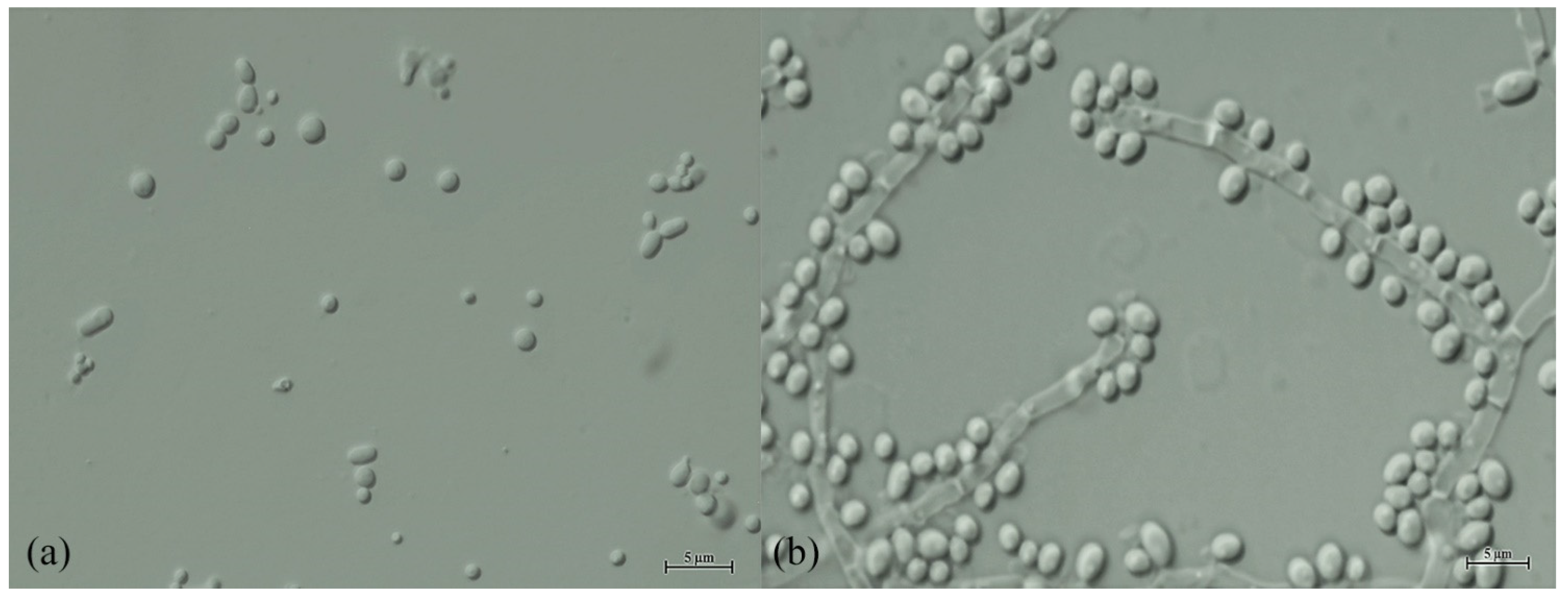
2.3. Phenotypic Characterization

The investigated yeasts were morphologically and physiologically characterized by standard methods described by Kurtzman et al. [23]. Yeasts were grown for three days in YM broth and YM agar at 25 °C for morphological study. Pseudo-hyphae and true hyphae formation were investigated on corn meal agar slide cultures at 25 °C for three days. Growth at different temperatures (15, 25, 30, 35, 37, 40, 42 and 45 °C) was determined in YM broth. The strains were examined individually or mixed in pairs for ascospore formation using different media including PDA, YM agar, YPD agar, corn meal agar, 5% malt extract agar, Gorodkowa agar, V8 agar, Fowell’s acetate agar [24] and yeast carbon base ammonium sulfate (YCBAS) agar [25] at 25 °C for up to twelve weeks with periodic microscopic inspection. Carbon and nitrogen source assimilation, carbohydrate fermentation, starch-like compounds production, and cycloheximide resistance tests were conducted in liquid media. A urea hydrolysis test was performed on a urea slant medium. Acid production and Diazonium Blue B (DBB) tests were conducted on solid medium in Petri dishes. All experiments were carried out with three replicates.

3. Results

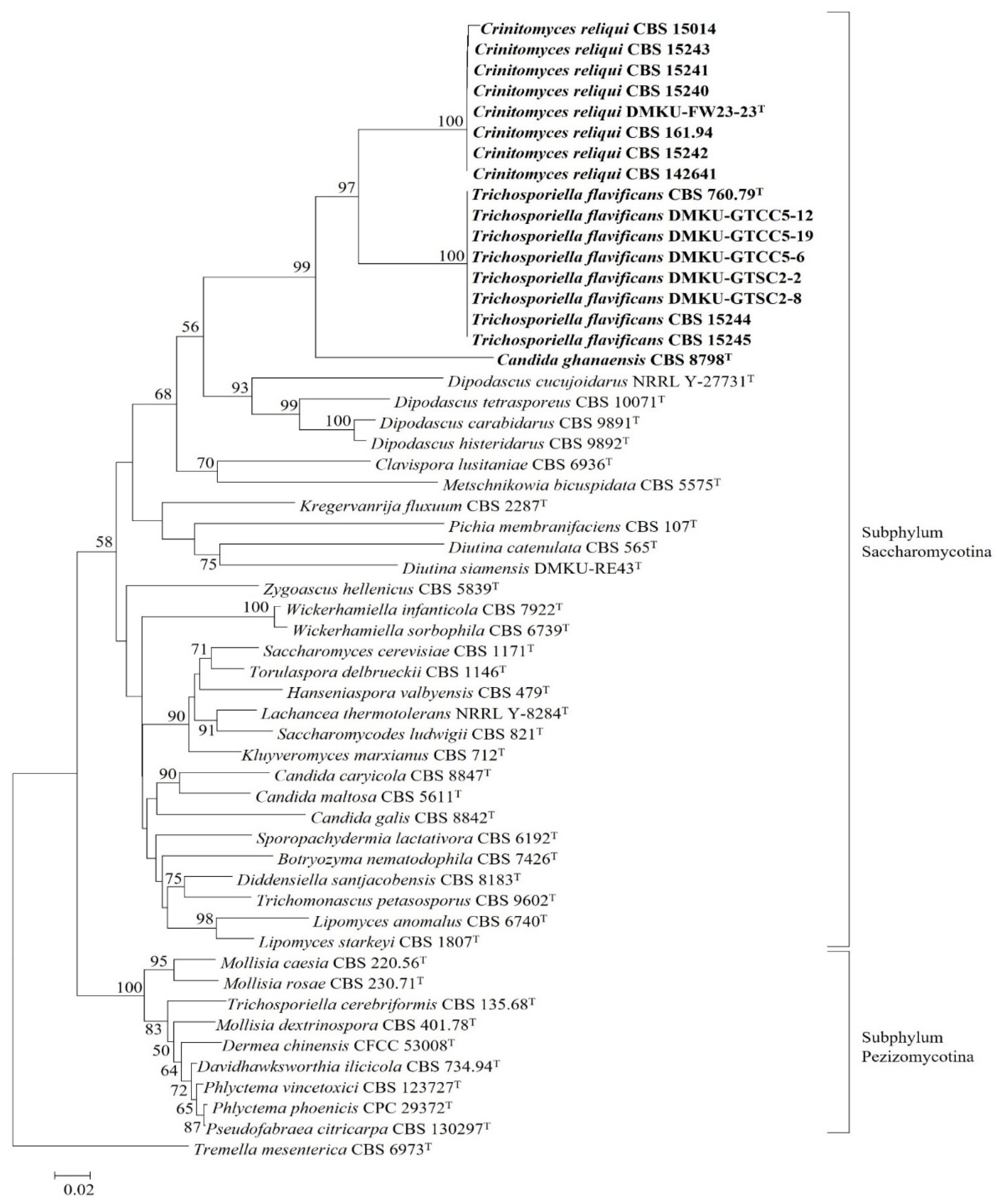
3.1. Species Delineation and Molecular Phylogeny

BLASTn search analysis of the D1/D2 domain of the LSU rRNA gene against the GenBank database was performed to identify the yeast strain DMKU-FW23-23 found during a study of yeast community in food waste. The result showed that the top two results from a BLASTn search hit with the currently recognized species Trichosporiella flavificans CBS 760.79T and Candida ghanaensis CBS 8798T, respectively. Surprisingly, these two species are described in different subphyla i.e., T. flavificans was placed in the subphylum Pezizomycotina and C. ghanaensis in the subphylum Saccharomycotina. The relationship between these two species was unexpected and needed to be clarified. Thus, a placement of the strains DMKU-FW23-23, T. flavificans CBS 760.79T and C. ghanaensis CBS 8798T was thoroughly investigated in this study. Seven additional strains, CBS 15240, CBS 15241, CBS 15242, CBS 15243, CBS 15014, CBS 161.94, and CBS 142641, that were similar to the strain DMKU-FW23-23, were found from BLASTn search analysis. Pairwise alignment revealed that the strain DMKU-FW23-23 and its companions differed from each other at 0–2 nucleotide substitutions without gaps in the D1/D2 domain of the LSU rRNA gene, while their ITS region showed no nucleotide substitutions and 0–1 gap (Table 3.).
All available strains with similar sequences to that of T. flavificans CBS 760.79T in the GenBank database, DMKU-GTSC2-8, DMKU-GTSC2-2, DMKU-GTCC5-6, DMKU-GTCC5-12, DMKU-GTCC5-19, CBS 15244, and CBS 15245, were subjected to physiological and molecular analyses. The results of the pairwise alignment of T. flavificans CBS 760.79T and its related strains are shown in Table 4. Identical sequences (0 nucleotide substitution with 0–1 gap) were found in the D1/D2 domain of the LSU rRNA gene and 0–3 nucleotide substitutions with 0–6 gaps were found in the ITS region among T. flavificans CBS 760.79T and its related strains.
To find the accurate taxonomic placement of the strain DMKU-FW23-23 and its companions, T. flavificans CBS 760.79T and C. ghanaensis CBS 8798T, a phylogenetic tree based on the D1/D2 domain of the LSU rRNA gene was constructed. In addition to the above-noted strains, related species were included in the analysis from the subphyla Saccharomycotina and Pezizomycotina. The results revealed that the strain DMKU-FW23-23 clustered with T. flavificans CBS 760.79T and C. ghanaensis CBS 8798T and their placements were found within the subphylum Saccharomycotina (Figure 1).
This result suggested that the assignment of T. flavificans to the genus Trichosporiella, which is nested in the subphylum Pezizomycotina, must be revised, and it should be transferred to the subphylum Saccharomycotina. In order to find its accurate placement within the subphylum Saccharomycotina, a phylogenetic tree based on a concatenated sequence of three genes including the small subunit (SSU) rRNA gene, the ITS region and the D1/D2 domain of the LSU rRNA gene was constructed (Figure 2 and Figure S1). The result demonstrated that the strain DMKU-FW23-23 and its companions formed a single lineage and were placed next to T. flavificans CBS 760.79T and also grouped together with C. ghanaensis CBS 8798T. These three species formed a distinct monophyletic clade that is clearly separated from other described yeast species. Hence, a novel yeast genus namely Crinitomyces is proposed to accommodate T. flavificans and C. ghanaensis which are reassigned as Crinitomyces flavificans and Crinitomyces ghanaensis, respectively. Moreover, the strain DMKU-FW23-23 and its companion strains are also proposed as a novel yeast species within this novel genus and the name Crinitomyces reliqui sp. nov. is proposed.
Phenotypic characters of the three yeast species: Crinitomyces reliqui sp. nov., Crinitomyces flavificans comb. nov. and Crinitomyces ghanaensis comb. nov. were compared and are summarized in Table 5.
A broad range of carbon sources was found to be assimilated, namely glucose, galactose, sorbose, cellobiose, maltose, sucrose, trehalose, melezitose, L-arabinose, D-xylose, erythritol, glucitol, mannitol, glycerol, ethanol, succinic acid, salicin and N-acetyl-glucosamine and only two carbon sources, inulin, and citric acid, were not assimilated by any of these yeasts. However, Crinitomyces ghanaensis CBS 8798T did not show fermentation ability. It should be noted that, by all strains of Crinitomyces flavificans, a yellow pigment was exuded onto the agar medium during cultivation. For morphological characteristics, yeast colonies of the three species are white to cream, convex and butyrous, with a dull surface. All of the three yeast species showed a hairy colony morphology (Figure 3) which is the origin of the genus name, Crinitomyces.

3.2. Ecology

The type strain of Crinitomyces reliqui DMKU-FW23-23T was isolated from domestic food waste in Thailand while the two related strains, CBS 161.94 and CBS 142641, were isolated from similar types of habitats, sewage sludge and wastewater in Republic of Poland and The Netherlands, respectively. However, the strain CBS 15,014 was isolated from soil and the four related strains were isolated from water surface of a river in The Netherlands and Hungary, respectively. These habitats showed a contrast in terms of amount and type of nutrients available. The occurrence of this species in food waste, sewage sludge and rivers raised the possibility that the water of the rivers might have been polluted at the time of sampling. Crinitomyces reliqui is suggested to be a cosmopolitan species because all eight strains of this species were found from four different countries.
The yeast Crinitomyces flavificans CBS 760.79T was isolated from washings of ion-exchange resin in a guanosine monophosphate manufacturing plant in Japan whereas its seven companion strains were isolated from food waste and water in different countries. Therefore, C. flavificans should also be claimed as a cosmopolitan species.
The isolation sources and geographical origin of all investigated strains are summarized in Table 1.

3.3. Taxonomy

Description of Crinitomyces V. Sakpuntoon, G. Péter, M. Groenew., D. Dlauchy, S. Limtong & N. Srisuk, gen. nov.
MycoBank number: 842461.
Crinitomyces (Cri.ni.to.my’ces. N.L. fem. n. Crinitomyces refers to the hairy colony appearance of yeast within the genus).
Cells are spherical or ellipsoidal and asexual reproduction proceeds by multilateral budding. Septate hyphae are produced. Ascospore formation is not observed. DBB reaction is negative, starch-like compounds are not produced, and urea hydrolysis is negative.
Phylogenetic placement: Saccharomycetales, Saccharomycotina, Ascomycota.
Type species: Crinitomyces flavificans (Nakase) V. Sakpuntoon, G. Péter, M. Groenew., D. Dlauchy, S. Limtong & N. Srisuk comb. nov.
Description of Crinitomyces reliqui V. Sakpuntoon, G. Péter, M. Groenew, D. Dlauchy, S. Limtong & N. Srisuk, sp. nov.
Crinitomyces reliqui (re.li.qu’i. L. fam. adj. reliquum of the residue; reliquum indicating that the type strain was isolated from residue of food or food waste).
After 3 days growth in YM broth at 25 °C, cells are spherical (1.5–3 μm) or ellipsoidal (1.5–2.0 × 2–3 μm). Colonies are white to cream, convex and butyrous, with a dull surface and filamentous margins (Figure 3c). True hyphae and branching lateral hyphae are observed on corn meal agar at 25 °C after 3 days (Figure 4b). Blasto-conidia are formed randomly from both hyphal types, globose to sub-globose, 1.8–3.0 μm. Ascospores are not found in individual cultures or in mixed cultures on PDA, YM agar, YPD agar, corn meal agar, 5% malt extract agar, Gorodkowa agar, Fowell’s acetate agar, V8 agar and YCBAS medium after 12 weeks at 25 °C. Glucose (viable), galactose (viable) and maltose (viable) are fermented. but lactose, sucrose, trehalose, melibiose and raffinose are not. Glucose, galactose, sorbose, cellobiose, lactose (weak), maltose (weak), melibiose (weak), sucrose (weak), trehalose, melezitose (weak), raffinose (weak), starch (variable), D-arabinose, L-arabinose, D-ribose, L-rhamnose, D-xylose, galactitol (weak), erythritol, D-glucitol (slow and weak), inositol (slow and weak), D-mannitol (slow and weak), glycerol, ribitol, ethanol, methanol (slow and weak), lactic acid, succinic acid, D-gluconic acid, α-Met-D-Glucoside, salicin (weak), N-Acetyl-D-Glucosamine, D-Glucono-5-lactone (weak), 2-Keto-D-gluconate (weak) and 5-Keto-D-gluconate (weak) are assimilated as the sole carbon sources, while inulin, citric acid, D-glucuronic acid and D-galacturonic acid are not assimilated. Ammonium sulfate, potassium nitrate (weak), sodium nitrite (weak), ethylamine hydrochloride, L-lysine and cadaverine dihydrochloride are utilized as sole nitrogen sources. Growth occurs in media containing 10% (w/v) sodium chloride/5% (w/v) glucose but not in 16% (w/v) sodium chloride/5% (w/v) glucose. Growth at 37 °C is positive for all strains except the strain CBS 15,014 of which growth occurs at 15 –30 °C. Growth is not observed in vitamin-free medium but variable results were found in medium supplemented with 0.01% (w/v) and 0.1% (w/v) cycloheximide. Acid production is variable. Urea hydrolysis, starch-like compounds production and diazonium blue B reaction are negative.
The holotype was isolated from domestic food waste in Bangkok, Thailand. The food waste sample was randomly collected via aseptic technique, and it was used for isolation process as previous described in materials and methods within 24 h. The obtained yeast colony was purified by cross-steaking on YM medium. After an initial BLASTn search analysis, additional representatives of the novel species were found. The strain was preserved at −80 °C in YM broth supplemented with 30% (v/v) glycerol. The holotype has been deposited and permanently preserved in a metabolically-inactive state in the Thailand Bioresource Research Centre (TBRC), Thailand, as TBRC 15054. An isotype has been permanently preserved in a metabolically-inactive state at the Department of Microbiology, Faculty of Science, Kasetsart University, Bangkok, Thailand as strain DMKU-FW23-23 and in the collection of the Portuguese Yeast Culture Collection (PYCC), Caparica, Portugal, as strain PYCC 9001. MycoBank number 842462.
New combinations.
Crinitomyces flavificans (Nakase) V. Sakpuntoon, G. Péter, M. Groenew., D. Dlauchy, S. Limtong & N. Srisuk, comb. nov.
MycoBank number: 842463.
Basionym: Candida flavificans, T. Nakase (1975). Antonie van Leeuwenhoek 41:202
Holotype: CBS 760.79, from washings of ion-exchange resin in a guanosine monophosphate manufacturing plant.
Note: Crinitomyces flavificans was first described as Candida flavificans based on an analysis of physiological and biochemical characteristics in 1975 [27]. Later, based on morphological characters it was reclassified as Trichosporiella flavificans [27]. However, the type species of Trichosporiella, T. cerebriformis, is nested in the subphylum Pezizomycotina. Based on the phylogenetic analyses carried out in this study, T. flavificans is reclassified here in Saccharomycotina as Crinitomyces flavificans. All phenotypic characters of the type strain, CBS 760.79, were re-examined in this study and results were found consistent with those of the first report. Nevertheless, some characters were differed among the companion strains and were then reported as “variable” as shown in the Table 5.
Crinitomyces ghanaensis (Kurtzman) V. Sakpuntoon, G. Péter, M. Groenew., D. Dlauchy, S. Limtong & N. Srisuk, comb. nov.
MycoBank number: 842464.
Basionym: Candida ghanaensis, C.P. Kurtzman (2001). Antonie van Leeuwenhoek 79:355.
Holotype: CBS 8798, from soil in Ghana.

4. Conclusions and Discussion

Candida flavificans CBS 760.79T was isolated from washing of the ion-exchange resin in a guanosine monophosphate manufacturing plant and classified based on DNA base composition, proton-magnetic-resonance spectrum of polysaccharide, and serological characteristics [27]. However, in 1985, it was reclassified as Trichosporiella flavificans CBS 760.79T due to a stronger coherence between hyphal cells and an absence of arthroconidia that split Trichosporiella from Candida [28]. Sequence analysis of this yeast genus has not been accomplished since then. In the era in which molecular study and sequencing technology play an important role in taxonomic study, yeast classification by the aforementioned analyses may not be sufficient and may also cause misidentifications.
Even if molecular methods and phylogenetic analyses have been used to identify yeast, unstable placement in the evolution line may occur, since they may be characterized based on short sequences and/or a small number of genes in phylogenetic analysis. Candida ghanaensis CBS 8798T isolated from soil in Ghana, was first described with a weakly supported phylogenetic placement based on the D1/D2 domain of the LSU of rRNA gene by Kurtzman [26]. Subsequent analysis of the SSU of the rRNA gene sequence revealed that C. ghanaensis had a weak and probably insignificant affinity with C. incommunis, but the highest matches in the GenBank database were found with members of the Dipodascus/Geotrichum clade [8]. However, a well-supported placement of this yeast species has not yet established and a phylogenetic reconstruction from additional data was required.
Accurate classification of yeasts may not be achieved by a single method. Although molecular methods are irreplaceable, yeast identification requires combined application of several approaches (polyphasic taxonomy). Similarly, multiple conspecific strains are more reliable to propose a new yeast species. Nevertheless, it will also be more reliable if conspecific strains are isolated from different samples and/or geographical regions.
In this study we described a new yeast species, Crinitomyces reliqui, which is closely related to T. flavificans and C. ghanaensis as they formed a well-supported clade in the phylogenetic trees and they also shared morphological and some physiological characteristics. We proposed here a novel yeast genus, Crinitomyces, to accommodate the novel species Crinitomyces reliqui as well as T. flavificans and C. ghanaensis, which were reassigned as Crinitomyces flavificans and Crinitomyces ghanaensis, respectively.

Supplementary Materials

The following are available online at https://www.mdpi.com/article/10.3390/jof8030224/s1, Figure S1: Phylogenetic tree based on concatenated sequences of the SSU rRNA gene, the ITS region and the D1/D2 domain of the LSU rRNA gene showing the placement of Trichosporiella flavificans, Candida ghanaensis and Crinitomyces reliqui within the subphylum Saccharomycotina. The phylogenetic tree was constructed using the maximum likelihood (ML) method with optimized models for 3 partitions (SSU, ITS region and LSU) in IQ-TREE. The numbers provided on branches are frequencies with which a given branch appeared in 1000 bootstrap replications. Bootstrap values of less than 50% are not shown. Deakozyma indianensis CBS 12903T was served as an outgroup species. Bar, 0.1 substitutions per site.

Author Contributions

Data curation, formal analysis, investigation, and writing—original draft preparation, V.S.; Supervision and validation, G.P. and M.G.; Methodology, D.D.; Supervision, S.L.; Conceptualization, funding acquisition, resources, supervision, writing—review and editing, N.S. All authors have read and agreed to the published version of the manuscript.

Funding

This work was supported by the Kasetsart University Research and Development Institute (KURDI) grant no. FF(KU)18.64 to N.S., the Royal Golden Jubilee PhD programme grant no. PHD/0070/2560 to V.S. This work was also supported by the Hungarian Ministry for Innovation and Technology and by the European Union and co-financed by the European Social Fund (grant agreement no. EFOP-3.6.3-VEKOP-16-2017-00005 to G.P. and D.D.).

Institutional Review Board Statement

Not applicable.

Informed Consent Statement

Not applicable.

Data Availability Statement

All sequence data are available in NCBI GenBank following the accession numbers in the manuscript.

Acknowledgments

We would like to thank Kasetsart University Research and Development Institute (KURDI) for the grant no. FF(KU)18.64, the Royal Golden Jubilee PhD programme, the Hungarian Ministry for Innovation and Technology and by the European Union and co-financed by the European Social Fund (grant agreement no. EFOP-3.6.3-VEKOP-16-2017-00005), UGSAS-GU via the “Microbiology Laboratory Station for IC-GU12” at Kasetsart University and International SciKU Branding (ISB), Faculty of Science, Kasetsart University.

Conflicts of Interest

The authors declare that there is no conflict of interest in terms of the publication.

References

  1. Boekhout, T.; Aime, M.C.; Begerow, D.; Gabaldon, T.; Heitman, J.; Kemler, M.; Khayhan, K.; Lachance, M.A.; Louis, E.J.; Sun, S.; et al. The evolving species concepts used for yeasts: From phenotypes and genomes to speciation networks. Fungal Divers. 2021, 109, 27–55. [Google Scholar] [CrossRef] [PubMed]
  2. Lucking, R.; Aime, M.C.; Robbertse, B.; Miller, A.N.; Ariyawansa, H.A.; Aoki, T.; Cardinali, G.; Crous, P.W.; Druzhinina, I.S.; Geiser, D.M.; et al. Unambiguous identification of fungi: Where do we stand and how accurate and precise is fungal DNA barcoding? IMA Fungus 2020, 11, 14. [Google Scholar] [CrossRef] [PubMed]
  3. Moncada, B.; Lücking, R.; Suárez, A. Molecular phylogeny of the genus Sticta (lichenized Ascomycota: Lobariaceae) in Colombia. Fungal Divers. 2014, 64, 205–231. [Google Scholar] [CrossRef]
  4. Lücking, R.; Forno, M.D.; Moncada, B.; Coca, L.F.; Vargas-Mendoza, L.Y.; Aptroot, A.; Arias, L.J.; Besal, B.; Bungartz, F.; Cabrera-Amaya, D.M.; et al. Turbo-taxonomy to assemble a megadiverse lichen genus: Seventy new species of Cora (Basidiomycota: Agaricales: Hygrophoraceae), honouring David Leslie Hawksworth’s seventieth birthday. Fungal Divers. 2017, 84, 139–207. [Google Scholar] [CrossRef]
  5. Merényi, Z.; Varga, T.; Hubai, A.G.; Pitlik, P.; Erős, Á.; Trappe, J.M.; Bratek, Z. Challenges in the delimitation of morphologically similar species: A case study of Tuber brumale agg. (Ascomycota, Pezizales). Mycol. Prog. 2017, 16, 613–624. [Google Scholar] [CrossRef]
  6. Daniel, H.M.; Lachance, M.A.; Kurtzman, C.P. On the reclassification of species assigned to Candida and other anamorphic ascomycetous yeast genera based on phylogenetic circumscription. Antonie Van Leeuwenhoek 2014, 106, 67–84. [Google Scholar] [CrossRef]
  7. Kurtzman, C.P.; Robnett, C.J.; Blackwell, M. Description of Teunomyces gen. nov. for the Candida kruisii clade, Suhomyces gen. nov. for the Candida tanzawaensis clade and Suhomyces kilbournensis sp. nov. FEMS Yeast Res. 2016, 16, fow041. [Google Scholar] [CrossRef] [Green Version]
  8. Lachance, M.; Boekhout, T.; Scorzetti, G.; Fell, J.W.; Kurtzman, C.P. Candida Berkhout The Yeasts, a Taxonomic Study; Elsevier: Amsterdam, The Netherlands, 1923; pp. 987–1278. [Google Scholar]
  9. Urbina, H.; Blackwell, M. Multilocus Phylogenetic Study of the Scheffersomyces Yeast Clade and Characterization of the N-Terminal Region of Xylose Reductase Gene. PLoS ONE 2012, 7, e39128. [Google Scholar] [CrossRef] [Green Version]
  10. Kurtzman, C.P.; Robnett, C.J. Three new anascosporic genera of the Saccharomycotina: Danielozyma gen. nov., Deakozyma gen. nov. and Middelhovenomyces gen. nov. Antonie Van Leeuwenhoek 2014, 105, 933–942. [Google Scholar] [CrossRef]
  11. Khunnamwong, P.; Lertwattanasakul, N.; Jindamorakot, S.; Limtong, S.; Lachance, M.A. Description of Diutina gen. nov., Diutina siamensis, f.a. sp. nov., and reassignment of Candida catenulata, Candida mesorugosa, Candida neorugosa, Candida pseudorugosa, Candida ranongensis, Candida rugosa and Candida scorzettiae to the genus Diutina. Int. J. Syst. Evol. Microbiol. 2015, 65, 4701–4709. [Google Scholar] [CrossRef]
  12. Kurtzman, C.P. Description of Martiniozyma gen. nov. and transfer of seven Candida species to Saturnispora as new combinations. Antonie Van Leeuwenhoek 2015, 108, 803–809. [Google Scholar] [CrossRef] [PubMed]
  13. Kurtzman, C.P. Description of Groenewaldozyma gen. nov. for placement of Candida auringiensis, Candida salmanticensis and Candida tartarivorans. Antonie Van Leeuwenhoek 2016, 109, 1041–1045. [Google Scholar] [CrossRef] [PubMed]
  14. Boontham, W.; Angchuan, J.; Boonmak, C.; Srisuk, N. Limtongozyma siamensis gen. nov., sp. nov., a yeast species in the Saccharomycetales and reassignment of Candida cylindracea to the genus Limtongozyma. Int. J. Syst. Evol. Microbiol. 2020, 70, 199–203. [Google Scholar] [CrossRef] [PubMed]
  15. Sakpuntoon, V.; Angchuan, J.; Boontham, W.; Khunnamwong, P.; Boonmak, C.; Srisuk, N. Grease Waste as a Reservoir of Lipase-Producing Yeast and Description of Limtongella siamensis gen. nov., sp. nov. Microorganisms 2019, 8, 27. [Google Scholar] [CrossRef] [Green Version]
  16. Miles, D.J.; Busser, K.; Stalder, C.; Higgins, D.R. Isolation of Nucleic Acids. In Pichia Protocols; Higgins, D.R., Cregg, J.M., Eds.; Humana Press: Totowa, NJ, USA, 1998; Volume 103, pp. 73–80. [Google Scholar]
  17. Rosa, C.A.; Lachance, M.A.; Teixeira, L.C.; Pimenta, R.S.; Morais, P.B. Metschnikowia cerradonensis sp. nov., a yeast species isolated from ephemeral flowers and their nitidulid beetles in Brazil. Int. J. Syst. Evol. Microbiol. 2007, 57, 161–165. [Google Scholar] [CrossRef] [Green Version]
  18. Kurtzman, C.; Robnett, C. Phylogenetic relationships among yeasts of the complex determined from multigene sequence analyses. FEMS Yeast Res. 2003, 3, 417–432. [Google Scholar] [CrossRef] [Green Version]
  19. Kurtzman, C.P.; Robnett, C.J. Identification and phylogeny of ascomycetous yeasts from analysis of nuclear large subunit (26S) ribosomal DNA partial sequences. Antonie Van Leeuwenhoek 1998, 73, 331–371. [Google Scholar] [CrossRef]
  20. Hall, T.A. BioEdit: A user-friendly biological sequence alignment editor and analysis program for Windows 95/98/NT. Nucl. Acids Symp. Ser. 1999, 41, 95–98. [Google Scholar]
  21. Kumar, S.; Stecher, G.; Tamura, K. MEGA7: Molecular Evolutionary Genetics Analysis version 7.0 for bigger datasets. Mol. Biol. Evol. 2016, 33, 1870–1874. [Google Scholar] [CrossRef] [Green Version]
  22. Felsenstein, J. Confidence limits on phylogenies: An approach using the bootstrap. Evolution 1985, 39, 783–791. [Google Scholar] [CrossRef]
  23. Kurtzman, C.P.; Fell, J.W.; Boekhout, T.; Robert, V. Methods for Isolation, Phenotypic Characterization and Maintenance of Yeasts. In The Yeasts, a Taxonomic Study, 5th ed.; Kurtzman, C.P., Fell, J.W., Boekhout, T., Eds.; Elsevier: Amsterdam, The Netherlands, 2011; pp. 87–110. [Google Scholar]
  24. Fowell, R.R. Sodium acetate agar as a sporulation medium for yeasts. Nature 1952, 170, 578. [Google Scholar] [CrossRef] [PubMed]
  25. Kurtzman, C.P.; Fell, J.W.; Boekhout, T. The Yeasts, a Taxonomic Study, 5th ed.; Elsevier: Amsterdam, The Netherlands, 2011; Volume 1. [Google Scholar]
  26. Kurtzman, C.P. Four new Candida species from geographically diverse locations. Antonie Van Leeuwenhoek 2001, 79, 353–361. [Google Scholar] [CrossRef] [PubMed]
  27. Nakase, T. Three new asporogenous yeasts found in industrial waste water. Antonie Van Leeuwenhoek 1975, 41, 201–210. [Google Scholar] [CrossRef] [PubMed]
  28. de Hoog, G.S.; Rantio-Lehtimaki, A.H.; Smith, M.T. Blastobotrys, Sporothrix and Trichosporiella: Generic delimitation, new species, and a Stephanoascus teleomorph. Antonie Van Leeuwenhoek 1985, 51, 79–109. [Google Scholar] [CrossRef] [PubMed]
Figure 1. Phylogenetic tree based on the D1/D2 domain of the LSU rRNA gene showing an overview placement of Trichosporiella flavificans, T. cerebriformis, Candida ghanaensis and the novel species Crinitomyces reliqui. The phylogenetic tree was constructed using the neighbor-joining (NJ) method. The numbers provided on branches are frequencies with which a given branch appeared in 1000 bootstrap replications. Bootstrap values of less than 50% are not shown. Tremella mesenterica CBS 6973T served as an outgroup species. Bar, 0.02 substitutions per site.
Jof 08 00224 g001
Figure 2. Phylogenetic tree based on concatenated sequences of the SSU rRNA gene, the ITS region and the D1/D2 domain of the LSU rRNA gene showing the placement of Trichosporiella flavificans, Candida ghanaensis and Crinitomyces reliqui within the subphylum Saccharomycotina. The phylogenetic tree was constructed using the neighbor-joining (NJ) method. The numbers provided on branches are frequencies with which a given branch appeared in 1000 bootstrap replications. Bootstrap values of less than 50% are not shown. Deakozyma indianensis CBS 12903T served as an outgroup species. Bar, 0.02 substitutions per site.
Jof 08 00224 g002
Figure 3. A hairy colony appearance of three yeast species on YM agar. (a) Crinitomyces flavificans CBS 760.79T; (b) Crinitomyces ghanaensis CBS 8798T and (c) Crinitomyces reliqui DMKU-FW23-23T sp. nov.
Jof 08 00224 g003
Figure 4. Morphology of Crinitomyces reliqui DMKU-FW23-23T (a) Cells in YM broth after 3 days at 25 °C (bar, 5 μm) and (b) True hypha formation on corn meal agar after incubated at 25 °C for 3 days (bar, 5 μm).
Jof 08 00224 g004
Table 1. Yeast strains and isolation sources investigated in this study.
YeastSource of IsolationGeographical Origin
Crinitomycesreliqui sp. nov.
DMKU-FW23-23TDomestic food waste trapThailand
CBS 15014Soil taken from 2 cm deep in UtrechtThe Netherlands
CBS 161.94Sewage sludge in KatowiceRepublic of Poland
CBS 142641Sediment from wastewater treatment facility in ZeewoldeThe Netherlands
CBS 15,240
(=NCAIM Y.01958)
Water of Danube Budapest
(Location 1, 47.484163; 19.054271)
Hungary
CBS 15,241
(=NCAIM Y.02184)
Water of Danube Budapest
(Location 2, 47.594721; 19.070331)
Hungary
CBS 15242Water of Danube Budapest
(Location 2, 47.594721; 19.070331)
Hungary
CBS 15,243
(=NCAIM Y.02185)
Water of Danube Budapest
(Location 3, 47.592204; 19.069164)
Hungary
Trichosporiella flavificans
CBS 760.79TWashings of ion-exchange resin in a guanosine monophosphate manufacturing plantJapan
DMKU-GTSC2-8Food waste trap of Faculty of Science, KU canteenThailand
DMKU-GTSC2-2Food waste trap of Faculty of Science, KU canteenThailand
DMKU-GTCC5-6Food waste trap of KU central canteen Thailand
DMKU-GTCC5-12Food waste trap of KU central canteen Thailand
DMKU-GTCC5-19Food waste trap of KU central canteen Thailand
CBS 15,244
(=NCAIM Y.02186)
Water of Danube Budapest
(Location 3, 47.592204; 19.069164)
Hungary
CBS 15245Water of Danube Budapest
(Location 3, 47.592204; 19.069164)
Hungary
Candida ghanaensis
CBS 8798TSoil in GhanaGhana
KU: Kasetsart University, Bangkok.
Table 2. The accession numbers of studied yeasts and reference sequences retrieved from the GenBank database.
YeastsStrainSSUITSD1/D2
Botryozyma nematodophilaCBS 7426TNG061133NR111167NG042439
Candida caryicolaCBS 8847T-NR077194NG055176
Candida galisCBS 8842T-NR151797NG058980
Candida ghanaensisCBS 8798TNG065532KY102101NG055180
Candida haemuloniiCBS 5149TNG063413NR130669JX459759
Candida maltosaCBS 5611T-NR138346KY106554
Candida tropicalisCBS 94TEU348785NR111250NG054834
Clavispora lusitaniaeCBS 6936TNG065595NR130677NG055408
CrinitomycesreliquiDMKU-FW23-23TOK275053MW720560OK298472
CrinitomycesreliquiCBS 15014OK275054GU195664OK298471
CrinitomycesreliquiCBS 161.94OK275055MG250346OK298463
CrinitomycesreliquiCBS 142641OK275056MG250347OK298464
CrinitomycesreliquiCBS 15,240 (NCAIM Y.01958)OK275057MZ331539OK298466
CrinitomycesreliquiCBS 15,241 (NCAIM Y.02184)OK275058MZ312239OK298468
CrinitomycesreliquiCBS 15242OK275059MZ312240OK298469
CrinitomycesreliquiCBS 15,243 (NCAIM Y.02185)OK275060MZ312241OK298470
Davidhawksworthia ilicicolaCBS 734.94T-NR154008NG067307
Deakozyma indianensisNRRL YB-1937TNG061171KJ476205NG064315
Debaryomyces hanseniiNRRL Y-7426TNG063420NR120016NG042634
Dermea chinensisCFCC 53008T-NR171069NG073667
Diddensiella santjacobensisCBS 8183TNG063433NR151808NG058985
Dipodascus albidusCBS 766.85TMK834548AY788342NG066154
Dipodascus carabidarusCBS 9891T-NR155144NG058292
Dipodascus cucujoidarusNRRL Y-27731T-NR111352NG055370
Dipodascus geniculatusCBS 184.80TNG064797AY788301NG066466
Dipodascus histeridarusCBS 9892T-NR111351NG042466
Dipodascus tetrasporeusCBS 10071TAB300502AB300502AB300502
Diutina catenulataCBS 565T-NR077200NG059158
Diutina siamensisDMKU-RE43T-KT336715KT336715
Geotrichum candidumCBS 772.71TJQ698930HE663404JQ689071
Hanseniaspora valbyensisCBS 479TNG063247NR111113NG042630
Kluyveromyces marxianusCBS 712T-NR111251NG042627
Kockiozyma suomiensisCBS 7251TNG062713NR155335NG055355
Kregervanrija fluxuumCBS 2287TNG063291NR111196NG042445
Lachancea thermotoleransNRRL Y-8284TNG061071NR111334NG042626
Limtongia smithiaeCBS 7407TNG062712NR138235NG055354
Lipomyces anomalusCBS 6740TNG062697KT923624NG055345
Lipomyces starkeyiCBS 1807T-NG055350NR077109
Magnusiomyces magnusiiNRRL Y-17563TMK834553AY788307MK834532
Metschnikowia bicuspidataCBS 5575TNG065596KY104192KY108455
Meyerozyma guilliermondiiCBS 2030TNG063363NR111247NG042640
Middelhovenomyces petrohuensisCBS 8173TNG063431NR156314NG055211
Middelhovenomyces tepaeCBS 5115TNG063435NR154200NG055181
Mollisia caesiaCBS 220.56T-MH857591MT026503
Mollisia dextrinosporaCBS 401.78T-NR119489MH872917
Mollisia rosaeCBS 230.71T-MH860088MH871865
Phlyctema phoenicisCPC 29372T-NR155690NG067319
Phlyctema vincetoxiciCBS 123727T-NR145310NG067282
Pichia membranifaciensCBS 107TNG064813NR111195NG042444
Pseudofabraea citricarpaCBS 130297T-NR154319NG069282
Saccharomyces cerevisiaeCBS 1171TNG063315NR111007NG042623
Saccharomycodes ludwigiiCBS 821TNG063254NR165986NG042629
Saprochaete chiloensisCBS 8187TNG070306AY788349MK834538
Saprochaete saccharophilaCBS 252.91TNG070310AY788309MK834545
Saturnispora disporaCBS 794TEF550358NR155832NG055103
Savitreea pentosicarensDMKU-GTCP10-8TNG073529NR172171NG073813
Scheffersomyces stipitisNRRL Y-7124TNG063362NR165947NG042637
Sporopachydermia lactativoraCBS 6192T-NR111310KY109772
Starmerella bombicolaNRRL Y-17069TJQ698924NR121483NG042648
Starmerella geocharesCBS 6870TNG065473KJ630497NG060806
Tortispora caseinolyticaCBS 7781TNG065577NR154482NG055343
Torulaspora delbrueckiiCBS 1146TNG061300NR111257NG058413
Tremella mesentericaCBS 6973T-NR155937NG069419
Trichomonascus petasosporusCBS 9602TNG062797NR155940NG055332
Trichosporiella cerebriformisCBS 135.68T-NR155940MH859089
Trichosporiella flavificansCBS 760.79TOK275050MH873011OK298462
Trichosporiella flavificansDMKU-GTSC2-8OK275046MN460331OK283398
Trichosporiella flavificansDMKU-GTSC2-2OK275045MN460330OK283396
Trichosporiella flavificansDMKU-GTCC5-6OK275047MN460342OK283393
Trichosporiella flavificansDMKU-GTCC5-12OK275048MN460340OK283395
Trichosporiella flavificansDMKU-GTCC5-19OK275049MN460339OK283397
Trichosporiella flavificansCBS 15,244 (NCAIM Y.02186)OK275052MG250348OK298465
Trichosporiella flavificansCBS 15245OK275051MZ331540OK298467
Trigonopsis variabilisCBS 1040TNG061132NR154506NG055341
Wickerhamiella domercqiaeCBS 4351TNG061104DQ911462NG055328
Wickerhamiella infanticolaCBS 7922T-NR155985NG058278
Wickerhamiella osmotoleransDMKU VGT1-14TMN192121MN194615MH141490
Wickerhamiella sorbophilaCBS 6739T-NR155987NG055325
Zygoascus hellenicusCBS 5839TNG063434NR111258AY447007
T: Type strain of species.
Table 3. Pairwise DNA sequence comparisons between the strain DMKU-FW23-23 and its related strains.
YeastsNucleotide Substitution (bp)/Gap (bp)/Percentage of Sequence Similarity (%)
SSUITSD1/D2
CBS 15,240 (NCAIM Y.01958)2/0/99.90/0/1000/0/100
CBS 15,241 (NCAIM Y.02184)0/0/1000/0/1000/0/100
CBS 15,243 (NCAIM Y.02185)0/0/1000/0/1000/0/100
CBS 152420/0/1000/1/99.90/0/100
CBS 150140/0/1000/1/99.92/0/99.6
CBS 161.940/0/1000/0/1000/0/100
CBS 1426410/0/1000/1/99.90/0/100
Table 4. Pairwise DNA sequence comparisons between Trichosporiella flavificans CBS 760.79T and its related strains.
YeastsNucleotide Substitutions (bp)/Gaps (bp)/Percentage of Sequence Similarity (%)
SSUITSD1/D2
T. flavificans DMKU-GTSC2-80/0/1000/0/1000/0/100
T. flavificans DMKU-GTSC2-21/0/99.91/0/99.80/0/100
T. flavificans DMKU-GTCC5-61/0/99.91/1/99.80/1/99.8
T. flavificans DMKU-GTCC5-120/0/1003/6/98.60/0/100
T. flavificans DMKU-GTCC5-190/0/1001/2/99.80/1/99.8
T. flavificans CBS 15,244 (NCAIM Y.02186)0/0/1000/0/1000/0/100
T. flavificans CBS 152450/0/1001/1/99.90/0/100
Table 5. Physiological characteristics of Crinitomyces reliqui in comparison to its closely related species.
Physiological Characteristics123
Fermentation
   Glucosev+-
   Galactosevv-
   Lactose---
   Maltosev--
   Melibiose---
   Sucrose---
   Trehalose-v-
   Raffinose---
Carbon assimilation
   D-glucose+++
   Galactose+++
   Sorbose+vv
   Cellobiose+++
   Lactosew+-
   Maltosew++
   Melibiosew--
   Sucrosewv+
   Trehalose+++
   Melezitosewv+
   Raffinosewv-
   Inulin---
   Starchvv-
   D-arabinose+v-
   L-arabinose+++
   D-ribose++-
   L-rhamnose+--
   D-xylose+++
   Galactitolw--
   Erythritol+++
   D-glucitols/wv+
   Inositols/w--
   D-mannitols/wv+
   Glycerol+++
   Ribitol+v-
   Ethanol+s/w+
   Methanols/ws/w-
   Citric acid---
   Lactic acid++-
   Succinic acid+++
   D-gluconic acid+v-
   D-glucuronic acid--nd
   D-galacturonic acid--nd
   α-Met-D-glucoside+-+
   Salicinw++
   N-acetyl-D-glucosamine+++
   D-Glucono-5-lactonew+nd
   2-Keto-D-gluconatewv-
   5-Keto-D-gluconatew--
Nitrogen assimilation
   (NH4)2SO4++nd
   KNO3wv-
   NaNO2wwnd
   Ethylamine-HCl++nd
   L-lysine++nd
   Cadaverine+++
Others
   Diazonium Blue B--nd
   Starch-like compounds ---
   Growth at vitamin free medium---
   Urea hydrolysis--nd
   Growth at 15 °C++nd
   Growth at 25 °C+++
   Growth at 30 °C+++
   Growth at 35 °Cv++
   Growth at 37 °Cv++
   Growth at 40 °C-+-
   Growth at 42 °C---
   Growth at 45 °C---
   0.1% Cycloheximide resistancev-+
   0.01% Cycloheximide resistancev-+
   Growth in medium supplemented with 50% glucose+vnd
   Growth in medium supplemented with 60% glucose++nd
   Growth in medium supplemented with 5% glucose +10%NaCl+++
   Growth in medium supplemented with 5% glucose +16%NaCl--nd
   Acid productionvvnd
Species: 1, Crinitomyces reliqui sp. nov. (DMKU-FW23-23T and seven additional strains); 2, Crinitomyces flavificans comb. nov. (CBS 760.79T and seven additional strains) and 3, Crinitomyces ghanaensis comb. nov. CBS 8798T [26] and data obtained from CBS. Abbreviation: +, positive; -, negative; s, slow positive; w, weak positive; v, variable (some strains are positive, others negative); nd, no data.
Publisher’s Note: MDPI stays neutral with regard to jurisdictional claims in published maps and institutional affiliations.

