Article

Sporobolomyces lactucae sp. nov. (Pucciniomycotina, Microbotryomycetes, Sporidiobolales): An Abundant Component of Romaine Lettuce Phylloplanes

1
Department of Botany and Plant Pathology, Purdue University, West Lafayette, IN 47907, USA
2
Research Group Mycology, Department of Biology, Ghent University, 9000 Ghent, Belgium
3
Faculty of Science, University of South Bohemia, 370 05 České Budějovice, Czech Republic
4
Division of Plant Industry, Florida Department of Agriculture and Consumer Services, Gainesville, FL 32608, USA
*
Author to whom correspondence should be addressed.
J. Fungi 2022, 8(3), 302; https://doi.org/10.3390/jof8030302
Submission received: 28 January 2022 / Revised: 28 February 2022 / Accepted: 7 March 2022 / Published: 16 March 2022

Abstract

:
Shifts in food microbiomes may impact the establishment of human pathogens, such as virulent lineages of Escherichia coli, and thus are important to investigate. Foods that are often consumed raw, such as lettuce, are particularly susceptible to such outbreaks. We have previously found that an undescribed Sporobolomyces yeast is an abundant component of the mycobiome of commercial romaine lettuce (Lactuca sativa). Here, we formally describe this species as Sporobolomyces lactucae sp. nov. (Pucciniomycotina, Microbotryomycetes, and Sporidiobolales). We isolated multiple strains of this yeast from commercial romaine lettuce purchased from supermarkets in Illinois and Indiana; additional isolates were obtained from various plant phylloplanes in California. S. lactucae is a red-pigmented species that is similar in appearance to other members of the genus Sporobolomyces. However, it can be differentiated by its ability to assimilate glucuronate and D-glucosamine. Gene genealogical concordance supports S. lactucae as a new species. The phylogenetic reconstruction of a four-locus dataset, comprising the internal transcribed spacer and large ribosomal subunit D1/D2 domain of the ribosomal RNA gene, translation elongation factor 1-α, and cytochrome B, places S. lactucae as a sister to the S. roseus clade. Sporobolomyces lactucae is one of the most common fungi in the lettuce microbiome.

1. Introduction

The genus Sporobolomyces was erected by Kluyver and van Niel (1924) [1] to accommodate asexual basidiomycetous yeasts, with Sporobolomyces salmonicolor as the type species [2,3]. Similar species typified by a sexual morph were placed in Sporidiobolus [3,4,5]. It is now known that yeasts previously placed in form genus Sporobolomyces can be found across the three subphyla of Basidiomycota [6,7] and even in Ascomycota [8]. Sporobolomyces salmonicolor, however, belongs to Sporidiobolales (Pucciniomycotina, Microbotryomycetes), and Sporidiobolus is now considered a synonym of Sporobolomyces [9]. Currently, 22 species of Sporobolomyces sensu stricto (s.s.) are accepted [3,10,11], although estimates are that the genus contains upwards of 60 species [12]. All known species in the genus produce an asexual morph and reproduce by ballistoconidia. Some species produce a sexual morph and pseudohyphae in addition to yeast cells [13].
Yeasts in the genus Sporobolomyces are known for their bright red, orange, or pink appearance in culture [2] and have been studied for a variety of applications. The red pigmentation of the yeasts is due to the production of carotenoids such as beta-carotene [14,15]. The carotenoid-producing capability of Sporobolomyces yeasts has been of interest to the field of biotechnology to develop commodities such as pigments [14,15,16]. Sporobolomyces roseus exhibits antimicrobial activity inhibiting the growth of Pseudomonas fluorescens and Staphylococcus aureus, two bacteria that are both known to infect humans as opportunistic pathogens [17]. As a biological control agent, S. roseus has been effective against Cochliobolus sativus (common root rot) [18].
Sporobolomyces yeasts grow in various habitats, such as aquatic systems, soil, and plant phylloplanes [2,7,10,12]. They are best known for their association with plant leaves, with many organisms from the genus first isolated from the phylloplane [12,19]. Sporobolomyces yeasts are cosmopolitan in this regard and are capable of growing on a wide variety of plants, including agricultural crops [12,20,21,22,23,24]. The yeasts of Sporidiobolales are known to inhabit vegetable surfaces with little, if any, association with food spoilage [25,26].
We have previously found that the yeasts of Sporidiobolales are represented in the phylloplane of commercially grown romaine lettuce [12], which is consistent with a previous study on the lettuce microbiome [23]. We then characterized the mycobiome (fungi of the microbiome) of romaine lettuce obtained in Urbina and Aime [12]. By conducting this characterization, we found that over 25% of the mycobiome is represented by a single Sporobolomyces species, which is previously undescribed [24]. Although basidiomycetous yeasts comprise a small fraction of the romaine lettuce phylloplane microbiome, Sporidiobolales yeasts are the most common fungi present [12] and are represented overwhelmingly by Sporobolomyces spp. [24]. Here, we describe the most abundant of these red yeasts, Sporobolomyces lactucae sp. nov., and discuss its ecology and natural range.

2. Materials and Methods

2.1. Lettuce Leaf Preparation and Culturing of Fungal Isolates

Lettuce leaf homogenization and culture plating were completed as described in previous work [12,24]. In brief, commercial lettuce was purchased from grocery stores in Illinois and Indiana. Lettuce leaves were homogenized in 225 mL of 100 µM phosphate buffer (5.4 g monosodium phosphate L−1 and 8.7 g disodium phosphate L−1). Aliquots were plated on yeast extract–peptone–glucose agar with 25 µg chloramphenicol mL−1 and 50 µg ampicillin mL−1 to inhibit bacterial growth (BD, Franklin Lakes, NJ, USA; Thermo Fisher, Waltham, MA, USA). Isolates collected in California were obtained by using the ballistospore drop method [27]. Sections from leaves collected in the field were secured with petroleum jelly to the inside of a Petri plate lid. Ballistospores that dropped from the leaf-inhabiting yeasts grew on either potato dextrose agar (PDA) or yeast malt agar (YMA) (BD; Thermo Fisher). Subculturing was repeated until axenic cultures were obtained and maintained on PDA. Back-up cultures for long-term preservation were prepared in 40% (v/v) glycerol for −80 °C storage and on PDA slants for 4 °C storage. Working cultures were incubated on PDA at room temperature (25 °C). Live cultures were deposited in the Agricultural Research Service Culture Collection (NRRL) and the Westerdijk Fungal Biodiversity Institute (CBS). All isolates obtained for this study and their origins are presented in Supplementary Table S1.

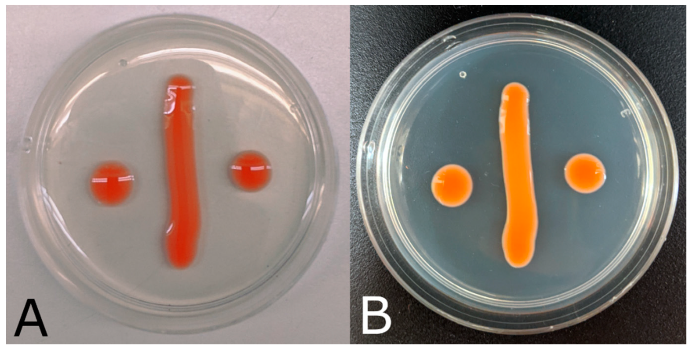
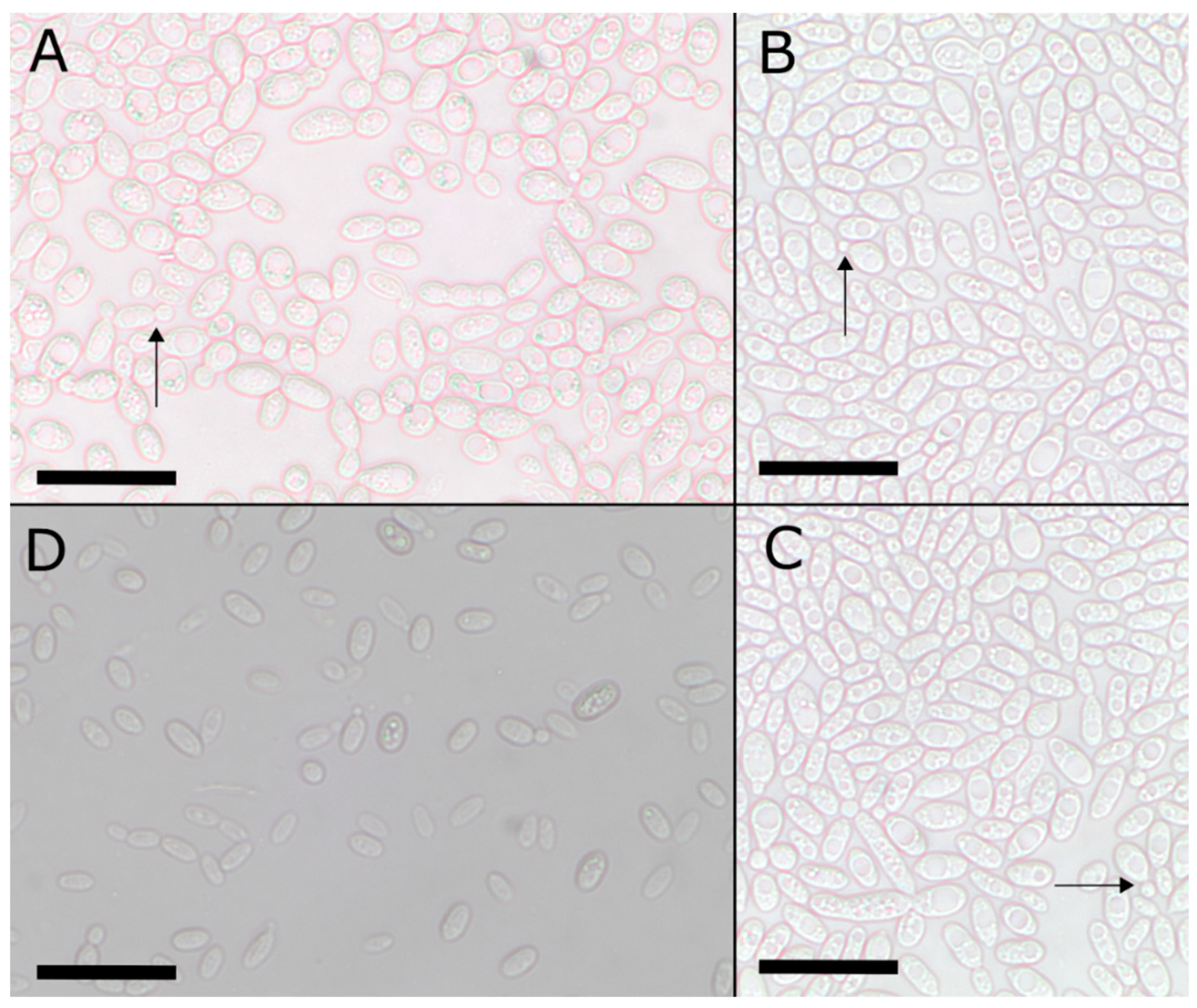
2.2. Morphological and Physiological Characterization

The morphological and physiological description was performed according to Suh et al. [28]; their protocols conform to the standard outlined in The Yeasts [29]. Culture morphology was observed on YMA, corn meal agar (CMA), and YM broth (BD; Thermo Fisher). After seven-day incubation, colonies were described in terms of elevation, margin, color (oac; [30]), form, and surface texture. Individual cells were examined under a compound microscope (Olympus BH-2; Tokyo, Japan). Micrographs were captured using an Olympus SC30 camera. A total of 30–180 cells were measured per isolate per treatment using Piximètre v5.10 (http://www.piximetre.fr/, accessed on 4 May 2021). Carbon and nitrogen assimilations were performed according to Suh et al. [28]. Positive assimilations marked with “+++” or “++” were reassigned as “+” while negative assimilations were assigned as “−”; weak growth was denoted as “(w)”, and delayed growth was denoted as “(d)”. This modified scale was used to normalize our data and allow for comparison across previously published data with varying scales.

2.3. DNA Extraction, PCR Amplification, and Sequencing

DNA was extracted using the Wizard Genomic DNA Purification Kit (Promega, Madison, WI, USA) following the manufacturer’s instructions. Alternatively, PCR amplifications were performed directly from colonies [27]. Various loci were sequenced to perform phylogenetic analyses as in Urbina and Aime [12]. The internal transcribed spacer (ITS) region was amplified using ITS1F and ITS4 primers [31,32]; the small ribosomal subunit (SSU) ribosomal RNA gene (rDNA) using NS1 and NS4 [31]; large ribosomal subunit (LSU) rDNA using LR0R and LR6 [33,34]; translation elongation factor 1-α (tef1) using EF1-983F and EF1-1567R [35]; and cytochrome B (cytb) using E1M4 and E2mr3 [36].
PCR protocols followed Toome et al. [37] for SSU, ITS, and LSU. For tef1, we used a touchdown PCR protocol as in Wang et al. [38]. For cytb, we followed Wang and Bai [39]. Purification and Sanger sequencing with amplification primers were outsourced to Gene-wiz, Inc. (South Plainfield, NJ, USA). Raw sequence reads were assembled and edited in Sequencher v. 5.2.3 (Gene Codes, Ann Arbor, MI, USA). Newly generated sequences were submitted to NCBI GenBank (accession numbers in Supplemental Table S1).

2.4. Phylogenetic Inferences and Species Concepts

Generated sequences of each locus were blasted against the NCBI GenBank standard nr/nt nucleotide database (http://ncbi.nlm.nih.gov/blast/Blast.cgi, accessed on 27 January 2022) to confirm identity [40]. For phylogenetic placement of our isolates, we downloaded ITS, LSU, tef1, and cytb ex-type sequences of Sporobolomyces from GenBank, following Urbina and Aime [12], Li et al. [11], and Tan et al. [41] as a guide for taxon selection to represent the 22 currently accepted species (Table 1). Rhodosporidiobolus microsporus and Rhodotorula babjevae were selected as outgroup taxa [12]. Sequences were aligned using MUSCLE v.3.8.1551 via the CIPRES Science Gateway [42,43]. The newly created multiple sequence alignment for each dataset was trimmed using TrimAI v.1.2.59 via CIPRES [43,44]. After trimming, single-locus trees were constructed using the Random Axelerated Maximum Likelihood (RAxML) v.8 program [45] available through CIPRES [43].
We applied a genealogical gene concordance hypothesis for species delimitation [54] by using information from multiple loci to establish overlapping phylogenies that are in consensus. Single-locus trees were constructed as above. Concordance was determined by hand. The SSU region was not informative and, thus, was not used in the multi-locus phylogenetic reconstruction.
The tef1 gene, cytb gene, LSU D1/D2 domain (28S rDNA region), and the ITS region were used for the construction of a four-locus phylogeny. The ITS and LSU D1/D2 domain regions are used here because they are widely used barcode regions for fungi, with LSU particularly suitable for yeasts. The genes tef1 and cytb were selected to represent nuclear and mitochondrial protein coding regions, respectively. The individual datasets were concatenated by using Mesquite v.3.61 [55]. Appropriate models for nucleotide substitution were selected using ModelFinder v1.6.12 [56] considering the Akaike Information Criterion. The models selected were TIM2 + F + R3 for ITS (−lnL = 2883.946), GTR + F + R2 for LSU (−lnL = 2077.917), GTR + F + R3 for tef1 (−lnL = 2235.566), and TVM + F + I + G4 for cytb (−lnL = 1704.151). Maximum likelihood was performed with IQ-TREE v.1.6.12 [57] under partitioned models [58]. Ultrafast bootstrapping was performed with 1000 replicates [59]. The reconstruction was visualized in FigTree v1.4.4 (http://tree.bio.ed.ac.uk/software/figtree/, accessed on 27 January 2022).

2.5. Environmental Determination of S. lactucae

To infer the broader distribution and habitats of S. lactucae, a second dataset was constructed of ITS sequences. The holotype strain of S. lactucae sp. nov. was pairwise aligned with environmental sequences deposited in GenBank that shared ≥98.0% identity. Environmental sequences were edited, aligned, and trimmed as above. We reconstructed an ITS-based phylogeny of the environmental sequences via IQ-TREE and inferred matching S. lactucae isolates based on this phylogeny. The final environmental dataset included 118 ITS sequences, 19 of which are Sporobolomyces s.s. type species, and was evaluated by using the K3Pu + F + G4 model (−lnL = 2533.283).

3. Results

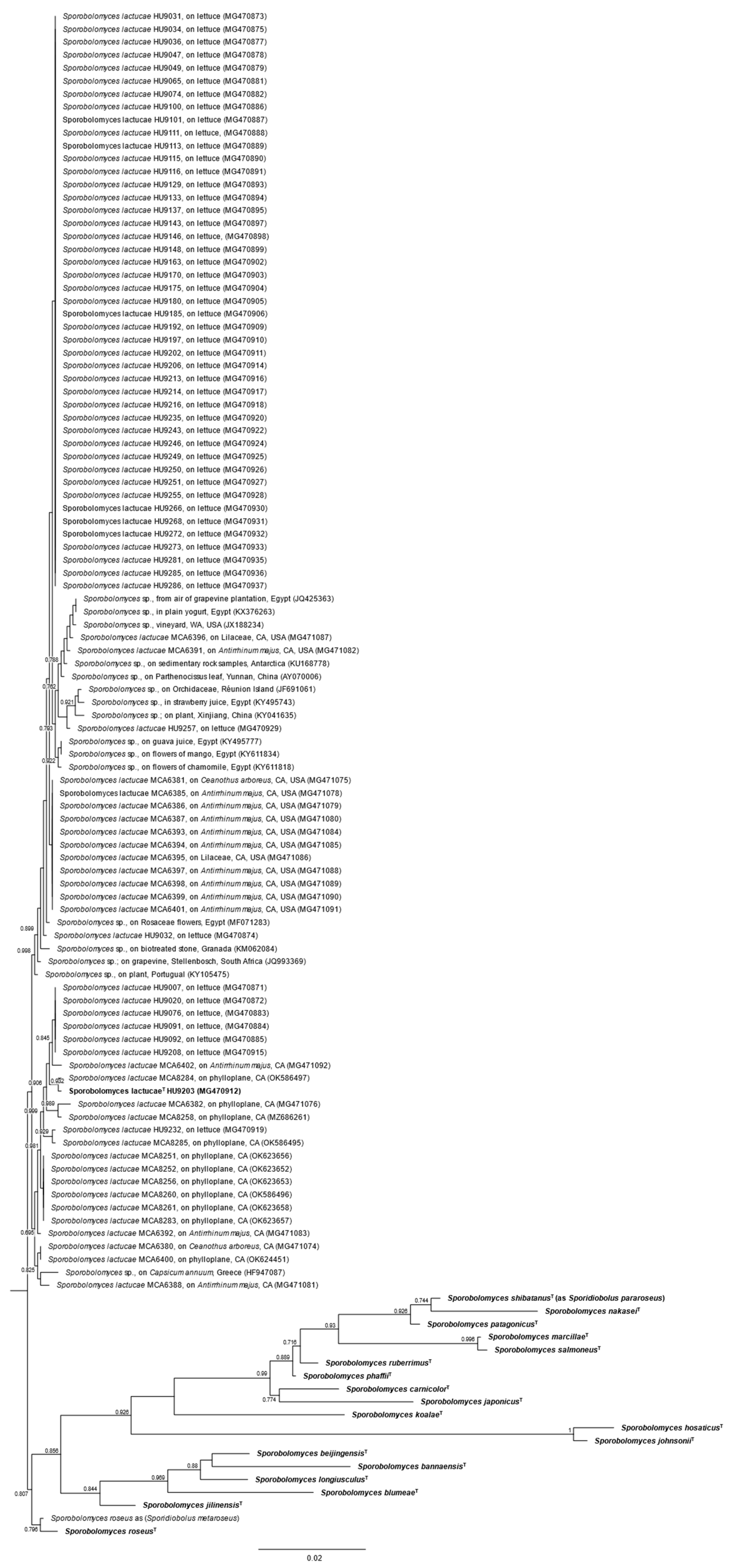
3.1. Phylogenetic Analyses

By performing gene genealogical concordance tests, each single-locus phylogenetic tree supported the delineation of S. lactucae as a new species. A congruence test of the four loci (ITS, LSU, tef1, and cytb) showed similar results with respect to our isolates. These single-locus phylogenies can be found in Supplemental Figures S1–S4. After determining concordance, a multi-locus phylogeny was constructed.
The final four-locus dataset (Figure 1) consisted of 32 taxa that are accepted as Sporobolomyces s.s.: 31 with ITS sequence data, 31 with LSU, 30 with tef1, and 28 with cytb. For ITS sequences, 610 characters constituted the dataset, of which 121 were parsimony-informative and 423 were constant. The LSU dataset consisted of 613 characters, with 85 parsimony-informative and 477 constant characters. The tef1 dataset comprised 473 characters: 85 were parsimony-informative and 359 were constant. The cytb dataset contained 414 characters, of which 60 were parsimony-informative and 293 were constant. Eight isolates form a well-supported, monophyletic lineage representative of S. lactucae: HU9111, HU9113, HU9170, HU9203, HU9214, HU9241, HU9243, and HU9244 (Figure 1).
In total, we recovered 66 S. lactucae isolates from commercial romaine lettuce. Additionally, we recovered 27 S. lactucae isolates from the phylloplanes of Rhamnaceae (Ceanothus arboreus), Plantaginaceae (Antirrhinum majus), and Lilaceae in two different years, all within the San Francisco Bay Area region of California (Figure 2; Supplemental Table S1). To better infer true S. lactucae environmental sequences, we reconstructed an ITS-based phylogeny. For our environmental dataset, we found 99 Sporobolomyces ITS sequences from ten studies or surveys (including the present study) that shared at least 98% sequence identity with S. lactucae.

3.2. Taxonomy

Sporobolomyces lactucae, Fatemi, Urbina & Aime, sp. nov., MycoBank MB 840687. Figure 3 and Figure 4. Ex-holotype identifiers: CBS 16795; NRRL Y-64010.
Etymology: lactucae (Latin), referring to the genus of the lettuce plant from which the holotype isolate was sourced.
Diagnosis: Similar to S. jilinensis and S. roseus but differing in the ability to assimilate glucoronate and D-glucosamine but not lactate or citrate.
Typification: USA, Illinois, Urbana-Champaign, from leaves of commercial Lactuca sativa (Asterales, Asteraceae), 9 May 2016, H. Urbina HU9203 (holotype PUL F27743 preserved as dried inert culture). Isotype PUL F27744 preserved as dried inert culture. Ex-holotype cultures at CBS (CBS 16795) and NRRL (Y-64010). Ex-holotype GenBank accession numbers: MG588994 (SSU); MG470912 (ITS); MG588947 (LSU); MG589082 (tef1); MG589041 (cytb).
Habitat and distribution: on leaf surfaces, particularly those of agricultural products, in mild or Mediterranean climates.
Description: In the asexual state, colonies are orange-pink in color (oac616) after 7 d incubation at 25 °C on PDA and YMA. Colonies are smooth and glistening, varying between circular and irregular with the entire margin. Colonies are raised in elevation. After 7 d incubation in YM broth, single cells appeared ellipsoidal, 5–11 µm × 3–5 µm, and uninucleate. On CMA, cells measured 5–10 µm × 3–6 µm. A single large vacuole forms in the cells. The formation of ballistoconidia was observed on CMA; new cells arise from sterigmata on mother cells. Pseudohyphae were not observed, but small chains of cells (usually about three cells) were rarely observed. No sexual stage was observed.
Fermentation of glucose was negative. Growth was observed on media containing yeast extract (1% w/v) and agar (2% w/v) with the following supplements: 50% w/v glucose or 60% w/v glucose. Negligible/weak growth was seen on the same media supplemented with 10% w/v sodium chloride and 16% w/v sodium chloride. Assimilation was positive for the following carbon compounds: glucose, galactose (weak, delayed), sucrose, maltose, cellobiose (weak, delayed), soluble starch (weak), glucono-1,5-lactone (weak), glucuronate, galacturonic acid (weak), ethanol (weak), and propane-1,2-diol (weak). Assimilation was negative for the carbon compounds lactose, inulin, myo-inositol, lactate, citrate, and methanol. Assimilation was positive for the nitrogen compounds potassium nitrate, sodium nitrate, ethylamine (weak), L-lysine, D-glucosamine, creatine (weak), creatinine (weak), and D-tryptophan (weak). Assimilation was negative for the nitrogen compound imidazole.
Additional materials: USA. INDIANA: Lafayette, commercial lettuce leaf, 30 April 2016, H. Urbina HU9007; 30 April 2016, H. Urbina HU9020; 30 April 2016, H. Urbina HU9031; 30 April 2016, H. Urbina HU9034; 30 April 2016, H. Urbina HU9035; 30 April 2016, H. Urbina HU9036; 2 May 2016, H. Urbina HU9047; 2 May 2016, H. Urbina HU9049; 6 May 2016, H. Urbina HU9062; 6 May 2016, H. Urbina HU9065; 6 May 2016, H. Urbina HU9074; 6 May 2016, H. Urbina HU9076; 2 May 2016, H. Urbina HU9091; 6 May 2016, H. Urbina HU9092; 6 May 2016, H. Urbina HU9100; 6 May 2016, H. Urbina HU9101; 2 May 2016, H. Urbina HU9111; 6 May 2016, H. Urbina HU9113; 6 May 2016, H. Urbina HU9115; 6 May 2016, H. Urbina HU9116; 6 May 2016, H. Urbina HU9128; 6 May 2016, H. Urbina HU9129; 6 May 2016, H. Urbina HU9133; 6 May 2016, H. Urbina HU9137; 6 May 2016, H. Urbina HU9143; 6 May 2016, H. Urbina HU9146; 6 May 2016, H. Urbina HU9148. USA. INDIANA: West Lafayette, commercial lettuce leaf, 8 May 2016, H. Urbina HU9152; 8 May 2016, H. Urbina HU9155; 8 May 2016, H. Urbina HU9163; 8 May 2016, H. Urbina HU9170; 8 May 2016, H. Urbina HU9175. USA. ILLINOIS: Urbana-Champaign, commercial lettuce leaf, 9 May 2016, H. Urbina HU9180; 9 May 2016, H. Urbina HU9185; 9 May 2016, H. Urbina HU9188; 9 May 2016, H. Urbina HU9192; 9 May 2016, H. Urbina HU9197; 9 May 2016, H. Urbina HU9202; 9 May 2016, H. Urbina HU9206; 9 May 2016, H. Urbina HU9208; 9 May 2016, H. Urbina HU9213; 9 May 2016, H. Urbina HU9214; 9 May 2016, H. Urbina HU9216; 9 May 2016, H. Urbina HU9233; 9 May 2016, H. Urbina HU9235. USA. ILLINOIS: Chicago, commercial lettuce leaf, 19 May 2016, H. Urbina HU9241; 19 May 2016, H. Urbina HU9243; 19 May 2016, H. Urbina HU9249; 19 May 2016, H. Urbina HU9250; 19 May 2016, H. Urbina HU9251; 19 May 2016, H. Urbina HU9255; 19 May 2016, H. Urbina HU9257; 19 May 2016, H. Urbina HU9266; 19 May 2016, H. Urbina HU9268; 19 May 2016, H. Urbina HU9272; 19 May 2016, H. Urbina HU9273; 19 May 2016, H. Urbina HU9279; 19 May 2016, H. Urbina HU9281; 19 May 2016, H. Urbina HU9285; 19 May 2016, H. Urbina HU9286. USA. CALIFORNIA: Berkeley, diseased leaf of Ceanothus arboreus (Rosales, Rhamnaceae), 5 August 2016, M.C. Aime MCA6380; 5 August 2016, MCA6381; healthy leaf of C. arboreus, 5 August 2016, M.C. Aime MCA6382; healthy leaf of Antirrhinum majus (Lamiales, Plantaginaceae), 5 August 2016, M.C. Aime MCA6385; diseased leaf of A. majus, 5 August 2016, M.C. Aime MCA6386; 5 August 2016, M.C. Aime MCA6387; healthy flower of A. majus, 5 August 2016, M.C. Aime MCA6391; 5 August 2016, M.C. Aime MCA6392; 5 August 2016, M.C. Aime MCA6393; 05 Aug 2016, M.C. Aime MCA6394; necrotic leaf (Liliaceae), 5 August 2016, M.C. Aime MCA6395; 5 August 2016, M.C. Aime MCA6396; diseased leaf of A. majus, 5 August 2016, M.C. Aime MCA6397; necrotic leaf of A. majus, 5 August 2016, M.C. Aime MCA6398; diseased leaf of A. majus, 5 August 2016, M.C. Aime MCA6399; phylloplane of undetermined host, 5 August 2016, M.C. Aime MCA6400; necrotic leaf of A. majus, 5 August 2016, M.C. Aime MCA6401; healthy flower of A. majus, 5 August 2016, M.C. Aime, MCA6402; Santa Cruz, phylloplane of undetermined host, 8 March 2019, M.C. Aime, MCA8251; phylloplane of undetermined host, 8 March 2019, M.C. Aime, MCA8252; phylloplane of undetermined host, 8 March 2019, M.C. Aime, MCA8256; phylloplane of undetermined host, 8 March 2019, M.C. Aime, MCA8258; phylloplane of undetermined host, 8 March 2019, M.C. Aime, MCA8260; phylloplane of undetermined host, 8 March 2019, M.C. Aime, MCA8261; phylloplane of undetermined host, 8 March 2019, M.C. Aime, MCA8283; phylloplane of undetermined host, 8 March 2019, M.C. Aime, MCA8284; phylloplane of undetermined host, 8 March 2019, M.C. Aime, MCA8285.
Notes: Because of the high similarity in colony morphology, colony color, and individual cell shape between yeasts of Sporobolomyces, assimilations are a more effective means of diagnosis. S. lactucae is weak in its assimilation of ethanol, but S. jilinensis and S. roseus more readily assimilate the compound [5,13,21]. Glucuronate and D-glucosamine are both substrates that S. roseus is unable to assimilate, while S. lactucae can. S. jilinensis is also unable to assimilate D-glucosamine (Table 2). Assimilations of galactose, cellobiose, soluble starch, and ethanol are all weak in S. lactucae, while the closely related species S. roseus can assimilate all four compounds (Table 2). S. lactucae is also capable of growth on high osmotic media (50% w/v glucose) while S. roseus experiences variable growth and S. jilinensis experiences no growth on the same type of media. Most S. lactucae cultures showed visible growth within one to two days of incubation, with full colony growth displayed by day seven (Figure 3).

4. Discussion

Sporobolomyces lactucae was the most frequently isolated yeast from phylloplanes of commercial romaine lettuce purchased from grocery stores in Illinois and Indiana, USA [24]. Of the thousands of phylloplane isolates of Sporidiobolales we have made throughout the world ([12]; M.C. Aime, unpubl.), we have recovered S. lactucae only from plant samples in the San Francisco and Monterrey Bay areas of California (Supplemental Table S2); identical sequences have been detected by others in areas across the world that have similar climates (Table 3). The majority of lettuce produced in the USA is grown in California [60], and we were able to trace the romaine lettuce origins for most of our samples to the Salinas Valley in California (Supplemental Table S3). It is well-documented that pathogenic microorganisms can spread through the production, distribution, and preparation of food, with greater risks of foodborne illnesses for foods consumed raw such as lettuce [61]. It stands to reason that commensal organisms are moved through our food distribution systems as well. In this case, S. lactucae isolated from lettuce purchased in the Midwest may very well have originated in California. The ability to trace commensal microbial organisms through food distribution may improve our ability to trace pathogenic outbreaks.
Much like other members of Sporidiobolales, S. lactucae appears to be widespread, occupying various habitats [12]. We found sequences consistent with S. lactucae in public databases from regions that experience a Mediterranean climate (Table 3). Half of the environmental samples were isolated in Egypt. The other half are distributed across Réunion Island (Indian Ocean), South Africa, Greece, Portugal, Granada, and Pullman (Washington, USA). Additionally, our environmental analysis indicates the potential presence of S. lactucae in Antarctica as well as Yunnan Province, China. Although the substrate from which environmental sequences were obtained vary, most of these originated from agricultural samples. Substrates were either agricultural crops, such as grape berries; in food products such as strawberry or guava juice; or flowers of agricultural or horticultural plants.

Supplementary Materials

The following supporting information can be downloaded at the following website: https://www.mdpi.com/article/10.3390/jof8030302/s1, Figure S1: Maximum likelihood tree of Sporobolomyces s.s. species using the cyt-b locus, Figure S2: Maximum likelihood tree of Sporobolomyces s.s. species using the ITS locus, Figure S3: Maximum likelihood tree of Sporobolomyces s.s. species using the LSU D1/D2 locus, Figure S4: Maximum likelihood tree of Sporobolomyces s.s. species using the tef-1 locus, Table S1: Isolates that were examined in the description of the new species Sporobolomyces lactucae. All HU strains were isolated via lettuce leaf homogenization and plated. All MCA strains were isolated via spore drop method, noted as SDC (spore drop culture) in the table below [27]. Holotype is denoted in bold, Table S2: Carbon and nitrogen assimilations and high osmotic pressure tests for all Sporobolomyces s.s. species included in phylogenetic analysis in this manuscript. Weak = w; delayed = d; variable = v, and Table S3: Lettuce origins of samples from which S. lactucae was isolated based on publicly available data. Lettuce samples were cross-referenced with brand and retailer. Suppliers of identified brands and/or retailers were determined via a public search engine.

Author Contributions

Conceptualization, S.F., D.H., H.U. and M.C.A.; methodology, S.F., D.H., H.U. and M.C.A.; software, not applicable; validation, S.F., D.H. and M.C.A.; formal analysis, S.F., D.H. and M.C.A.; investigation, S.F., H.U., S.B., M.L.H. and M.C.A.; resources, H.U., M.L.H. and M.C.A.; data curation, S.F., D.H., H.U., S.B., M.L.H. and M.C.A.; writing—original draft preparation, S.F.; writing—review and editing, S.F., D.H., H.U. and M.C.A.; visualization, S.F., D.H. and M.C.A.; supervision, D.H. and M.C.A.; project administration, M.C.A.; funding acquisition, M.C.A. All authors have read and agreed to the published version of the manuscript.

Funding

This work was supported by the Centre for Food Safety Engineering at Purdue University funded by the U.S. Department of Agriculture, Agricultural Research Service, under project 8072-42000-077-00D. Support was also received from the USDA National Institute of Food and Agriculture Hatch, project 1010662.

Institutional Review Board Statement

Not applicable.

Informed Consent Statement

Not applicable.

Data Availability Statement

Sporobolomyces lactucae cultures were deposited in the USDA-ARS Collection (NRRL) and the Westerdijk Fungal Biodiversity Institute (CBS). Sequence data are available via GenBank (NCBI), and aligned data sets are available on TreeBase (Project number 29521; http://purl.org/phylo/treebase/phylows/study/TB2:S29521, accessed on 27 January 2022).

Acknowledgments

The authors thank Ethan Eckert for assistance with cultures, Jingyu (Lucy) Liu for assistance with sequence generation, and the members of the Aime Lab and anonymous reviewers for helpful suggestions on earlier drafts of the manuscript.

Conflicts of Interest

The authors declare no conflict of interest.

References

  1. Kluyver, A.J.; van Niel, C.B. Über Spiegelbilder erzeugende Hefearten und die neue Hefegattung Sporobolomyces. Centbl. Bakt. ParasitKde 1924, 63, 19. [Google Scholar]
  2. Hamamoto, M.; Boekhout, T.; Nakase, T. Sporobolomyces Kluyver & van Niel (1924). In The Yeasts: A Taxonomic Study, 5th ed.; Kurtzman, C.P., Fell, J.W., Boekhout, T., Eds.; Elsevier: Burlington, MA, USA, 2011; Volume 3, pp. 1929–1990. [Google Scholar]
  3. Wang, Q.-M.; Yurkov, A.M.; Göker, M.; Lumbsch, H.T.; Leavitt, S.D.; Groenewald, M.; Theelen, B.; Liu, X.-Z.; Boekhout, T.; Bai, F.-Y. Phylogenetic classification of yeasts and related taxa within Pucciniomycotina. Stud. Mycol. 2015, 81, 149–189. [Google Scholar] [CrossRef] [PubMed] [Green Version]
  4. Nyland, G. Studies on Some Unusual Heterobasidiomycetes from Washington State. Mycologia 1949, 41, 686–701. [Google Scholar] [CrossRef]
  5. Valério, E.; Gadanho, M.; Sampaio, J.P. Reappraisal of the Sporobolomyces roseus species complex and description of Sporidiobolus metaroseus sp. nov. Int. J. Syst. Evol. Microbiol. 2008, 58, 736–741. [Google Scholar] [CrossRef] [Green Version]
  6. Aime, M.C.; Matheny, P.B.; Henk, D.A.; Frieders, E.M.; Nilsson, R.H.; Piepenbring, M.; McLaughlin, D.J.; Szabo, L.J.; Begerow, D.; Sampaio, J.P.; et al. An overview of the higher level classification of Pucciniomycotina based on combined analyses of nuclear large and small subunit rDNA sequences. Mycologia 2006, 98, 896–905. [Google Scholar] [CrossRef]
  7. Wang, Q.-M.; Groenewald, M.; Takashima, M.; Theelen, B.; Han, P.-J.; Liu, X.-Z.; Boekhout, T.; Bai, F.-Y. Phylogeny of yeasts and related filamentous fungi within Pucciniomycotina determined from multigene sequence analyses. Stud. Mycol. 2015, 81, 27–53. [Google Scholar] [CrossRef] [Green Version]
  8. Haelewaters, D.; Peterson, R.A.; Nevalainen, H.; Aime, M.C. Inopinatum lactosum gen. & comb. nov., the first yeast-like fungus in Leotiomycetes. Int. J. Syst. Evol. Microbiol. 2021, 71, 004862. [Google Scholar] [CrossRef]
  9. Aime, M.C.; Castlebury, L.A.; Abbasi, M.; Begerow, D.; Berndt, R.; Kirschner, R.; Marvanová, L.; Ono, Y.; Padamsee, M.; Scholler, M.; et al. Competing sexual and asexual generic names in Pucciniomycotina and Ustilaginomycotina (Basidiomycota) and recommendations for use. IMA Fungus 2018, 9, 75–89. [Google Scholar] [CrossRef]
  10. Lorenzini, M.; Zapparoli, G.; Azzolini, M.; Carvalho, C.; Sampaio, J.P. Sporobolomyces agrorum sp. nov. and Sporobolomyces sucorum sp. nov., two novel basidiomycetous yeast species isolated from grape and apple must in Italy. Int. J. Syst. Evol. Microbiol. 2019, 69, 3385–3391. [Google Scholar] [CrossRef]
  11. Li, A.-H.; Yuan, F.-X.; Groenewald, M.; Bensch, K.; Yurkov, A.M.; Li, K.; Han, P.-J.; Guo, L.-D.; Aime, M.C.; Sampaio, J.; et al. Diversity and phylogeny of basidiomycetous yeasts from plant leaves and soil: Proposal of two new orders, three new families, eight new genera and one hundred and seven new species. Stud. Mycol. 2020, 96, 17–140. [Google Scholar] [CrossRef]
  12. Urbina, H.; Aime, M.C. A closer look at Sporidiobolales: Ubiquitous microbial community members of plant and food biospheres. Mycologia 2018, 110, 79–92. [Google Scholar] [CrossRef] [PubMed]
  13. Kurtzman, C.P.; Fell, J.W.; Boekhout, T. The Yeasts, a Taxonomic Study, 5th ed.; Elsevier: Burlington, MA, USA, 2011; Volume 3. [Google Scholar]
  14. Davoli, P.; Mierau, V.; Weber, R.W.S. Carotenoids and Fatty Acids in Red Yeasts Sporobolomyces roseus and Rhodotorula glutinis. Appl. Biochem. Microbiol. 2004, 40, 392–397. [Google Scholar] [CrossRef]
  15. Mannazzu, I.; Landolfo, S.; da Silva, T.L.; Buzzini, P. Red yeasts and carotenoid production: Outlining a future for non-conventional yeasts of biotechnological interest. World J. Microbiol. Biotechnol. 2015, 31, 1665–1673. [Google Scholar] [CrossRef] [PubMed] [Green Version]
  16. Wei, C.; Wu, T.; Ao, H.; Qian, X.; Wang, Z.; Sun, J. Increased torulene production by the red yeast, Sporidiobolus pararoseus, using citrus juice. Prep. Biochem. Biotechnol. 2020, 50, 66–73. [Google Scholar] [CrossRef] [PubMed]
  17. McCormack, P.J.; Wildman, H.G.; Jeffries, P. Production of antibacterial compounds by phylloplane-inhabiting yeasts and yeastlike fungi. Appl. Environ. Microbiol. 1994, 60, 927–931. [Google Scholar] [CrossRef] [PubMed] [Green Version]
  18. Bashi, E.; Fokkema, N.J. Environmental factors limiting growth of Sporobolomyces roseus, an antagonist of Cochliobolus sativus, on wheat leaves. Trans. Br. Mycol. Soc. 1977, 68, 17–25. [Google Scholar] [CrossRef]
  19. Di Menna, M.E. Yeasts from the leaves of pasture plants. N. Z. J. Agric. Res. 1959, 2, 394–405. [Google Scholar] [CrossRef]
  20. Hamamoto, M.; Nakase, T. Ballistosporous yeasts found on the surface of plant materials collected in New Zealand. 1. Six new species in the genus Sporobolomyces. Antonie Van Leeuwenhoek 1995, 67, 151–171. [Google Scholar] [CrossRef]
  21. Wang, Q.-M.; Bai, F.-Y. Four new yeast species of the genus Sporobolomyces from plant leaves. FEMS Yeast Res. 2004, 4, 579–586. [Google Scholar] [CrossRef] [Green Version]
  22. Barata, A.; Malfeito-Ferreira, M.; Loureiro, V. The microbial ecology of wine grape berries. Int. J. Food Microbiol. 2012, 153, 243–259. [Google Scholar] [CrossRef]
  23. Hunter, P.J.; Pink, D.A.C.; Bending, G.D. Cultivar-level genotype differences influence diversity and composition of lettuce (Lactuca sp.) phyllosphere fungal communities. Fungal Ecol. 2015, 17, 183–186. [Google Scholar] [CrossRef]
  24. Haelewaters, D.; Urbina, H.; Brown, S.; Newerth-Henson, S.; Aime, M. Isolation and Molecular Characterization of the Romaine Lettuce Phylloplane Mycobiome. J. Fungi 2021, 7, 277. [Google Scholar] [CrossRef] [PubMed]
  25. Andrews, J.H.; Harris, R.F. The Ecology and Biogeography of Microorganisms on Plant Surfaces. Annu. Rev. Phytopathol. 2000, 38, 145–180. [Google Scholar] [CrossRef]
  26. Nguyen-The, C.; Carlin, F. The microbiology of minimally processed fresh fruits and vegetables. Crit. Rev. Food Sci. Nutr. 1994, 34, 371–401. [Google Scholar] [CrossRef]
  27. Albu, S.; Toome, M.; Aime, M.C. Violaceomyces palustris gen. et sp. nov. and a new monotypic lineage, Violaceomycetales ord. nov. in Ustilaginomycetes. Mycologia 2015, 107, 1193–1204. [Google Scholar] [CrossRef] [PubMed]
  28. Suh, S.-O.; Zhang, N.; Nguyen, N.; Gross, S.; Blackwell, M. Lab Manual for Yeast Study; Louisiana State University: Baton Rouge, LA, USA, 2008. [Google Scholar]
  29. Kurtzman, C.P.; Fell, J.W.; Boekhout, T.; Robert, V. Methods for Isolation, Phenotypic Characterization, and Maintenance of Yeasts. In The Yeasts: A Taxonomic Study, 5th ed.; Kurtzman, C.P., Fell, J.W., Boekhout, T., Eds.; Elsevier: Burlington, MA, USA, 2011; pp. 87–110. [Google Scholar]
  30. Online Auction Color Chart Co. The Online Auction Color Chart: The New Language of Color for Buyers and Sellers; Online Auction Color Chart Co.: Palo Alto, CA, USA, 2004. [Google Scholar]
  31. White, T.J.; Bruns, T.D.; Lee, S.B.; Taylor, J.W. Amplification and direct sequencing of fungal ribosomal RNA genes for phy-logenetics. In PCR Protocols: A Guide to Methods and Applications; Academic Press: San Diego, CA, USA, 1990; pp. 315–322. [Google Scholar]
  32. Gardes, M.; Bruns, T.D. ITS primers with enhanced specificity for basidiomycetes—Application to the identification of mycorrhizae and rusts. Mol. Ecol. 1993, 2, 113–118. [Google Scholar] [CrossRef] [PubMed]
  33. Vilgalys, R.; Hester, M. Rapid genetic identification and mapping of enzymatically amplified ribosomal DNA from several Cryptococcus species. J. Bacteriol. 1990, 172, 4238–4246. [Google Scholar] [CrossRef] [Green Version]
  34. Hopple, J.S., Jr. Phylogenetic Investigations in the Genus Coprinus Based on Morphological and Molecular Characters. Ph.D. Thesis, Duke University, Durham, NC, USA, 1994. [Google Scholar]
  35. Rehner, S.A.; Buckley, E. A Beauveria phylogeny inferred from nuclear ITS and EF1-α sequences: Evidence for cryptic diversification and links to Cordyceps teleomorphs. Mycologia 2005, 97, 84–98. [Google Scholar] [CrossRef] [PubMed]
  36. Biswas, S.K.; Yokoyama, K.; Nishimura, K.; Miyaji, M. Molecular phylogenetics of the genus Rhodotorula and related basidiomycetous yeasts inferred from the mitochondrial cytochrome b gene. Int. J. Syst. Evol. Microbiol. 2001, 51, 1191–1199. [Google Scholar] [CrossRef] [Green Version]
  37. Toome, M.; Roberson, R.W.; Aime, M.C. Meredithblackwellia eburnean gen. et sp. nov., Kriegeriaceae fam. nov. and Kriegeriales ord. nov.—Toward resolving higher-level classification in Microbotryomycetes. Mycologia 2013, 105, 486–495. [Google Scholar] [CrossRef]
  38. Wang, Q.M.; Theelen, B.; Groenewald, M.; Bai, F.-Y.; Boekhout, T. Moniliellomycetes and Malasseziomycetes, two new classes in Ustilaginomycotina. Persoonia-Mol. Phylogeny Evol. Fungi 2014, 33, 41–47. [Google Scholar] [CrossRef] [PubMed] [Green Version]
  39. Wang, Q.-M.; Bai, F.-Y. Molecular phylogeny of basidiomycetous yeasts in the Cryptococcus luteoluslineage (Tremellales) based on nuclear rRNA and mitochondrial cytochromebgene sequence analyses: Proposal of Derxomyces gen. nov. and Hannaella gen. nov., and description of eight novel Derxomyces species. FEMS Yeast Res. 2008, 8, 799–814. [Google Scholar] [CrossRef] [PubMed] [Green Version]
  40. Johnson, M.; Zaretskaya, I.; Raytselis, Y.; Merezhuk, Y.; McGinnis, S.; Madden, T.L. NCBI BLAST: A better web interface. Nucleic Acids Res. 2008, 36, W5–W9. [Google Scholar] [CrossRef] [PubMed]
  41. Tan, Y.P.; Bishop-Hurley, S.L.; Marney, T.S.; Shivas, R.G. Nomenclatural novelties. Index Fungorum 2021, 503, 1–8. Available online: http://www.indexfungorum.org/Publications/Index%20Fungorum%20no.503.pdf (accessed on 27 January 2022).
  42. Edgar, R.C. MUSCLE: Multiple sequence alignment with high accuracy and high throughput. Nucleic Acids Res. 2004, 32, 1792–1797. [Google Scholar] [CrossRef] [PubMed] [Green Version]
  43. Miller, M.A.; Pfeiffer, W.; Schwartz, T. Creating the CIPRES Science Gateway for inference of large phylogenetic trees. In Proceedings of the 2010 Gateway Computing Environments Workshop (GCE), New Orleans, LA, USA, 14 November 2010; Institute of Electrical and Electronics Engineers: Piscataway, NJ, USA, 2010; pp. 1–8. [Google Scholar] [CrossRef] [Green Version]
  44. Capella-Gutiérrez, S.; Silla-Martínez, J.M.; Gabaldón, T. trimAl: A tool for automated alignment trimming in large-scale phylogenetic analyses. Bioinformatics 2009, 25, 1972–1973. [Google Scholar] [CrossRef] [PubMed]
  45. Stamatakis, A. RAxML version 8: A tool for phylogenetic analysis and post-analysis of large phylogenies. Bioinformatics 2014, 30, 1312–1313. [Google Scholar] [CrossRef] [PubMed]
  46. Schoch, C.L.; Robbertse, B.; Robert, V.; Vu, D.; Cardinali, G.; Irinyi, L.; Meyer, W.; Nilsson, R.H.; Hughes, K.; Miller, A.N.; et al. Finding needles in haystacks: Linking scientific names, reference specimens and molecular data for Fungi. Database 2014, 2014, bau061. [Google Scholar] [CrossRef] [PubMed]
  47. Takashima, M.; Nakase, T. Four new species of the genus Sporobolomyces isolated from leaves in Thailand. Mycoscience 2000, 41, 357–369. [Google Scholar] [CrossRef]
  48. Bai, F.Y. Reclassification of the Sporobolomyces roseus and Sporidiobolus pararoseus complexes, with the description of Sporobolomyces phaffii sp. nov. Int. J. Syst. Evol. Microbiol. 2002, 52, 2309–2314. [Google Scholar] [CrossRef] [PubMed]
  49. Vu, D.; Groenewald, M.; Szöke, S.; Cardinali, G.; Eberhardt, U.; Stielow, B.; de Vries, M.; Verkleij, G.J.M.; Crous, P.W.; Boekhout, T.; et al. DNA barcoding analysis of more than 9000 yeast isolates contributes to quantitative thresholds for yeast species and genera delimitation. Stud. Mycol. 2016, 85, 91–105. [Google Scholar] [CrossRef] [PubMed]
  50. Fell, J.W.; Scorzetti, G.; Statzell-Tallman, A.; Pinel, N.; Yarrow, D. Recognition of the basidiomycetous yeast Sporobolomyces ruberrimus sp. nov. as a distinct species based on molecular and morphological analyses. FEMS Yeast Res. 2002, 1, 265–270. [Google Scholar] [CrossRef] [PubMed] [Green Version]
  51. Satoh, K.; Makimura, K. Sporobolomyces koalae sp. nov., a basidiomycetous yeast isolated from nasal smears of Queensland koalas kept in a Japanese zoological park. Int. J. Syst. Evol. Microbiol. 2008, 58, 2983–2986. [Google Scholar] [CrossRef] [PubMed]
  52. Libkind, D.; Gadanho, M.; Van Broock, M.; Sampaio, J.P. Sporidiobolus longiusculus sp. nov. and Sporobolomyces patagonicus sp. nov., novel yeasts of the Sporidiobolales isolated from aquatic environments in Patagonia, Argentina. Int. J. Syst. Evol. Microbiol. 2005, 55, 503–509. [Google Scholar] [CrossRef] [PubMed] [Green Version]
  53. Maria, J.S. Sporobolomyces marcillae nov. spec., isolated from the air. Arch Mikrobiol. 1958, 32, 29–31. [Google Scholar] [CrossRef] [PubMed]
  54. Taylor, J.W.; Jacobson, D.J.; Kroken, S.; Kasuga, T.; Geiser, D.M.; Hibbett, D.S.; Fisher, M. Phylogenetic Species Recognition and Species Concepts in Fungi. Fungal Genet. Biol. 2000, 31, 21–32. [Google Scholar] [CrossRef] [PubMed] [Green Version]
  55. Maddison, W.P.; Maddison, D.R. Mesquite: A Modular System for Evolutionary Analysis, Version 3.61. 2019. Available online: https://www.mesquiteproject.org (accessed on 27 January 2022).
  56. Kalyaanamoorthy, S.; Minh, B.Q.; Wong, T.K.F.; Von Haeseler, A.; Jermiin, L.S. ModelFinder: Fast model selection for accurate phylogenetic estimates. Nat. Methods 2017, 14, 587–589. [Google Scholar] [CrossRef] [PubMed] [Green Version]
  57. Nguyen, L.-T.; Schmidt, H.A.; Von Haeseler, A.; Minh, B.Q. IQ-TREE: A Fast and Effective Stochastic Algorithm for Estimating Maximum-Likelihood Phylogenies. Mol. Biol. Evol. 2015, 32, 268–274. [Google Scholar] [CrossRef] [PubMed]
  58. Chernomor, O.; Von Haeseler, A.; Minh, B.Q. Terrace Aware Data Structure for Phylogenomic Inference from Supermatrices. Syst. Biol. 2016, 65, 997–1008. [Google Scholar] [CrossRef] [Green Version]
  59. Hoang, D.T.; Chernomor, O.; Von Haeseler, A.; Minh, B.Q.; Vinh, L.S. UFBoot2: Improving the Ultrafast Bootstrap Approximation. Mol. Biol. Evol. 2018, 35, 518–522. [Google Scholar] [CrossRef] [PubMed]
  60. USDA. Agricultural Statistics 2020; National Agricultural Statistics Service: Washington, DC, USA. Available online: https://usda.library.cornell.edu/concern/publications/j3860694x?locale=en (accessed on 13 April 2021).
  61. FDA. Guide to Minimize Microbial Food Safety Hazards for Fresh Fruits and Vegetables; US Department of Health and Human Services: Washington, DC, USA, 1998.
  62. Moulas, C.; Petsoulas, C.; Rousidou, K.; Perruchon, C.; Karas, P.; Karpouzas, D.G. Effects of Systemic Pesticides Imidacloprid and Metalaxyl on the Phyllosphere of Pepper Plants. BioMed Res. Int. 2013, 2013, 969750. [Google Scholar] [CrossRef] [PubMed] [Green Version]
  63. Martos, F.; Munoz, F.; Pailler, T.; Kottke, I.; Gonneau, C.; Selosse, M.-A. The role of epiphytism in architecture and evolutionary constraint within mycorrhizal networks of tropical orchids. Mol. Ecol. 2012, 21, 5098–5109. [Google Scholar] [CrossRef] [PubMed]
  64. Setati, M.E.; Jacobson, D.; Andong, U.-C.; Bauer, F.F. The Vineyard Yeast Microbiome, a Mixed Model Microbial Map. PLoS ONE 2012, 7, e52609. [Google Scholar] [CrossRef] [PubMed] [Green Version]
  65. Bourret, T.B.; Grove, G.G.; VanDeMark, G.J.; Henick-Kling, T.; Glawe, D.A. Diversity and molecular determination of wild yeasts in a central Washington State vineyard. N. Am. Fungi 2013, 8, 1–32. [Google Scholar] [CrossRef] [Green Version]
  66. Jroundi, F.; González-Muñoz, M.T.; Sterflinger, K.; Piñar, G. Molecular Tools for Monitoring the Ecological Sustainability of a Stone Bio-Consolidation Treatment at the Royal Chapel, Granada. PLoS ONE 2015, 10, e0132465. [Google Scholar] [CrossRef] [PubMed]
Figure 1. Phylogeny of Sporobolomyces s.s. reconstructed from a four-locus dataset. Only ex-type strains were included in this analysis, and the holotype selected for S. lactucae sp. nov. is highlighted in bold. Threshold for maximum likelihood bootstrap values was 70.
Jof 08 00302 g001
Figure 2. Phylogeny of environmental Sporobolomyces lactucae sequences reconstructed from ITS dataset. Sporobolomyces s.s. ex-type sequences are indicated with T.
Jof 08 00302 g002
Figure 3. Sporobolomyces lactucae. (A) Morphology of S. lactucae after 7-day incubation at ambient conditions on PDA, photographed against a white background. (B) The same plate photographed against a black background.
Jof 08 00302 g003
Figure 4. Sporobolomyces lactucae, micromorphological characteristics. Scale bar = 20 µm. Clockwise from top left: (AC) Cells of S. lactucae at 400× magnification, incubated for 7 days on CMA as a Dalmau culture at ambient conditions (25 °C). Ballistoconidia arising from sterigmata are denoted with arrows. (D) Cells of S. lactucae grown in YM broth for 7 days in ambient conditions (25 °C). Cells were mounted on 2% potassium hydroxide for microscopy.
Jof 08 00302 g004
Table 1. Isolates used in the phylogenetic reconstruction of Sporobolomyces s.s. Four loci were selected in this analysis: cytb, tef1, LSU D1/D2 domain, and ITS. Type strains are designated with T.
GenusSpeciesAuthorityStraintef1LSUITScytbSource
Rhodosporidiobolusmicrosporus(Higham ex Fell, Blatt, and Statzell) Q.M. Wang, F.Y. Bai, M. Groenew., and Boekhout 2015CBS 7041 TKJ707817NG_042344NR_073290KJ707724[7,46]
Rhodotorulababjevae(Golubev) Q.M. Wang, F.Y. Bai, M. Groenew., and Boekhout 2015CBS 7808 TNG_042339NR_077096[46]
SporobolomycesbannaensisF.Y. Bai and J.H. Zhao 2003CBS 9204 TKJ707934NG_068721NR_073345KJ707581[7,46]
SporobolomycesbeijingensisF.Y. Bai and Q.M. Wang 2004CGMCC 2.2365 TKJ707919AY364837NR_137663KJ707588[7,21]
SporobolomycesblumeaeM. Takash. and Nakase 2000CBS 9094 TKJ707926KY109742NR_137641KJ707673[7,47]
SporobolomycescarnicolorYamasaki and H. Fujii ex F.Y. Bai and BoekhoutCBS 4215 TKJ707912NG_067316NR_137659KJ707707[7,48]
SporobolomycescellobiolyticusQ.M. Wang, F.Y. Bai and A.H. Li (2020)CGMCC 2.5675 TMK849110MK050406MK050406MK848982[11]
SporobolomycesellipsoideusQ.M. Wang, F.Y. Bai and A.H. Li (2020)CGMCC 2.5619 TMK849088MK050409MK050409MK848957[11]
SporobolomycesjaponicusIizuka and Goto 1965CBS 5744 TKJ707932KY109745NR_155844KJ707578[7,49]
SporobolomycesjilinensisF.Y. Bai and Q.M. Wang 2004CGMCC 2.2301 TKJ707913NG_068244NR_137664KJ707583[21]
Sporobolomycesjohnsonii(Nyland) Q.M. Wang, F.Y. Bai, M. Groenew., and Boekhout 2015CBS 5470 TKJ707931NG_042343NR_077090[7,50]
SporobolomyceskoalaeSatoh and Makimura 2008CBS 10914 TKJ707850NG_067317NR_137556KJ707604[7,51]
SporobolomyceslactucaeHU 9214MG589084MG588949MG470917MG589043[12]
SporobolomyceslactucaeFatemi, Urbina, Haelew., and Aime 2022HU 9203 TMG589082MG588947MG470912MG589041[12]
SporobolomyceslactucaeHU 9170MG589079MG588944MG470903MG589039[12]
SporobolomyceslactucaeHU 9113MG589077MG588942MG470889MG589037[12]
SporobolomyceslactucaeHU 9241MG589086MG588951MG470921MG589045[12]
SporobolomyceslactucaeHU 9243MG589087MG588952MG470922MG589046[12]
SporobolomyceslactucaeHU 9111MG589076MG588941MG470888MG589036[12]
SporobolomyceslactucaeHU 9244MG589088MG588953MG470923MG589047[12]
Sporobolomyceslongiusculus(Libkind, Van Broock, and J.P. Samp.) Q.M. Wang, F.Y. Bai, M. Groenew., and Boekhout 2015PYCC 5818 TKJ707929NG_068720NR_155773KJ707668[7,52]
SporobolomycesmarcillaeSanta María 1958JCM 6883 TKJ707933KJ707725[53]
SporobolomycesmusaeY.P. Tan, Marney, and R.G. Shivas 2021BRIP 28276 TOK483137OK483138[41]
SporobolomycespatagonicusLibkind, Van Broock, and J.P. Samp. 2005CBS 9657 TKJ707928KY109759NR_137666KP216520[7,52]
SporobolomycesphaffiiF.Y. Bai, M. Takash., and Nakase 2002CGMCC 2.2137 TKJ707918NG_068245NR_137660KJ707577[7,48]
SporobolomycesprimogenomicusQ.M. Wang and F.Y. Bai (2020)JCM 8242 TMK848998MK050417MK050417MK848872[11]
SporobolomycesreniformisQ.M. Wang, F.Y. Bai, and A.H. Li (2020)CGMCC 2.5627 TMK849096MK050408MK050408MK848965[11]
SporobolomycesroseusKluyver and C.B. Niel 1924CBS 486 THM014022NG_069417NR_155845KJ707569D.A. Henk unpubl.; [7,49]
SporobolomycesruberrimusYamasaki and H. Fujii ex Fell, Pinel, Scorzetti, Statzell, and Yarrow 2002CBS 7500 THM014017NG_067252NR_136959KJ707643D.A. Henk unpubl.; [50]
SporobolomycessalmoneusDerx 1930CGMCC 2.2195 TKJ707920KY109767KY105530KJ707580[7]
Sporobolomycessalmonicolor(B. Fisch. and Brebeck) Kluyver and C.B. Niel 1924JCM 1841 TKJ707923NG_056268NR_149325KJ707701[7]
Sporobolomycesshibatanus(Okun.) Verona and Cif.CBS 491 THM014019NG_067256NR_155770D.A. Henk unpubl.; [50]
Table 2. Assimilation and physiological results of S. lactucae and closely related species S. jilinensis and S. roseus. Weak assimilation is denoted with “w”; delayed growth with “d”; and variable growth with “v”.
SpeciesS. lactucaeS. jilinensisS. roseus
Type StrainHU 9203 (CBS 16795)CGMCC 2.2301CBS 486
ReferenceThis Paper[21][13]
CARBON ASSIMILATIONSGlucose+++
Galactose+ (w, d)++
Sucrose+++
Maltose+++
Cellobiose+ (w, d)-+
Lactose---
Inulin---
Soluble starch+ (w)++
myo-Inositol---
Glucono-1,5-lactone+ (w)n/an/a
Glucuronate+n/a-
Galacturonic Acid(w)n/a-
Lactate--(as DL-lactic acid)+
Citrate--(as citric acid)+
Methanol---
Ethanol(w)++
NITROGEN ASSIMILATIONSPropane-1,2-diol(w)n/an/a
K Nitrate+++
Na Nitrite+++
Ethylamine(w)-n/a
L-lysine++n/a
Cadaverine(w)+n/a
Creatine(w)n/an/a
Creatinine(w)n/an/a
D-glucosamine+--
Imidazole-n/an/a
D-tryptophan(w)n/an/a
OTHER10% (w/v) NaCl(w)n/an/a
16% (w/v) NaCl(w)n/an/a
50% (w/v) Glucose+-v
60% (w/v) Glucose+n/an/a
Table 3. Environmental sequences of S. lactucae included in ecological determination. ITS sequences were aligned and trimmed; a phylogenetic reconstruction of the ITS sequences was made.
Accession NumberGenbank IdentificationStrain IdentificationOur IdentificationPercent IdentificationLocalitySubstrateReference
AY070006Sporobolomyces sp.AS 2.2108Sporobolomyces lactucae99.82%Yunnan, Chinawilting leaf of Parthenocissus sp.[48]
HF947090Sporobolomyces sp. (as Sporidiobolus sp.)Sporobolomyces lactucae99.65%Greecephylloplane of Capsicum annuum[62]
JF691061AtractiellalesSporobolomyces lactucae99.46%Réunion Islandorchid roots[63]
JQ425363Sporobolomyces sp. (as Sporidiobolus sp.)JPS-2007aSporobolomyces lactucae99.65%Egyptair, grapevine plantationZ.S.M. Soliman unpubl.
JQ993369Sporobolomyces roseusIWBT-Y808Sporobolomyces lactucae99.47%South Africawine grape berries[64]
JX188234Sporobolomyces sp. (as Sporidiobolus sp.)JPS-2007aSporobolomyces lactucae99.82%Pullman, WA, USAon Vitis vinifera[65]
KM062084Sporobolomyces sp.2H-7Sporobolomyces lactucae99.65%Granadastone (biotreated)[66]
KU168778Sporobolomyces roseus (as Sporidiobolus metaroseus)T11-22Sporobolomyces lactucae99.82%AntarcticarockS. Barahona et al. unpubl.
KX376263Sporobolomyces roseus (as Sporidiobolus metaroseus)AUMC 10722Sporobolomyces lactucae99.65%EgyptyogurtZ.S.M. Soliman unpubl.
KY105475Sporobolomyces roseus (as Sporidiobolus metaroseus)CBS 10225Sporobolomyces lactucae99.82%Portugalplant[49]
KY495743Sporobolomyces roseusAUMC 10775Sporobolomyces lactucae99.12%Egyptstrawberry juiceZ.S.M. Soliman unpubl.
KY495777Sporobolomyces roseusAUMC 11209Sporobolomyces lactucae99.82%Egyptguava juiceZ.S.M. Soliman unpubl.
KY611818Sporobolomyces roseusAUMC 11213Sporobolomyces lactucae99.47%Egyptflower, chamomileZ.S.M. Soliman unpubl.
KY611834Sporobolomyces roseusAUMC 11233Sporobolomyces lactucae99.65%Egyptflower, mango (Mangifera indica)Z.S.M. Soliman unpubl.
MF071283Sporobolomyces roseus (as Sporidiobolus metaroseus)AUMC 11218Sporobolomyces lactucae99.64%Egyptflower of Rosaceae plantZ.S.M. Soliman unpubl.
Publisher’s Note: MDPI stays neutral with regard to jurisdictional claims in published maps and institutional affiliations.

