Article

Polyphasic Systematics of the Fungicolous Genus Cladobotryum Based on Morphological, Molecular and Metabolomics Data

by
Nikola Milic
1,2,
Anastasia C. Christinaki
3,
Dimitra Benaki
4,
Aimilia A. Stavrou
3,†,
Nikolaos Tsafantakis
2,
Nikolas Fokialakis
2,*,
Vassili N. Kouvelis
3,* and
Zacharoula Gonou-Zagou
1,*
1
Section of Ecology and Systematics, Department of Biology, National and Kapodistrian University of Athens, Panepistimiopolis, 157 84 Athens, Greece
2
Section of Pharmacognosy and Chemistry of Natural Products, Department of Pharmacy, National and Kapodistrian University of Athens, Panepistimiopolis, Zografou, 157 71 Athens, Greece
3
Section of Genetics and Biotechnology, Department of Biology, National and Kapodistrian University of Athens, Panepistimiopolis, 157 84 Athens, Greece
4
Section of Pharmaceutical Chemistry, Department of Pharmacy, National and Kapodistrian University of Athens, Panepistimiopolis, Zografou, 157 71 Athens, Greece
*
Authors to whom correspondence should be addressed.
Current address: GenDx, Yalelaan 48, 3584 CM Utrecht, The Netherlands.
J. Fungi 2022, 8(8), 877; https://doi.org/10.3390/jof8080877
Submission received: 29 July 2022 / Revised: 16 August 2022 / Accepted: 17 August 2022 / Published: 20 August 2022
(This article belongs to the Special Issue Fungal Diversity in Europe)

Abstract

:
(1) Background: Species of the anamorphic genus Cladobotryum, are known for their fungicolous lifestyle, making them important mycopathogens in fungiculture. Many morphological, ecological, and molecular phylogenetic studies of the genus have been done to date, but taxonomic uncertainties and challenges still remain. Fungal secondary metabolites, being vastly diverse, are utilised as an extra tool in fungal systematics. Despite being studied for their potentially bioactive compounds, Cladobotryum species are insufficiently investigated regarding metabolomics. (2) Methods: The aim of this study is the identification of Greek strains of Cladobotryum by integrating morphological data, ITS-based phylogeny, and 1H NMR-based metabolomics into a polyphasic approach. (3) Results: Twenty-three strains, isolated from sporophores of macromycetes inhabiting diverse Greek ecosystems, were morphologically identified as Cladobotryum apiculatum, C. fungicola, C. mycophilum, C. varium, C. verticillatum, and Hypomyces rosellus (anamorph C. dendroides), whereas seven strains, which produced red-pigmented metabolites, presented an ambiguous taxonomic position at the species level. Molecular phylogenetics and metabolomics corroborated the morphological findings. (4) Conclusions: Thorough morphological study, ITS region-based phylogeny, and NMR-based metabolomics contribute complementarily to resolving the genus Cladobotryum systematics.

1. Introduction

Species encompassed within the ascomycetous genus, Cladobotryum Nees, represent a fraction of anamorphs (conidial or asexual states) of the pleomorphic hypocrealean genus, Hypomyces (Fr.) Tul. and C. Tul. (Ascomycota, Pezizomycotina, Sordariomycetes, Hypocreomycetidae, Hypocreales, Hypocreaceae). Teleomorphs (sexual states) of Cladobotryum outside the genus Hypomyces are not hitherto reported, whereas Hypomyces is associated with several other anamorphic genera, some of which are now merged into Cladobotryum [1,2]. For many Hypocreales, including the genus Cladobotryum, the teleomorph–anamorph interconnections remain unresolved [3,4,5]. However, in some Cladobotryum species, the teleomorph was observed in culture—or rarely in nature—and typified as Hypomyces [6,7,8,9,10,11,12]. According to the International Nomenclature Code for Algae, Fungi, and Plants (ICN), and following the “one fungus, one name” principle, a priority species name must be defined and established in current use for taxa having names for both morphs [13,14]. There were proposals for the displacement of Cladobotryum in favor of Hypomyces [3,15], however the matter seems to still be pending.
Hypocrealean fungi establish a plethora of biological interactions in the environment [4]. However, the ecological hallmark of many Hypocreales—and especially of the genus Cladobotryum (and its teleomorph Hypomyces)—is a fungicolous (mycetophilous or mycophilic sensu Rudakov [16]) lifestyle [2,17,18,19,20]. Fungicolous fungi are characterised by the development of a stable association with other fungi (hosts on which they grow) [20,21]. The host fungi of the genus Cladobotryum are mainly macromycetes, principally the sporophores of basidiomycetes, but also those of ascomycetes, and this kind of fungal–fungal interaction appears to vary in terms of host specificity [20,22,23]. Finally, some Cladobotryum species are important mycopathogens of cultivated mushrooms, causing serious pathological conditions to basidiomes, called “cobweb disease”, thus having negative implications on fungiculture [24,25,26,27]. On the other hand, this ecological feature opened new horizons for the exploitation of the genus Cladobotryum as a potential biocontrol agent against fungal phytopathogens [28]. The micromorphology of the anamorphic state is characterised by (sub)verticillately-branched conidiophores from which subulate conidiogenous cells emerge, producing mainly septate hyaline conidia in various manners, and usually by formation of non-dehiscent, thick-walled resting sclerotioid structures [29]. In the teleomorphic state, mostly two-celled, apiculate ascospores are produced within perithecia [1]. Multiple morphological and ecological studies of the genus Cladobotryum (and the teleomorphic Hypomyces) have been made to date, giving rise to a convenient categorisation of its species into host-based groups (i.e., agaricicolous, aphyllophoricolous, polyporicolous, and boleticolous), thereby emphasising the preference for specific groups of basidiomycetes [6,30,31,32]. Nevertheless, taxonomic difficulties surrounding the genus Cladobotryum are rooted in its micromorphology. As is the case with other hypocrealean anamorphs [1], the pleomorphism that characterises this genus, in addition to overlapping of the taxonomic characters among distinct species and the micromorphological variability, which is often observed within a single species, make its taxonomy a challenging feat.
An early attempt at the utilisation of chemical characters in the systematics of fungicolous fungi—in particular, the genus Hypomyces and its anamorphic states—underlined the production of pigmented secondary metabolites (SMs) and their possible taxonomic value [7]. For instance, skyrin, emodin, rugulosin, bikaverin, rosellisin, and particularly the red-hued metabolite, aurofusarin, have been associated with various species of Cladobotryum/Hypomyces [7,33,34]. A potential chemotaxonomic significance of some of those pigmented compounds (e.g., aurofusarin) was also implied from previous phylogenetic studies [2,35,36]. Finally, the biological activity of Cladobotryum SMs was also reported, indicating a biotechnological potential of the genus [37,38,39,40,41,42,43,44].
Genus Cladobotryum is insufficiently studied to date when it comes to molecular genotyping and large-scale biochemical/metabolic phenotyping or metabolomics. Considering the former approach, mainly the ribosomal RNA ITS region (ITS1-5.8S-ITS2) has been used, followed by the rarely used molecular markers, 28S and a combination of rpb1, rpb2, tef1, and FG1093 [2,35,36,45,46]. Regarding the second approach, in fungal chemotaxonomy and metabolomics, various sophisticated chromatographic techniques, often coupled with spectral analysis, are used, thereby contributing to a polyphasic approach in systematics by extracting data from multiple sources, such as morphology, genome, (bio)chemistry [47,48,49,50,51,52,53,54,55,56]. Although NMR spectroscopy was used for the structural characterisation and elucidation of compounds produced by Cladobotryum/Hypomyces [38,39,40,41,42,43,44], to the best of our knowledge, the aim of the already published studies implementing this analytical technique has not been targeted to taxonomic investigations of the genus so far, per se. The structure of aurofusarin was elucidated decades ago using NMR from Fusarium, Hypomyces rosellus, and its anamorphic state, Cladobotryum dendroides [57,58,59,60].
Cladobotryum dendroides and C. mycophilum, and their teleomorphs H. rosellus and Hypomyces odoratus, accordingly, are the most widely-distributed species in Europe, causing cobweb disease in cultivated and wild mushrooms [61,62,63,64,65]. Species with few reports from Europe are C. varium (teleomorph Hypomyces aurantius), C. verticillatum, C. apiculatum, C. fungicola (teleomorph Hypomyces semitranslucens), and C. penicillatum [6,7,8,9,10,22,29,30,31,35,36,45]. The appearance of the species, C. tenue and C. rubrobrunnescens, seems to be restricted to very few countries, i.e., Germany and Estonia for both, and Spain and Israel for each one, respectively [7,35]. Almost all species records are from Northern Europe, whereas very few and sporadic references are available from Southern Europe. Species delimitation, diversity, phylogeography, and host associations of red-pigmented, temperate, and tropical Cladobotryum/Hypomyces are addressed in comprehensive works [2,35,36].
The aim of this study is the identification of Greek strains of the fungicolous genus, Cladobotryum, by integrating morphological, molecular phylogenetic, and metabolomic data into a polyphasic systematic approach. Coupling of NMR-based metabolomics with morphology and ITS-based phylogenetics was initially piloted in a preliminary study of Greek Cladobotryum spp. [66]. The present paper represents the first in-depth, comprehensive study of the genus Cladobotryum in Greece and its fungicolous diversity. The fact that macromycetes (both basidiomycetes and ascomycetes) from diverse Greek habitats have often been encountered as hosts of other fungi is especially motivating and offers a fertile ground for further research. Consequently, this study also represents the country’s first official report of fungal hosts of Cladobotryum species.

2. Materials and Methods

2.1. Fungal Material and Culture Conditions

The total number of investigated strains of the genus Cladobotryum amounts to thirty-two, thirty of which were isolated from various fungal hosts inhabiting diverse Greek habitats in different sites, while the other two strains represent ex-type cultures obtained from Westerdijk Fungal Biodiversity Institute (Utrecht, The Netherlands) as reference and comparison material (Table 1). All specimens and strains are deposited at the Mycetotheca ATHUM in the Dried Specimen Collection and Culture Collection of Fungi of the National and Kapodistrian University of Athens (NKUA, Athens, Greece), respectively. The same ATHUM number is assigned to both the specimens and the cultures.
For the morphological study, the isolates were cultured in Petri dishes (Ø 90 mm) on potato dextrose agar (PDA, BD Difco) and incubated at 23 °C in a natural day/night photoperiod, while for the molecular analysis they were grown in potato dextrose broth and incubated in a shaker (200 rpm), under the same conditions. All strains intended for the metabolomic analysis were grown in triplicates in the same conditions as in the morphological study and sent for analysis on the sixteenth day of growth.

2.2. Morphological Study

Both the macro- and micromorphology of the fungal colonies were thoroughly investigated for a period of at least four weeks. Macromorphological characteristics that were taken into consideration include colony obverse colour, texture and growth, and colony reverse colour. Additionally, a colour reaction test—regarded as negative or positive—was conducted by applying potassium hydroxide solution (KOH 3%) on actively growing mycelia. Micromorphology was meticulously studied, with emphasis given to anamorphic reproductive structures (conidiophores, conidiogenous cells, and conidia), vegetative structures (sclerotioid), and, if produced, on teleomorphs (perithecia and ascospores). Where possible, conidiogenesis was also observed and described. Conidia sizes are followed by the mean quotient of length and width (Qm). In the micromorphological study of the reference and the unidentified strains, the slide culture method was also applied [67]. Microphotographs were obtained using an AxioImager A1 (Carl Zeiss AG, Oberkochen, Germany) Differential Interference Contrast (DIC) microscope. Microscopic mounts were stained using phloxine B, lactophenol cotton blue, KOH 3%, or Melzer’s reagent. Conidia were measured using AxioVision Rel. 4.8, Zen 3.2 (blue edition) and 3.4 (blue edition) (Carl Zeiss AG, Oberkochen, Germany) and a statistical analysis of spore measurements was conducted using Microsoft Excel and GraphPad Prism 6.0.1 and 8.0.1 for Windows (GraphPad Software, San Diego, CA, USA). For visualisation of the data obtained from the statistical spore analysis, the RAWGraphs visualisation platform [68] was used. The Inkscape 1.1.2 vector graphics editor [69] was used for the assemblage of the figures, customisation of charts, and illustration of the simplified diagrammatic depictions.
In order to visualise the data derived from conidial measurements (being one of the crucial taxonomic characters), a customised bubble-type chart was chosen to illustrate three conidial dimensions (mean values of)—conidial lengths, conidial widths, and conidial length/with ratios (Qm). The third variable (Qm) is represented—instead of using usual bubbles or circles—via ellipsoidal shapes (not to scale) at each data point, and each one is approximated by the mean length and width values corresponding to each species/strain and are coloured by applying an automated diverging (two-hue) colour scale. A diverging colour scale is chosen to visualise phenomena going towards two different directions or extremes. That is, if the shape of Cladobotryum conidia is approximated to an ellipsis, in the case of our specimens, the conidia diversify from a broader ellipsoidal to a narrower ellipsoidal shape.

2.3. Molecular Phylogenetic Analyses

2.3.1. DNA Extraction and PCR Reaction

Mycelia were collected by vacuum filtration and the total DNA isolation was performed using 50 mg of ground fungal material, as previously described [70]. The isolated DNA samples were used for PCR amplification of the ITS1-5.8S-ITS2 region of the Internal transcribed spacer (ITS) using the previously published primers, 18ITS1 [5′-GTCCCTGCCCTTTGTA-3′] and 28ITS2 [5′-CCTGGTGGTTTCTTTTCC-3′] [71].
PCR amplification reactions were performed with a KAPA Taq PCR Kit (KAPA Biosystems, Wilmington, MA, USA) in a PTC-200 Gradient Peltier Thermal Cycler (MJ Research, Waltham, MA, USA), according to the manufacturer’s instructions. The amplification protocol for the ITS region was: 3 min at 95 °C; 35 cycles of 30 s at 95 °C, 60 s at 48 °C, 2 min at 72 °C; and a final extension 5-min incubation at 72 °C. PCR amplicons were purified and cleaned using the PCR cleanup kit (NEB, Monarch PCR and DNA Cleanup Kit). All ITS amplicons were sequenced in both directions and assessed using the program SeqMan of Lasergene Suite 11 (DNASTAR Inc., Madison, WI, USA) [72]. The final sequences were deposited into GenBank. (Acc. Nos. OM993297–OM993326).

2.3.2. Phylogenetic Tree Reconstruction

In addition to the 29 ITS sequences produced in this study, 63 publicly available sequences of species belonging to the genera Cladobotryum and Hypomyces were used for determining the phylogenetic relationships within and among these genera (Table 2). Fusarium sp. ATHUM 10261 (C1032B) was used as an outgroup in this analysis (Acc. No. OM 993315) since it belongs to the order Hypocreales. The alignment was created using the E-INS-i method, as implemented in the multiple sequence alignment program MAFFT [73,74]. Alignment parameters were set to default.
The phylogenetic tree was constructed using both the Neighbor Joining (NJ) method, through PAUP4 [75], and Bayesian Inference (BI) method, through MrBayes [76]. In the NJ method, the reliability of nodes was evaluated using 10,000 bootstrap iterations. In the BI analysis, the most suitable evolutionary model was determined using the program, JmodelTest (ver. 2.0), and it was GTR + G + I, when the BIC Information Criterion was applied [77,78]. For the BI approach, 4 independent MCMCMC analyses were performed, using a sampling set adjustment for every 100,000 generations with a total of 10 million generations. The resulting phylogenetic tree was edited in FigTree v1.4.3 (http://tree.bio.ed.ac.uk/software/figtree/, accessed on 28 July 2022).

2.4. NMR-Based Metabolomics

2.4.1. Metabolite Extraction

The experimental design presented in this study was based on previously suggested protocols with some modifications [79,80]. The aim was to capture both endo- and exometabolome of the fungi under study, therefore pieces of each culture (mycelium together with agar medium underneath) were used in the extraction process.
Three pieces of a specified size (approximately 7 mm in diameter) of each 16-day-old culture triplicate underwent quenching by adding cold methanol (−20 °C), which accompanied ultrasound-assisted extraction in a solvent system created by adding an equal volume of ethyl acetate (MeOH:EtOAc, 2:2 mL) for one hour. The quenching/extraction step was conducted in a separate test tube for each triplicate. The obtained extracts were filtrated and concentrated with Rotavapor® R-215 (BÜCHI Labortechnik AG, Flawil, Switzerland). The dried extracts were subsequently re-dissolved in a solvent system of methanol, chloroform, and water (MeOH:CHCl3:H2O, 1:1:0.9 mL). The re-dissolved extracts were vortexed and then separated into polar (hydroalcoholic, MeOH/H2O) and non-polar (chloroformic, CHCl3) phases via centrifugation (Thermo Fisher Scientific, Waltham, MA, USA) at 4300 rpm. Afterwards, the polar phases were dried using a nitrogen evaporator REACTI-THERM III #TS-18824 Heating Module (Thermo Fisher Scientific, Waltham, MA, USA) and the extracts were kept at −80 °C until NMR analysis. The sample preparation step for the 1H NMR spectroscopy required the re-dissolution of the extracts in deuterated methanol (MeOD) containing tetramethylsilane (TMS) as an internal standard for the calibration of the chemical shift axis. The protocol is depicted in Figure S1 (Supplementary Materials).

2.4.2. NMR Spectroscopy

The 1H NMR spectra of the strains were acquired on a BRUKER Avance III 600 MHz Spectrometer (BRUKER, Karlsruhe, Germany) equipped with a PABBI z-gradient probe and an automatic sample changer of 60 holders (B-ACS 60). A fully automated procedure including insertion of the sample in the magnet, temperature stabilization (300 K), field homogeneity optimization, pulse calibration, spectra acquisition, Fourier transformation, phase correction, and chemical shift axis calibration were accomplished by IconNMR software based on TopSpin v. 2.1. For each sample, a 1H 1D NMR spectrum with water suppression using a combination of 1D NOE pulse sequence with presaturation (noesygppr1D, Bruker library) and a J-resolved pseudo-2D spectrum were recorded. The 1H 1D NMR spectra were recorded for a spectral width of 20 ppm with 64k data points, resulting in a 2.66 s acquisition time, 128 repetitions, and a mixing time of 100 ms. J-resolved spectra were recorded with 12k data points over a spectral width of 16 ppm in the 1H axis and 40 points in the indirect dimension of 78.12 Hz width.

2.4.3. Statistical Analysis

High-resolution 1H 1D NMR spectra, from 9.60 to 0.04 ppm, were segmented into 0.02 ppm-width buckets using the software package, AMIX Statistics v3.9.14 (Analysis of MIXtures, Bruker BioSpinGmbH, Karlsruhe, Germany), and the corresponding integrals were exported as an excel worksheet in order to be subjected to statistical analysis. Solvent trace regions were excluded from the analysis (residual water signal: 4.92–4.76 ppm and methanol signal and satellites: 3.36–3.28, 3.44–3.40, and 3.20–3.18 ppm, respectively) and data were normalised to total intensity by assigning the unit to the sum of the spectrum buckets. A multivariate analysis was performed in the SIMCA v. 14.1 (Umetrics, Umeå, Sweden) environment, which aimed to unravel hidden cluster formations. Principal component analysis (PCA) and the supervised projection to latent structure discriminant analysis (PLS-DA) were applied and the generated models were evaluated from the explained and the predicted fractions of the total data variation. Additionally, a permutation test of 100 random changes were applied for the validation of the PLS-DA models.

3. Results

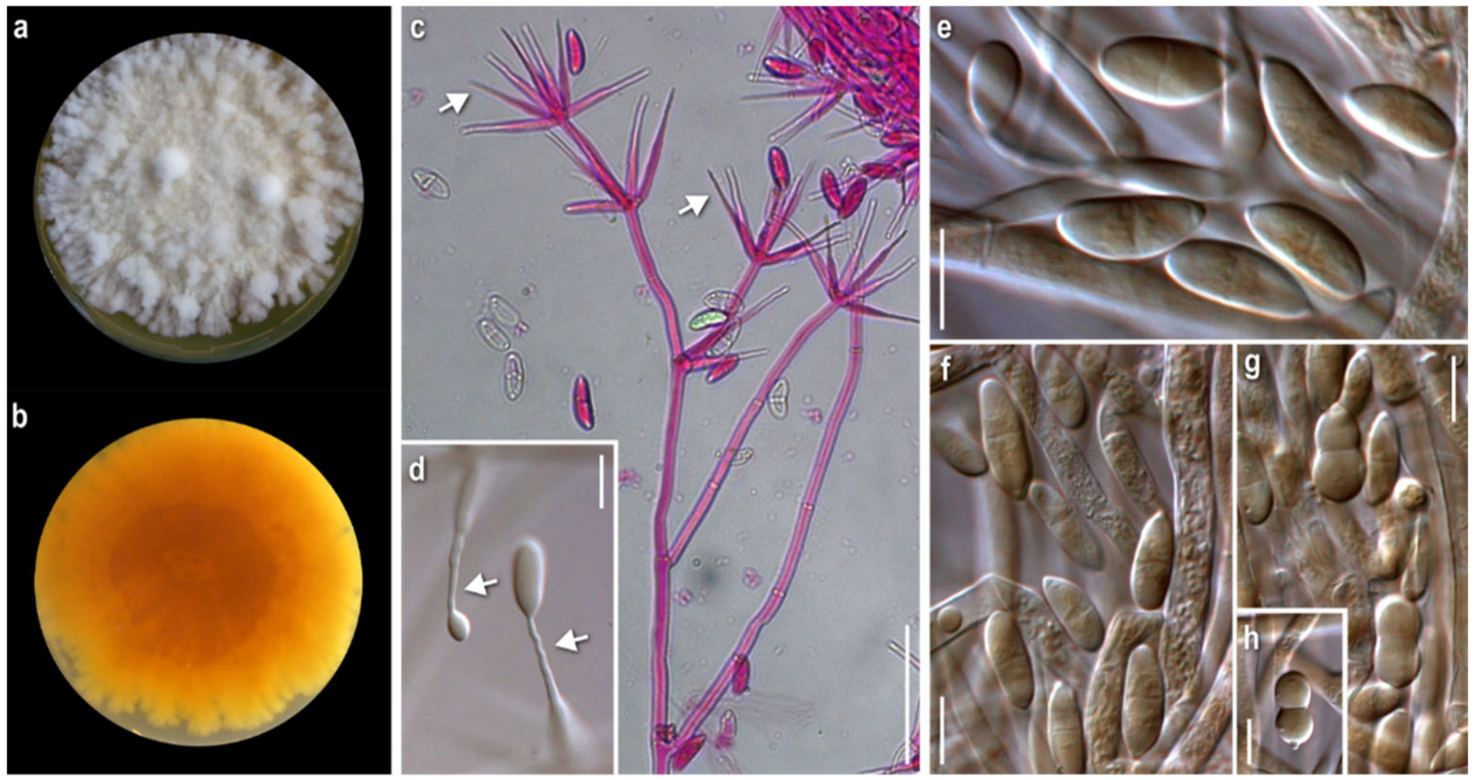
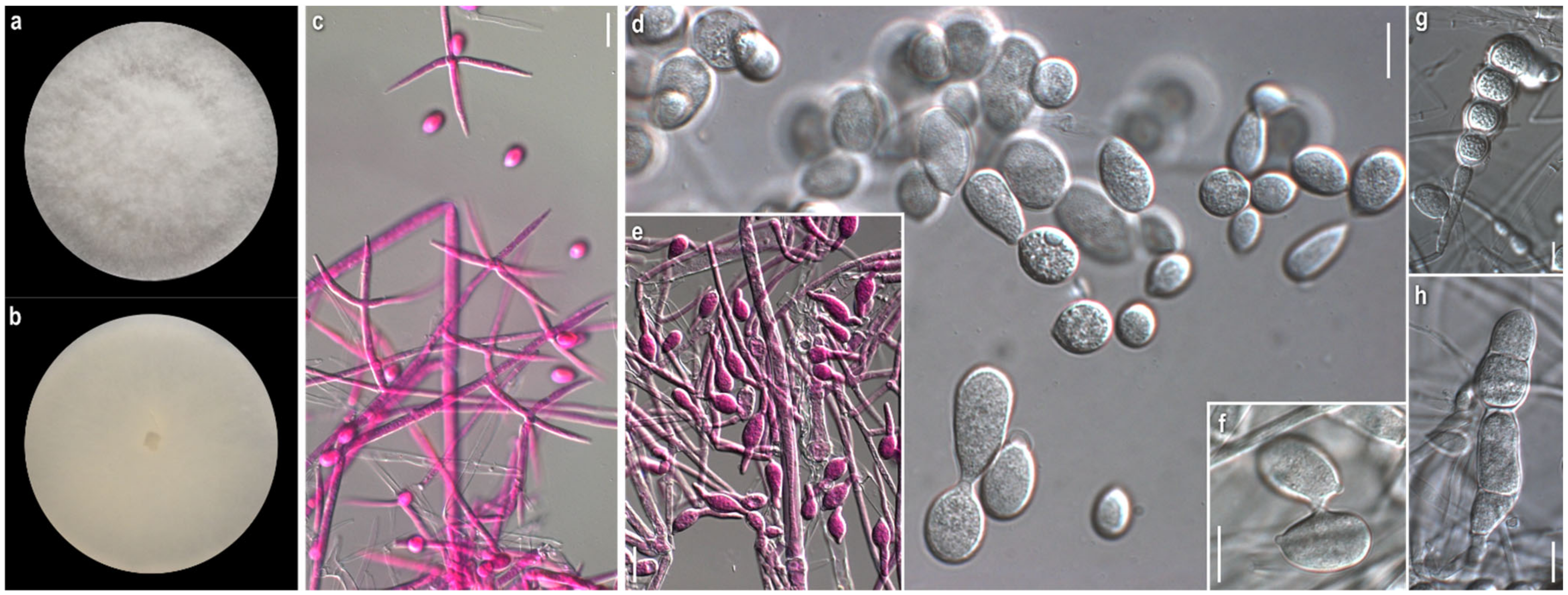
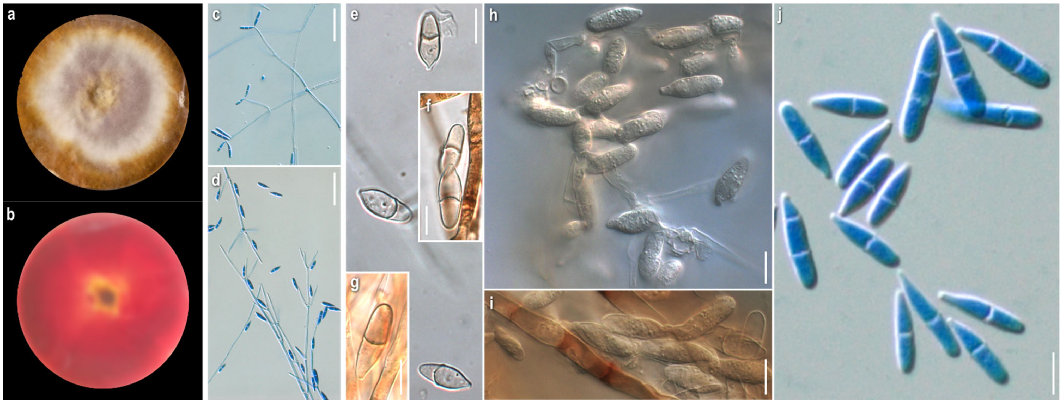
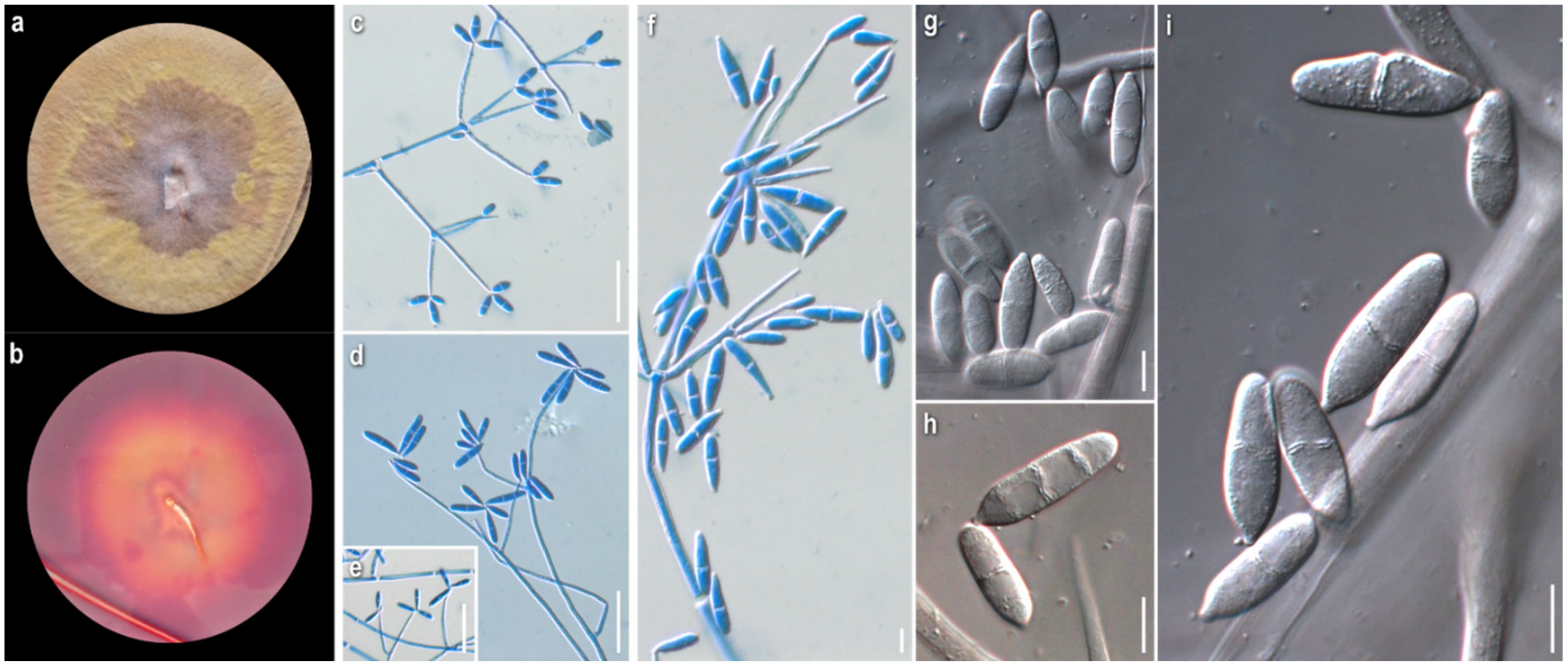
3.1. Morphological Study

From the morphological study of the 30 strains, 23 were assigned to the following six species viz. C. fungicola, C. apiculatum, C. verticillatum, C. varium, C. mycophilum, and H. rosellus (anamorph C. dendroides), while the remaining 7 could not be identified at the species level. For each of the six identified species and the two reference strains grown on PDA, colony obverse and reverse observations, conidial data, and short comments are given below. Analytical descriptions of these species can be found in Text S1 (Supplementary Materials). Furthermore, all seven unidentified strains are described in detail and are grouped as “Unidentified Red-Pigmented” Strains (URPs).
Cladobotryum fungicola (G.R.W. Arnold) Rogerson and Samuels, Mycologia 85 (2): 262 (1993)
Strain examined: Greece: Fthiotida: Mesochori, Mt. Oiti, in Quercus frainetto, Q. pubescens, and Q. coccifera forest with Abies cephalonica (sporadically), on basidiome of Cortinarius sp., 2008, coll./isol. Z. Gonou-Zagou, ATHUM 6855.
Mycelium: floccose to cottony; white, with prominent tufts; relatively symmetrically spreading, with irregular margin (Figure 1a). Colony reverse: pale buff to yellow, progressively turning intensely yellow to bright orange (Figure 1b). Conidia: ellipsoidal to cylindrical or fusiform; (13)15–18(20) × 5–6.5 μm, Qm 2.8; mainly two-celled, rarely one-celled; sometimes slightly curved or swollen at base or intensely constricted at septa; hyaline, smooth (Figure 1e–h).
Comments: The micromorphological characters of our strain fit perfectly with the original description of the anamorphic state [11], where the species is described as Sibirina fungicola. The long, zigzag-shaped apical part of conidiogenous cells is prominent and characteristic (Figure 1c,d). They are vaguely illustrated by Põldmaa and Samuels [31], though they are not mentioned in the text or in the original description.
Hosts of Cladobotryum fungicola recorded by now in the available literature belong to various genera of basidiomycetes, mainly aphyllophoraceous [22]. As far as our knowledge goes, this is the first record of the genus Cortinarius as its host.
Cladobotryum apiculatum (Tubaki) W. Gams and Hooz., Persoonia 6 (1): 97 (1970)
Strain examined: Greece: Karditsa: Ag. Nikolaos, Mt. Zigourolivado, in Fagus sylvatica forest, on basidiome of Russula sp., 2009, coll. P. Delivorias, isol. A. Liakouri, ATHUM 6907.
Mycelium: cottony, compactly to aerial; white to off-white; symmetrically spreading, with regular margin (Figure 2a). Colony reverse: white, progressively turning off-white, buff to ochre; sometimes visible brownish granules corresponding to sclerotioid structures (Figure 2b). Conidia: ovoid, ellipsoidal to broadly cylindrical or clavate; (11)13–28(33) × (3)5–9.5(11) μm, Qm 3; one-celled, very rarely two-celled; quite often swollen at base; hyaline, smooth; many easily germinating from both edges (Figure 2e,f). Sclerotioid structures: present (Figure 2g,h).
Cladobotryum verticillatum (Link) S. Hughes, Canadian Journal of Botany 36 (6): 750 (1958).
Strains examined: Greece: Arkadia: Megalopoli, Mt. Lykaio, in Quercus sp. forest, on basidiome of Lactarius subumbonatus, 2009, coll. M. Triantafyllou, isol. Z. Gonou-Zagou, ATHUM 6850; Megalopoli, Mt. Lykaio, in Quercus sp. forest, on basidiome of Lactarius subumbonatus, 2010, coll. M. Triantafyllou, isol. A. Liakouri ATHUM 6920; Megalopoli, Mt. Lykaio, in Quercus sp. forest, on basidiome of Lactarius subumbonatus, 2010, coll. M. Triantafyllou, isol. A. Liakouri ATHUM 6921.
Mycelium: felt-like to cottony; white to off-white; symmetrically spreading, with regular margin (Figure 3a). Colony reverse: white, progressively turning off-white to very pale buff (Figure 3b). Conidia: subglobose to ovoid or broadly ellipsoidal; 11–24(27) × (6)8–11(13) μm, Qm 2.2; one-celled; extremely rarely two-celled; hyaline, smooth; many easily germinating from both edges; few joined at their base or laterally (Figure 3e–f). Sclerotioid structures: present (Figure 3g,h).
Comments: Our strains of C. apiculatum and C. verticillatum are quite similar macromorphologically in culture. In terms of micromorphology, the most notable difference between the two species is the presence of mainly one-celled, mostly subglobose to ovoid conidia in C. verticillatum, in contrast to one- to rarely two-celled, ovoid to broadly elongated conidia in C. apiculatum. The measurements of the micromorphological characters fit with the various descriptions [8,29,30].
Both species have a preference to russuloid basidiomes, as expected [22]. Cladobotryum apiculatum occurs on Russula sp. and all C. verticillatum strains were isolated from the basidiomycete Lactarius subumbonatus, which is reported, to the best of our knowledge, for the first time as a host of the genus Cladobotryum [22].
Cladobotryum varium Nees, System der Pilze und Schwämme: 56, t. 4:54 (1817)
Strains examined: Greece: Attiki: Mt. Parnitha, in Abies cephalonica forest, on basidiome of Clitocybula familia, 2008, coll./isol. Z. Gonou-Zagou, ATHUM 6845; Mt. Parnitha, in Abies cephalonica forest, on basidiome of Cortinarius sp., 2009, coll./isol. Z. Gonou-Zagou, ATHUM 6856; Mt. Parnitha, in Abies cephalonica forest, on basidiome of Panellus sp., 2009, coll./isol. Z. Gonou-Zagou, ATHUM 6846; Eurytania: near Krikello, in Abies boresii-regis forest, on basidiome of Inocybe sp., 1998, coll./isol. Z. Gonou-Zagou, ATHUM 6514; near Krikello, in Abies borisii-regis forest, on polypore basidiome, 1998, coll./isol. Z. Gonou-Zagou, ATHUM 8003; Mt. Tymfristos, in Abies borisii-regis forest with Juniperus oxycedrus (sporadically), on basidiome of Ganoderma sp., 2004, coll./isol. Z. Gonou-Zagou, ATHUM 8002; Karditsa: Ag. Nikolaos, Mt. Zigourolivado, in Fagus sylvatica forest, on agaricoid basidiome, 2009, coll. P. Delivorias, isol. A. Liakouri ATHUM 6908; Rodopi: Frakto virgin forest on polypore basidiome, 2000, coll. Th. Angelopoulos, isol. Z. Gonou-Zagou ATHUM 7996; No data: on polypore hymenophore, 1997, coll./isol. Z. Gonou-Zagou, ATHUM 7995.
Mycelium: slightly felt-like or cottony to powdery or with floccose to powdery tufts radially or irregularly arranged; white, sometimes yellow at centre; usually when mature with bright yellow-orange irregular patches; symmetrically spreading, with regular margin (Figure 4a). Colony reverse: buff, progressing towards various yellow hues with ochre and orange undertones (Figure 4b). Conidia: ellipsoidal to shorty cylindrical; (10)13–17(20) × (5)6–8(9.5) μm, Qm 2.1; mainly two-celled, rarely one-celled or three-celled; sometimes constricted at septa; base with small apiculus or truncate, sometimes with a ring-like formation; many produced in a slanting manner; hyaline, smooth; predominantly single, sometimes forming chains, often branched (Figure 4g,h). Sclerotioid structures: present (Figure 4i).
Comments: Our isolates exhibit inter- and intrastrain variability. Nevertheless, the colonies of C. varium are distinguished macromorphologically from those of all other studied species by their remaining white, powdery mycelium; the irregular orange patches, usually formed on their surface—a feature not referred to in the existing descriptions—and the buff-yellow-orange reverse. Microscopically, the conidia are mostly ellipsoidal and two-celled, and in the strains, ATHUM 6846 and 4856, form characteristic long chains, some of which branched. This uncommon feature was also observed by Matsushima in only one strain (MFC 1816 = CBS 675.77) [81] and was referred to by Rogerson and Samuels [6]. Conidial connections via anastomoses in the strain, C. varium ATHUM 6846, were also observed [82]. In addition, the heterogeneity of conidiogenous cells is apparent in our strains, especially in colonies of ATHUM 8002 and ATHUM 6908, where the cells, when maturing, become gradually shorter and from their truncated apices two types of conidia are gradually produced—ellipsoidal conidia truncated at the base and cylindrical conidia with both sides truncated—revealing a probable retrogressive mode of conidiogenesis (Figure 4d–f). This characteristic ontogeny of the conidia is described by Cole and Kendrick [83]. In any case, the conidiogenesis observed is similar only to that of C. mycophilum.
The basidiomycetes Clitocybula familia, Ganoderma sp., Inocybe sp., and Panellus sp., from which our strains were isolated, as far as our knowledge goes, are not reported as hosts of C. varium [22].
Cladobotryum mycophilum (Oudem.) W. Gams and Hooz., Persoonia 6 (1): 102 (1970)
Strains examined: Greece: Attiki: cultivated Agaricus bisporus, 2010, coll./isol. Z. Gonou-Zagou, ATHUM 8001. Eurytania: Ag. Nikolaos, in Platanus orientalis forest with Quercus sp., Abies cephalonica and Castanea sativa (sporadically), on basidiome of Mycena sp., 2010, coll./isol. Z. Gonou-Zagou, ATHUM 8000; Karditsa: Belakomitis, in Abies borisii-regis forest, on basidiome of Inocybe sp., 1999, coll./isol. Z. Gonou-Zagou, ATHUM 7994. Magnisia: Mt. Pilio, in Castanea sp. forest, on basidiome of Hypholoma sp., 2009, coll. P. Delivorias, isol. A. Liakouri ATHUM 6906.
Mycelium: felt-like at first, becoming fibrous, floccose to cottony; white, yellow, buff to ochre, progressing towards various red hues with brownish and purplish undertones; when mature with white powdery tufts, aerial or superficial, irregularly or radially arranged; symmetrically or asymmetrically spreading, with regular margin; sometimes distinct, minute brownish granular formations; rarely production of perithecia (Figure 5a). Colony reverse: yellow, buff, progressing towards ochre, pinkish hues, and various red hues with brownish undertones (Figure 5b). Conidia: ovoid to broadly ellipsoidal to ellipsoidal or cylindrical; mainly two-celled, many three-celled, few four-celled, and rarely one-celled; some curved; variably sized, two-celled (16)20–28(32) × (7)9–11.5(13), Qm’ 2.1, 3-4-celled 25–35(39) × 9–12 μm, Qm″ 3.1, Qm = (Qm′ + Qm″)/2 = 2.6; hyaline, smooth; at base with apiculi sometimes indistinct or truncate; characteristic production in a slanting manner indicative of retrogressive conidiogenesis; mature conidia with hyaline pore-like protrusions, some joined in twos or forming short chains; production of secondary conidia on a conidiogenous cell emerging directly from a conidium (Figure 5g–k). Sclerotioid structures: present (Figure 5l). Teleomorph: only in strain ATHUM 8001; Perithecia: mostly subglobose to pyriform, reddish to purplish brown; on ostiole either slightly pigmented droplets or light yellowish mass of ascospores; partially immersed in the subiculum (Figure 5m). Ascospores: fusiform to oblong, apiculate; 25–30(32) × 4–5.5(6.5) μm (incl. apiculi); two-celled, sometimes with constriction at median septum; many curved; apiculi rather conical, acute to rarely obtuse; 4.5–5.5(6.5) μm; slightly verrucose (Figure 5n).
Comments: All strains studied exhibited intra- and interstrain variability as far as macro- and micromorphology is concerned—i.e., colony texture (including formations such as anamorphic sporulation, sclerotioid aggregations, ascigerous structures) and colour and production of pigmented metabolites, and their corresponding microscopic characters. Nevertheless, our descriptions—and the flunctuations of the characters—fit perfectly with the protologue of Hypomyces odoratus [10], where both sexual and asexual reproduction were described from culture. According to our observations, the anamorph is characterised by mainly two-celled conidia, but also three- to four- celled specimens, varying in size and shape, which are produced retrogressively from gradually shortening conidiogenous cells. Conidiogenesis in C. mycophilum is similar to that of C. varium and very characteristic of the retrogressive way of production. It is worth noting that secondary conidia seem to be produced from mature conidia and most probably the hyaline protrusions of the latter indicate the place of their blastic formation. Joined conidia were depicted in a drawing by Arnold [10], though this trait was not mentioned in the text. Sexual reproduction was observed in the strain, ATHUM 8001, which was isolated from a cultivated basidioma of Agaricus bisporus. Perithecia were formed in the very first cultures, with the strain losing this ability with time. The teleomorphs seem to be produced only in culture and are described very few times [8,9,10,46], while the fungus appears as an anamorph in nature. A species of the genus Hypholoma, from which one of the studied strains was isolated, to the best of our knowledge, is not hitherto recorded as a host of C. mycophilum [22].
Hypomyces rosellus (Alb. and Schwein.) Tul. and C. Tul., Annales des Sciences Naturelles Botanique 13: 12 (1860)
Anamorph: Cladobotryum dendroides (Bull.) W. Gams and Hooz., Persoonia 6 (1): 103 (1970)
Strains examined: Greece: Attiki: Athens “National Gardens”, on basidiome of Flammulina velutipes, 2008, coll./isol. Z. Gonou-Zagou, ATHUM 6847; Mt. Parnitha, in Abies cephalonica forest, on basidiome of Tricholoma sp., 2008, coll./isol. Z. Gonou-Zagou ATHUM 6849; Mt. Parnitha, in Abies cephalonica forest, on basidiome of Hohenbuehelia sp., 2000, coll./isol. Z. Gonou-Zagou ATHUM 7998. Eurytania: Ag. Nikolaos, in Platanus orientalis forest with Quercus sp., Abies cephalonica and Castanea sativa (sporadically), on ascocarp of Helvella lacunosa, 2010, coll./isol. Z. Gonou-Zagou, ATHUM 7999. Karditsa: Ag. Nikolaos, Mt. Zigourolivado, in Fagus sylvatica forest, on polypore basidiome, 2009, coll. P. Delivorias, isol. A. Liakouri ATHUM 6909. Xanthi: Mt. Leivaditis, in Fagus sylvatica forest with Juniperus communis, on basidiome of Polyporus varius, 2009, coll. A. Sergentani, isol. Z. Gonou-Zagou, ATHUM 6848.
Mycelium: felt-like to floccose, somewhat powdery, or with somewhat radially arranged floccose-powdery tufts; whitish, buff, progressing towards ochre, pinkish hues, and various red hues with purplish and brownish undertones; symmetrically spreading, with regular margin (Figure 6a). Colony reverse: buff to ochre, progressing towards ochre and various red hues with brownish and purplish undertones (Figure 6b). Conidia: ovoid, broadly ellipsoidal to mainly ellipsoidal or cylindrical; (16)21.5–28(35) × (8.5)10–11.5(13) μm, Qm 2.3; two-, three- or four-celled, with prominent apiculus; hyaline, smooth; few slightly curved; mature conidia sometimes constricted at septa; some joined in twos at base or forming short bendy chains (Figure 6h–k). Sclerotioid structures: not observed.
Comments: Progressive pigmentation of both the colony and substrate from ochre into red hues is one of the main macromorphological traits of the pigment (aurofusarin)-producing C. dendroides. Sympodial conidiogenesis is the main diagnostic micromorphological character of all strains of C. dendroides (Figure 6d–g), setting it apart from other Cladobotryum species. In addition, the long, three- to four- celled conidia contribute to the distinction of the species. The hosts of the Greek specimens of C. dendroides, belonging to the basidiomycetous species, Flammulina velutipes and Hohenbuehelia sp., as well as to the ascomycetous Helvella lacunosa, are not recorded so far as hosts of C. dendroides, as far as our knowledge goes [22].
Cladobotryum rubrobrunnescens Helfer, Libri Botanici 1: 55 (1991)
Specimen examined: Germany, on Inocybe sp., 1989, A. Resinger, CBS 176.92, ex-type strain.
Mycelium: felt-like to compact cottony; whitish, yellow, buff, progressing towards ochre and various red hues with pinkish and purplish undertones; when mature copper-coloured, either in scattered patches or at margin; symmetrically spreading, with regular margin (Figure 7a). Colony reverse: buff, progressing towards ochre and various red hues with pinkish, purplish, or brownish undertones (Figure 7b). Conidia: ellipsoidal or fusiform to cylindrical to bacilliform, rarely constricted at septa; in slide culture (21)22–26 (29) × (3.5)4–5 (5.5) μm, Qm 5.4, in petri dish culture (14)17–26(29) × (5.5)6–8.5(9.5) μm, Qm 2.9; mainly two-celled, sometimes the basal cell swollen, also one-celled or rarely three- or four-celled; hyaline, smooth; sometimes mature conidia joined in twos (Figure 7e–j). Sclerotioid structures: not observed.
Cladobotryum tenue Helfer, Libri Botanici 1: 57 (1991)
Specimen examined: Germany: Regesburg-Keilberg, on agaric., 1986, H. Besl, CBS 152.92, ex-type strain.
Mycelium: floccose to sparsely cottony; whitish, yellow, buff, progressing towards pale ochre to pinkish, rarely to various red hues; symmetrically spreading, with regular margin (Figure 8a). Colony reverse: buff, progressively turning from ochre to bright terracotta and various red hues with pinkish and purplish to brownish undertones (Figure 8b). Conidia: ellipsoidal to clavate, cylindrical to bacilliform, rarely constricted at septa; in slide culture (21)22–28.5(36) × (3.5)4.5–5.5(6) μm, Qm 5.5, in petri dish culture 23–34.5(38) × 4–6, Qm 6.4; mainly two-celled, rarely one- or three-celled, hyaline, smooth (Figure 8f–l). Sclerotioid structures: present (Figure 8m).
Comments: There are only the protologue descriptions of C. rubrobrunnescens and C. tenue ever made so far [7]. In terms of macro- and micromorphology, our observations on PDA culture generally comply with the original descriptions, where the medium malt peptone agar (MPA) was used. Some fluctuations in spore shapes and sizes can be attributed to the species variability due to the different culture medium and conditions used. In slide culture of C. rubrobrunnescens, the conidia are distinctively much narrower. As the main diagnostic microscopic features of the two species in petri dish culture, the longer and narrower conidia can be considered, as well as the simpler conidiophores of C. tenue.
Cladobotryum sp.
Strain examined: Greece: Fthiotida: Mesochori, Mt. Oiti, in Quercus frainetto, Q. pubescens and Q. coccifera forest with Abies cephalonica (sporadically), on basidiome of Laccaria laccata, 2008, coll./isol. Z. Gonou-Zagou ATHUM 6851.
Mycelium: uneven in terms of texture and pigmentation, patchy-like with cottony and compactly felt-like areas, irregularly distributed; ranging from white-yellowish to buff, pale ochre, pink, progressing towards dark red with pinkish undertones; asymmetrically spreading, with irregular margin (Figure 9a). KOH reaction: positive. Growth rate: relatively low. Colony reverse: buff, progressively turning from ochre to various red hues with purplish undertones (Figure 9b). Conidiophores: very long, delicate, subverticillate, unbranched or sparsely branched, unilaterally or dichotomously at conidiophore apices, sometimes emerging almost orthogonally to conidiophore axis; septate; hyaline (Figure 9c–e). Conidiogenous cells: elongated, delicate, narrow-cylindrical to almost filiform, subulate, with obtuse apices; arranged in verticils at conidiophore apices, 1–4 per verticil; aseptate, rarely septate; hyaline (Figure 9c–e). Conidiogenesis: blastic; a single to multiple conidiogenous loci per conidiogenous cell and a single conidium or up to 5(6) conidia, mainly 3, in a wreath-shaped arrangement (Figure 9c–e). Conidia: in petri dish culture ellipsoidal to cylindrical or fusiform to rarely bacilliform; (17.5)19–28(32) × (5.5)6.5–8(10) μm, Qm 3.2; mainly two-celled, rarely three- or four-celled; in slide culture narrowly ellipsoidal to cylindrical or bacilliform; (23)26–33(41) × (4.5)6–7(8.5) μm, Qm 4.8; mainly two-celled, rarely three-celled; hyaline, smooth; basal apiculi central or slightly eccentric (Figure 9f–i). Sclerotioid structures: rarely present, mainly as short chains of globose to ellipsoidal cells, thick-walled.
Cladobotryum sp.
Strain examined: Greece: Fthiotida: Mesochori, Mt. Oiti, in Quercus frainetto, Q. pubescens and Q. coccifera forest with Abies cephalonica (sporadically), on basidiome of Lactarius salmonicolor, 2008, coll./isol. Z. Gonou-Zagou, ATHUM 6852.
Mycelium: cottony to compactly felt-like; white at first, then buff, progressing towards pinkish hues; sporadically white aerial loose tufts; symmetrically spreading, with irregular margin (Figure 10a). Growth rate: moderate. Colony reverse: buff, progressively turning from terracotta to various red hues with brownish and purplish undertones (Figure 10b). Conidiophores: very long, unbranched to sparsely branched in a unilateral manner at conidiophore apices (Figure 10c–f). Conidiogenous cells: elongated, narrow-cylindrical, subulate, with obtuse apices; arranged in verticils 1–4(5) per verticil, mainly 3–4 at the apex; aseptate, rarely septate; hyaline (Figure 10c–f). Conidiogenesis: blastic; a single to multiple conidiogenous loci per conidiogenous cell and a single conidium or up to 6 conidia, mainly 3, in a wreath-shaped arrangement (Figure 10c–f). Conidia: in petri dish culture ovoid, mainly ellipsoidal to cylindrical; (15.5)19–26(28) × 6–10 μm Qm 2.8; sometimes slightly swollen at base and constricted in septal area; mainly two-celled, also three-celled, but rarely four-celled; hyaline, smooth; many germinated; quite often in maturity disintegration of one cell (out of the two) or separation of the two cells; many variably connected in twos or threes at their base or side, rarely forming short chains; possible secondary production of conidia from germinated cell of conidium; in slide culture fusiform to narrowly ellipsoidal to cylindrical; (14)17–23(30) × 5–8 μm, Qm 3.2; mainly two-celled. (Figure 10f–k). Sclerotioid structures: present, mainly as chains of globose to ellipsoidal cells, thick-walled.
Cladobotryum sp.
Strain examined: Greece: Fthiotida: Mesochori, Mt. Oiti, in Quercus frainetto, Q. pubescens and Q. coccifera forest with Abies cephalonica (sporadically), on basidiome of Russula sp., 2008, coll./isol. Z. Gonou-Zagou, ATHUM 6853.
Mycelium: cottony to loosely felt-like; whitish, buff, progressing towards pale ochre to pinkish hues; when mature, secondary whitish cottony at centre (Figure 11a). KOH reaction: positive. Growth rate: relatively high. Colony reverse: buff, progressively turning from ochre to orange and red hues with pinkish undertones (Figure 11b). Conidiophores: very long, delicate, unbranched or sparsely branched in a unilateral manner at conidiophore apices or solitary, shorter, emerging orthogonally from very long hyphae; septate; hyaline (Figure 11c–e). Conidiogenous cells: elongated, delicate, narrow-cylindrical to almost filiform, subulate, with obtuse apices; arranged in verticils, 1–4(5) per verticil, mainly 3–4 at the apex, sometimes emerging orthogonally to conidiophore axis or in a dichotomous manner at the apex; aseptate, sometimes septate; hyaline (Figure 11c–e). Conidiogenesis: blastic; a single to multiple conidiogenous loci per conidiogenous cell and a single conidium or up to 3 conidia in a wreath-shaped arrangement (Figure 11c–e). Conidia: in petri dish culture ellipsoidal to cylindrical or fusiform; (15)18–29(32) × (5.5)6.5–9(10.5) μm, Qm 2.9; mainly two-celled, sometimes restricted at septum or basal cell swollen, also three-celled, very rarely four-celled; sparsely curved; quite often joined in twos or threes at their base or laterally; in slide culture narrowly ellipsoidal to cylindrical or rarely bacilliform; (15.5)19.5–26.5(32) × (5)5.5–6.5(7) μm, Qm 3.9; mainly two-celled, very rarely three- to four-celled; hyaline, smooth (Figure 11f–h). Sclerotioid structures: present, cells globose to elongated, slightly thick-walled, forming monilioid chains, usually branched (Figure 11i).
Cladobotryum sp.
Strain examined: Greece: Karditsa: Ag. Nikolaos, Mt. Zigourolivado, in Fagus sylvatica forest, on basidiome of Lactarius sp., 2009, coll. P. Delivorias, isol. A. Liakouri ATHUM 6904.
Mycelium: compactly felt-like to cottony, with white tufts; buff, progressing towards pale ochre and pinkish hues; asymmetrically spreading, with irregular margin (Figure 12a). KOH reaction: negative or faintly positive. Growth rate: slow to moderate. Colony reverse: buff, progressively turning from ochre to red hues with purplish undertones (Figure 12b). Conidiophores: very long, delicate, unbranched or very sparsely branched in a unilateral manner at conidiophore apices or solitary, shorter, emerging from very long hyphae; septate; hyaline (Figure 12c–e). Conidiogenous cells: elongated, sometimes very elongated, delicate, narrow-cylindrical to almost filiform, subulate; arranged in verticils, 1–3(4) per verticil, mainly 2–3 at the apex, sometimes emerging orthogonally to conidiophore axis or in a dichotomous manner at the apex; aseptate; hyaline; (Figure 12c–e). Conidiogenesis: blastic; a single to multiple conidiogenous loci per conidiogenous cell and a single conidium or up to 2 conidia in a V-shaped arrangement (Figure 12c–e). Conidia: in petri dish culture ellipsoidal to cylindrical; (14.5)17.5–25(31) × (2.5)4.5–7.5(8.5) μm, Qm 3.7; mainly two-celled, rarely one-celled; basal cell often swollen; sometimes restricted at septum; in slide culture narrowly ellipsoidal to cylindrical or bacilliform; (16.5)19.5–26(32) × (3.5)4.5–5.5(6) μm, Qm 4.7, mainly two-celled, rarely one- to four-celled; hyaline, smooth; (Figure 12f–j). Sclerotioid structures: not observed.
Cladobotryum sp.
Strain examined: Greece: Attiki: Mt. Parnitha, in Abies cephalonica forest, on basidiome of Tricholoma atrosquamosum, 2009, L. Tsampiras, A. Liakouri ATHUM 6912.
Mycelium: felt-like to compactly cottony; whitish, buff, progressing towards pale ochre to brownish with pinkish hues; symmetrically spreading, with regular margin (Figure 13a). KOH reaction: negative or faintly positive. Growth rate: relatively slow to moderate. Colony reverse: buff, progressively turning from ochre to bright and dark orange-red hues (Figure 13b). Conidiophores: very long, unbranched, sometimes shorter, emerging orthogonally from very long hyphae; septate; hyaline (Figure 13c,d). Conidiogenous cells: very elongated, narrow-cylindrical to filiform, subulate, with obtuse apices; in verticils at conidiophores apices, 1–3 per verticil; aseptate, hyaline (Figure 13c,d). Conidiogenesis: blastic; a single to multiple conidiogenous loci per conidiogenous cell and a single conidium or up to 3 conidia (Figure 13c,d). Conidia: in petri dish culture fusiform to cylindrical or bacilliform, rarely ellipsoidal; (17.5)20–24.5(28) × (4.5)5–6.5(7) μm, Qm 3.8; in slide culture cylindrical to filiform; (21)24–38.5(44.5) × 3.5–5 μm, Qm 7.1; one-celled or two-celled; hyaline, smooth; (Figure 13e–g). Sclerotioid structures: not observed.
Cladobotryum sp.
Strain examined: Greece: Attiki: Mt. Parnitha, in Abies cephalonica forest, on basidiome of Lactarius salmonicolor, 2009, L. Tsampiras, A. Liakouri ATHUM 6913.
Mycelium: uneven in terms of texture and pigmentation, with cottony and compactly felt-like areas irregularly distributed, ranging from white to buff, pale ochre, pink; asymmetrically spreading, with irregular margin (Figure 14a). KOH reaction: negative. Growth rate: relatively slow. Colony reverse: buff, progressively turning from ochre to terracotta and various red hues with brownish undertones (Figure 14b). Conidiophores: very long, unbranched or sparsely branched in a very irregular and somewhat unilateral or dichotomous manner at conidiophore apices, sometimes shorter, emerging almost orthogonally from very long hyphae; septate; hyaline (Figure 14c). Conidiogenous cells: very narrow-cylindrical to almost filiform, subulate, with obtuse apices; arranged in verticils at conidiophores apices, 1–4 per verticil; aseptate; hyaline (Figure 14c). Conidiogenesis: blastic; a single or multiple conidiogenous loci per conidiogenous cell and a single conidium or up to 2 conidia in a V-shaped arrangement (Figure 14c). Conidia: in petri dish culture ellipsoidal to cylindrical, fusiform to bacilliform; (17.5)24–34.5(43.5) × (4)5–7(10), Qm 4.6; mainly two-celled, but also one-celled; with or without restriction in the septal area; many germinated; in slide culture ellipsoidal to cylindrical, fusiform to bacilliform or filiform; (19.5)22.5–34.5(51.5) × (3)4.5–5.5(6.5) μm, Qm 6.4; mainly two-celled, but also one-celled; hyaline, smooth; (Figure 14d–f). Sclerotioid structures: not observed.
Cladobotryum sp.
Strain examined: Greece: Attiki: Mt. Parnitha, in Abies cephalonica forest, on basidiome of Cortinarius sp., 2009, coll. L. Tsampiras, isol. A. Liakouri ATHUM 6914.
Mycelium: uneven in terms of texture and pigmentation, with compactly felt-like and floccose to cottony areas irregularly distributed, ranging from whitish, buff, pale ochre to red hues with pinkish undertones; asymmetrically spreading, with irregular margin (Figure 15a). KOH reaction: positive. Growth rate: relatively low. Colony reverse: buff, progressively turning from ochre to various red hues with pinkish undertones (Figure 15b). Conidiophores: very long, unbranched or very sparsely branched in a somewhat unilateral manner at conidiophore apices; septate; hyaline (Figure 15c,d). Conidiogenous cells: very narrow-cylindrical to almost filiform, subulate, with obtuse apices; in verticils on conidiophores apices, 1–3 per verticil; aseptate; hyaline (Figure 15c,d). Conidiogenesis: blastic; a single or multiple conidiogenous loci per conidiogenous cell and a single conidium or up to 2 conidia in a V-shaped arrangement (Figure 15c,d). Conidia: in petri dish culture ovoid, ellipsoidal to cylindrical; (16)17.5–22.5(26.5) × (5)7–8.5(10) μm, Qm 2.7; mainly two-celled, rarely one-celled or three–four-celled; many with restriction in the median septum; some joined in twos, or more, at their base or laterally; in slide culture fusiform to narrowly ellipsoidal or cylindrical; (12.5)15.5–20.5(22.5) × 5–8 μm, Qm 2.8; mainly two-celled (Figure 15e,f). Sclerotioid structures: rarely globose to elongated cells, thick walled, forming short chains.
Comments: All unidentified strains are characterised by the final reddish hues of both the mycelium and the colony reverse. They were isolated from various agaricoid basidiomes that come from three different geographic locations and habitats. Their macroscopic characters seem quite similar, that is, the colony is more or less felty to cottony, and yellow, buff to pinkish, while their microscopic ones exhibit similarities, and many overlaps as well as differences. They differ clearly from our strains of C. dendroides and C. mycophilum and exhibit similarities with C. rubrobrunnescens and C. tenue [7]. The strains, ATHUM 6912, 6913, 6914—coming from the same collecting site—are similar in terms of colony macromorphology, but the strain, ATHUM 6914, is differentiated considering spore shape and size from the other two that have narrower and longer spores. In addition, the strain, ATHUM 6914, shares resemblances in micro- and macromorphology with the strain, ATHUM 6852, which is isolated from a different site. The strains, ATHUM 6851 and ATHUM 6853, as well as ATHUM 6904, can be grouped together considering their micromorphological similarities, although they individually exhibit some deviations. It is worth noting that all the above strains were also cultured in slides, where the spores always appeared narrower and, in most cases, quite longer than the corresponding ones in the petri dish.
Figure 16 provides clear visual information on the grouping of the species according to their conidial morphology when grown in petri dish cultures with PDA. On one extreme, there are conidia approaching broader ellipsoidal shapes, as seen in the species C. varium, C. verticillatum, C. mycophilum, and C. dendroides; all of them belong to different size categories, with C. varium being distinctly smaller, while the two latter being clearly larger and resembling one another. On the other extreme, there are conidia exhibiting narrower ellipsoidal shape, as seen in the unidentified strain ATHUM 6913, and especially in the ex-type species C. tenue. In between are all the other species/strains, with C. fungicola being closer to a broader ellipsoidal shape and noticeably smaller, therefore clearly apart from the rest of the same ‘shape category’. Conidia of the strains, ATHUM 6904 and ATHUM 6912, are somewhere in-between the two extremes—considering both their shape and length—in comparison to the rest of the strains that are grouped together as a ‘cluster’.

3.2. Molecular Analyses

A phylogenetic approach based on the ITS region (ITS1-5.8S-ITS2) was implemented to clarify the relationships among the 29 examined Greek strains and the 2 ex-type strains (C. rubrobrunnescens and C. tenue) (Figure 17). In order to resolve any phylogenetic ambiguity, 63 publicly available sequences were also included, representing all known ITS sequences of the Cladobotryum species to date. Discrimination between the species is observed with high support (Posterior Probability value from 80 to 100%). Although the strains are well grouped at the species level, there is great intraspecific variability in most cases. As expected, Greek strains are distributed across the phylogenetic tree. In detail, C. fungicola is grouped with H. semitranslucens, which is the ex-type containing both states of reproduction (asexual/anamorphic and sexual/teleomorphic) with 100% support since their ITS sequences are identical. These species, with the currently available data, diverged in earlier evolutionary stages [84]. Cladobotryum varium and its associated teleomorph H. aurantius strains are grouped as a monophyletic clade next to C. heterosporum, for which a red pigment production is referred to in the available literature [36]. If the pigments, rugulosin, skyrin, and emodin, for the currently examined species of the genus Cladobotruym are considered, then the metabolite rugulosin seems to be a feature of the species, C. varium and C. fungicola, while skyrin and emodin can be found mainly in C. varium and probably in the cluster of C. heterosporum and C. amazoense [7,85]. Even though species C. apiculatum and C. verticillatum are sister clades with 87% PP support value, metabolite norbikaverin is produced only in C. verticillatum, and this latter species is grouped with the respective teleomorph species H. armeniacus with high support (99% PP value). Pigment bikaverin is produced in C. varium as described earlier [33]. The remaining species exhibiting red pigmentation, presumably due to aurofusarin, diverged later [7,36,59,60,85]. Cladobotryum mycophilum along with H. odoratus form a monophyletic clade that is basal to the rest of the examined species producing aurofusarin. The metabolites, rosellisin and rosellisin-aldehyde, are particularly produced by C. dendroides strains, which are grouped as two subclusters that are basal to the C. tenue and C. rubrobrunnescens clade, and also in C. varium [86,87]. Greek Cladobotryum spp. strains (URPs) are clustered along with C. tenue and present a similarity with C. rubrobrunnescens as a basal clade, but their relationship is unclear since the support values (PP) of these topologies are non-significant. However, ITS-based phylogeny shows the cluster of a large group that contains C. tenue, C. rubrobrunnescens, and the Greek Cladobotryum spp. strains (Figure 17).

3.3. NMR-Based Metabolomics/Chemotaxonomy

In order to employ a more integrative approach to the taxonomic investigation of Greek strains of the genus Cladobotryum, in conjunction with morphological and molecular analyses, an attempt to utilise 1H NMR-based metabolomics as a chemotaxonomic tool was made. Spectroscopic data were transformed to arithmetic values and subjected to multivariate statistical analysis.
The score plot of the unsupervised principal component analysis (PCA) is presented in Figure 18a. The generated model with 15 components and with R2X(cum) = 0.78 and Q2(cum) = 0.54 satisfactorily described the data variation. Three clusters were observed by visual inspection of the generated model, and, more specifically, Cluster I is comprised of the strains corresponding to the species C. apiculatum, C. verticillatum, and a small number of C. mycophilum strains; Cluster II encompasses the strains corresponding to the species C. varium and C. fungicola; while Cluster III involves the rest of the C. mycophilum strains, as well as those corresponding to the species C. dendroides, C. rubrobrunnescens, C. tenue, and the URPs. Cluster I is less populated and tight. Clusters II and III are more populated and the distribution of their samples along the PC1 axis suggests a higher variance of their spectral characteristics. Nevertheless, Clusters II and III are clearly separated along the PC2 axis.
Further analysis was performed using the species information as a Y variable with the aim to reveal species-specific spectral characteristics responsible for the clustering. The score plot of Projection to Latent Structures (PLS-DA) is shown in Figure 18b. The created model had 7 components with R2X(cum) = 0.57 and Q2(cum) = 0.48, while the R2Y(cum) = 0.68. The model was validated through a permutation test (100 permutations; Figure S2, Supplementary Materials). The supervised analysis based on species corroborates the observations of the PCA, suggesting an existing intraspecies chemical similarity, thereby further separating C. fungicola strains from those corresponding to C. varium (Figure 18b). It is interesting to note that on the second axis, macroscopically-observed pigmentation differences of the species/isolates are projected, with the red-pigmented ones being located above and the non-red being located below x-axis.
A more thorough investigation was performed for the red-pigmented species, which aimed at a more detailed description of the spectroscopic profile of the URPs, and tried to possibly unravel the similarities and differences hidden within a wider context. The PCA and PLS-DA score plots are shown in Figure 19a,b, respectively. The model that resulted from PCA had 10 principal components and described 76% of the variability, while predicting a lower share (51%). The score plot suggests the spectral similarity of C. mycophilum and C. dendroides species. URPs are differentiated from C. mycophilum and C. dendroides along the 1st axis, indicating the existence of species-specific spectral characteristics. However, in the centre of the T2 Hotteling ellipse, samples from all strains are overlapped and only C. rubrobrunnescens is situated in the 2nd axis, at the margins of the ellipse. The supervised analysis with 6 components and R2X(cum) = 0.58, R2Y(cum) = 0.87, and Q2(cum) = 0.66 distinguishes C. mycophilum from C. dendroides species in the 2nd component, while in the 1st component of the model, the difference of URPs with the rest of the samples is projected (Figure 19b). Cladobotryum rubrobrunnescens and C. tenue ex-type strains are located at two edges of C. dendroides strains, exhibiting a more distant spectroscopic profile when analysed within the red-pigmented strains. Similarly, with the unsupervised analysis, a sub-group of URPs is found together with C. tenue isolates in close vicinity of C. dendroides strains. The model was validated using 100 random permutations (Figure S2, Supplementary Materials). The Variable Importance in Projection scores (VIPs) of the 1st component—the axis separating URPs from the other examined red-pigmented species—reveals sugar moieties as the major characteristic of URPs.
Τhe 1H NMR spectra of the hydroalcoholic extracts of the isolates are presented in Figure 20 and grouped according to the results obtained from the PCA analysis (Figure 18a) to facilitate visual inspection. More precisely, panel a shows the 1H NMR spectra of the isolates corresponding to the species C. mycophilum, C. verticillatum, C. apiculatum, and C. fungicola; panel b shows the isolates of the species C. varium; and panel c shows the isolates of the species C. dendroides, C. tenue, C. rubrobrunnescens, and the URPs. The investigated isolates exhibited rich metabolic fingerprints with dominant resonances characteristic of sugar moieties (6.00–3.00 ppm, with anomeric protons observed at 5.50–4.50 ppm as doublets), as well as polyols (3.90–3.60 ppm), among which mannitol appeared to be the prevailing one. Glucose, both in α and β configuration, was observed in a relatively high concentration in all C. varium strains when compared to other isolates. In C. dendroides, and especially in URPs, glucose also had a prominent presence. Acetic acid appeared as a prominent singlet at 1.89 ppm with a high inter- and intraspecies variability. In the majority of the spectra, an additional characteristic singlet at 2.15 ppm is indicative of the presence of N-acetylated compounds. Additional singlets related to choline derivatives (3.25–3.20 ppm) were detected close to the region where deuterated methanol and its satellites resonate. However, a relatively low concentration of these molecules allowed only the observation of N-trimethyl resonances—which are peaks of low diagnostic value—compared to the rest of the protons of choline derivatives, which exhibited characteristic complex patterns that, otherwise, would have facilitated peak annotation. The presence of a strong, featureless peak around 1.20 ppm, in combination with a triplet in the region of 0.95–0.85 ppm, provided evidence for the presence of long fatty acid moieties (methylene groups at 1.20 ppm and end-methyls at 0.80 ppm). These features are observed in several samples (one C. mycophilum strain, C. fungicola, two C. varium strains, few C. dendroides, and URPs). Additionally, in the same spectral region, the characteristic methyl doublet of alanine at 1.48 ppm and lactic acid at 1.31 ppm were also observed. Finally, among Cladobotryum spp., a wide distribution of a trans double bond region (7.80–6.60 ppm, JHH ~ 15 Hz) was observed, which is potentially explained by the presence of a characteristic group of fungal secondary metabolites that are structurally related to rosellisin. Lastly, out of six isolates of C. dendroides, two were able to produce a significant amount of phomalactone, characterised by a detailed analysis of 1D and 2D NMR spectra (Figure S3, Supplementary Materials).

4. Discussion

4.1. Contribution of Data Derived from Morphology and Notes on Ecology

The studied strains of the species C. fungicola, C. apiculatum, C. verticillatum, C. varium, C. mycophilum and H. rosellus/C. dendroides—due to their distinguishable morphological characters in culture, as well as their ecological features—can be identified with certainty. The colony texture, its obverse and reverse colours, and the morphology of the reproductive structures, either asexual (conidiophores, conidiogenous cells and conidia) or sexual (perithecia and ascospores)—when available—are diagnostic. Some morphological differences could be attributed to the different growth media and conditions used for culturing, since all our strains were grown on PDA, in contrast to a variety of media used in the already referred descriptions of Cladobotryum species. As observed from our study, conidia of a strain can vary in shape and size when studied from natural substrates or different growth media or culturing methodologies (slide, solid, and liquid cultures). Even though these deviations can be ascribed to the morphological variability within the genus due to environmental factors, in some cases they may amplify the ambiguity in the species discrimination and taxonomy. It is important when comparing character states to keep constant the factors that can affect the fungal development.
In contrast, the morphological characters of the ‘URP’ Cladobotryum strains cannot contribute to a valid species identification. They exhibit a great amount of similarity, with many micromorphological features overlapping and bearing resemblance to the species C. rubrobrunnescens and C. tenue, a trait verified by the ITS-based phylogenetic analysis. To clarify the identity of these strains, a more thorough study comprising a multi-loci molecular phylogenetic analysis is needed.
A very interesting feature observed in many strains is the formation of variously interconnected conidial chains. In some cases, this phenomenon featured the production of a secondary conidium, emerging directly from a mature primary conidium. In other cases, conidial anastomosis was a probable mechanism behind the chain formation—a claim supported by the observed structures, such as anastomosis-related short tubes and bridges. The phenomenon of chains of fused conidia was first depicted in a drawing of H. rosellus by Tulasne and Tulasne [91]. It was also observed in a strain of C. varium by Arnold and Yurchenko [82]. Roca et al. [92,93,94] described the process that takes place during the fusion of fungal spores and involves the formation and interaction of specialised hyphae, called conidial anastomosis tubes (CATs). CATs can be formed directly from the conidia; they might be induced by conidial density or starvation and reveal gene exchange, thus mediating heterokaryosis or recombination when sexual reproduction is rare or missing.
As mentioned previously, it seems that there is a taxonomic and nomenclatural issue regarding the combination of anamorphs and their teleomorphs that, in most cases, is resolved by isolations containing both states, either ex-types or other strains [6,7,8,9,10,11,12]. Molecular analyses of such isolations compared to those of solely anamorphic or teleomorphic strains can corroborate the interconnections. Interestingly, our strain of C. mycophilum ATHUM 8001 could be useful for such a comparison since it represents a holomorph containing both the teleomorph and anamorph. Therefore, the establishment of the current names is needed according to the ICN, following the “one fungus, one name” principle. In the case of most Cladobotryum/Hypomyces species listed in the mycological online databases MycoBank (https://www.mycobank.org, accessed on 28 July 2022) and Index Fungorum (http://www.indexfungorum.org, accessed on 28 July 2022), no such combination is mentioned, and the anamorph and teleomorph names are treated separately, with the only exception being C. dendroides/H. rosellus.
All identified species, besides C. apiculatum and C. verticillatum, do not exhibit a host preference, inhabiting mainly agaricoid basidiomes, whereas C. apiculatum and C. verticillatum are characterised by their preference for russuloid basidiomycetes.

4.2. Contribution of Data Derived from Molecular Phylogenetic Analyses

The phylogenetic analysis shows that the examined strains isolated from Greek habitats are spread across the phylogenetic tree among strains that originated from tropical or other climates. Both sequence and metabolic data suggest that the cluster including the species, C. fungicola, diverged early phylogenetically and presents an “ancestral”-like status (Figure 17). The appearance of red pigmentation, presumably due to aurofusarin and related pigment production [7], possibly occurred as one major evolutionary event at the divergence of C. apiculatum against the earlier diverging species (Figure 17). However, the exclusion of the possible reappearance of these metabolites in many different evolutionary events cannot be overruled. Additionally, the metabolite bikaverin occurs in the C. varium species and the C. verticillatum species [7,36]. URPs strains belonging to the C. tenue/rubrobrunnescens clade do not provide any morphological or phylogenetic information, which could contribute to the species identification as either of the ex-type strain of these two species, and thus, further studies have to be performed in the future for clarifying, beyond any doubt, the taxonomic status of these strains.

4.3. Contribution of Data Derived from NMR-Based Metabolomics

A multivariate analysis of 1H NMR spectral data represents a potent chemotaxonomic tool. It offers a holistic representation and a detailed characterization of biological systems by incorporating a large amount of metabolic/metabolome-derived information at a specific time point, regarding both the chemical composition and relative concentration of single components. The importance and the complexity hidden in minor compounds could play a decisive role in the identification process regarding fungi in comparison to the approaches focused on monitoring specific classes of metabolites [95]. The unsupervised principal component analysis (PCA) is indeed used in complex systems to provide an insight into the existence of clustering tendencies and the detection of outliers. On the other hand, supervised discriminant analyses (PLS-DA) and orthogonal PLS-DA (o-PLS-DA), are based on given clustering information and generate models to explain class separability.
Despite being studied for their potentially bioactive compounds, the species of the genus Cladobotryum are—to the best of our knowledge—unexplored in the context of large-scale metabolic profiling, regardless of the analytical technique. The results obtained from the chemotaxonomic analysis in the present study—conducted on the basis of 1H NMR data—were in line with those acquired via molecular and morphological approaches. The idea of the natural classification of species via application of polyphasic approaches—including those that incorporate multivariate analysis—has been successfully applied over the years in various fungi [51,53,54,96,97].
During the fingerprinting analysis of 1H NMR data, two major doublets at 4.47 ppm and 5.10 ppm have been identified and assigned to the pyranose forms of β- and α-glucose units, respectively. Sugar moieties (Figure 20) are related to the cultivation medium and their different abundances reflect the ability of each species to take advantage of available nutrients and to adapt to the existing cultivation conditions. A chemotaxonomic diagnostic character of a metabolite is reflected in its wide but not universal distribution, and a high degree of consistency regarding a variety of exogenous factors, such as carbon source (i.e., growth medium) [48]. It is worth noting that previous findings [50] argue that primary metabolites (PMs) can be as successfully utilised in fungal chemotaxonomy as secondary metabolites (SMs) have been—a context within which they have often been overlooked and characterised as compounds of lower differentiation power compared to SMs, thus being deemed unsuitable for taxonomic purposes [47,48,49]. It is, however, recognised that some of the primary metabolites (e.g., polyols, carbohydrates, lipids)—being a response to ecophysiological environmental factors—exhibit a certain potential to be species-specific [47]. Indeed, during our investigation using 1H NMR-based metabolomics, we found a clear separation of tested strains of the genus Cladobotryum with respect to their primary metabolites. It is well-known that optimal fungal growth—and hence the assimilation of nutrients and the production of both primary and secondary metabolites—is strongly influenced by the medium used and growth conditions [98,99]. There is an intrinsic interconnection and interdependence between fungal primary and secondary metabolism, since pathways for SMs are directly influenced by those of PMs [98,100]. In addition, the production of certain secondary metabolites is context-dependent or only occurs during a particular life stage of a fungus or under the influence of a particular stimulus, resulting in some SM pathways remaining silent [98]. Therefore, in cases of limited production of SMs, our findings show that—when it comes to the genus Cladobotryum—primary metabolites could serve as a valuable chemotaxonomic tool/marker.
Furthermore, the metabolomic analysis offered the possibility to obtain valuable chemical information related to URPs since the ITS-based and morphological approaches—although recognizing the distinguishing features of the URPs—did not make any further differentiations within this group. In fact, URPs, which were represented by the same cluster even in the PCA analysis, showed similarities in the trans double bond region of the 1H NMR spectra, as well as in the content of primary metabolites (Figure 20c). The distribution of the trans double bond region was also observed in several other fungal isolates (C. rubrobrunnescens and C. tenue) belonging to the same cluster, Cluster III of the PCA (Figure 20c). In its most basic sense, this fact may describe the tight similarity of URPs in terms of certain genetic elements with the species being successfully characterized by molecular and morphological evidence (i.e., C. tenue, C. rubrobrunnescens, C. dendroides). The wide distribution of the trans double bond region could be explained by the presence of a characteristic group of secondary metabolites that are structurally related to rosellisin, an acetogenin α-pyrone, which was previously isolated from H. rosellus, the teleomorph of Cladobotryum dendroides [7]. Rosellisin derivatives are a class of fungal-derived SMs containing a trans double bond system, which are widely present and isolated from Cladobotryum species [7,86,90]. However, the distribution could notably vary among species. For instance, beside the comparable distribution of common trans double bond NMR signals at 7.71 ppm and 6.71 ppm between the URPs and the rest of the strains belonging to Cluster III (Figure 20c), a significantly lesser abundance of signals at 7.40 ppm and 6.76 ppm in the URPs—in combination with the prominent presence of doublets at 7.60 ppm and 6.60 ppm—could explain the common clustering of URPs during PCA analysis, as well as their diversification with respect to the rest of Cladobotryum spp.
In conclusion, this study clearly showed that all different approaches, i.e., morphological analyses, molecular typing of single locus, but, most importantly, multi-loci comparisons and metabolomics, need to be combined to safely identify and characterise the different Cladobotryum species.

Supplementary Materials

The following supporting information can be downloaded at: https://www.mdpi.com/article/10.3390/jof8080877/s1, Text S1: Analytical Species Descriptions; Figure S1: Simplified diagram of the extraction protocol followed in the present study; Figure S2: Permutation tests, applying 100 random changes, of the PLS-DA models of (a) all the examined Cladobotryum species presented in Figure 18b and (b) the red-pigmented species presented in Figure 19b; Figure S3: (a) 1H NMR, (b) COSY, (c) HSQC, (d) HMBC spectra of phomalactone in MeOD. File S1: Phylogenetic tree matrix. References [2,3,6,7,8,9,10,11,12,22,23,29,30,31,35,36,46,81,82,101,102,103] are cited in the Supplementary Materials. Figure 1c,d, Figure 2c,d, Figure 3c, Figure 4c–f, Figure 5c–f, Figure 6c–g, Figure 7c,d, Figure 8c–e, Figure 9c–e, Figure 10c–f, Figure 11c–e, Figure 12c–e, Figure 13c,d, Figure 14c and Figure 15c,d are cited in the Supplementary Materials.

Author Contributions

Conceptualization, Z.G.-Z., V.N.K. and N.F.; formal analysis, A.C.C., A.A.S., D.B., N.M. and N.T.; investigation, Z.G.-Z., A.C.C., A.A.S., N.M. and N.F.; resources, Z.G.-Z., V.N.K. and N.F.; data curation, A.C.C. and A.A.S.; writing—original draft preparation, N.M.; writing—review and editing, Z.G.-Z., A.C.C., V.N.K., D.B., N.T. and N.F., visualization, Z.G.-Z., A.C.C., D.B., N.M. and N.T.; project administration, Z.G.-Z.; funding acquisition, Z.G.-Z., V.N.K. and N.F.; supervision, Z.G.-Z., V.N.K. and N.F. All authors have read and agreed to the published version of the manuscript.

Funding

This work was supported by “Special Account for Research Grants” of National and Kapodistrian University of Athens under Research Program with no 16673.

Institutional Review Board Statement

Not applicable.

Informed Consent Statement

Not applicable.

Data Availability Statement

The strains used in this study are deposited in the Culture Collection of the Mycetotheca ATHUM (NKUA), the sequences produced were deposited into GenBank (Acc. Nos. OM993297–OM993326) and the tree matrix is provided as Supplementary Data (File S1).

Conflicts of Interest

The authors declare no conflict of interest.

References

  1. Samuels, G.J.; Seifert, K.A. Taxonomic Implications of Variation among Hypocrealean Anamorphs. In Pleomorphic Fungi: The Diversity and Its Taxonomic Implications; Sugiyama, J., Ed.; Elsevier: Tokyo, Japan, 1987; pp. 29–56. ISBN 978-0-444-98966-6. [Google Scholar] [CrossRef]
  2. Põldmaa, K. Generic Delimitation of the Fungicolous Hypocreaceae. Stud. Mycol. 2000, 45, 83–94. [Google Scholar]
  3. Rossman, A.Y.; Seifert, K.A.; Samuels, G.J.; Minnis, A.M.; Schroers, H.-J.; Lombard, L.; Crous, W.; Põldmaa, K.; Cannon, P.F.; Summerbell, R.C.; et al. Genera in Bionectriaceae, Hypocreaceae, and Nectriaceae (Hypocreales) Proposed for Acceptance or Rejection. IMA Fungus 2013, 4, 41–51. [Google Scholar] [CrossRef] [PubMed]
  4. Rehner, S.A.; Samuels, G.J. Molecular Systematics of the Hypocreales: A Teleomorph Gene Phylogeny and the Status of Their Anamorphs. Can. J. Bot. 1995, 73, 816–823. [Google Scholar] [CrossRef]
  5. Sutton, B.C.; Hennebert, G.L. Interconnections amongst Anamorphs and Their Possible Contribution to Ascomycete Systematics. In Ascomycete Systematics; Hawksworth, D.L., Ed.; Springer: Boston, MA, USA, 1994; pp. 77–100. ISBN 978-1-4757-9292-8. [Google Scholar] [CrossRef]
  6. Rogerson, C.T.; Samuels, G.J. Polyporicolous Species of Hypomyces. Mycologia 1993, 85, 213–272. [Google Scholar] [CrossRef]
  7. Helfer, W. Pilze Auf Pilzfruchtkörpern: Untersuchungen Zur Ökologie, Systematik Und Chemie; Libri botanici, Band 1; IHW-Verlag: Eching, Germany, 1991. [Google Scholar]
  8. Gams, W.; Hoozemans, A.C.M. Cladobotryum-Konidienformen von Hypomyces-Arten. Pers.-Mol. Phylogeny Evol. Fungi 1970, 6, 95–110. [Google Scholar]
  9. Põldmaa, K. The Genus Hypomyces and Allied Fungicolous Fungi in Estonia. I. Species Growing on Aphyllophoralean Basidiomycetes. Folia Cryptogam. Est. 1999, 34, 15–31. [Google Scholar]
  10. Arnold, G. Über Eine Neue Hypomycetazee, Hypomyces odoratus Arnold Sp. Nov. Čes. Mykol. 1964, 18, 144–146. [Google Scholar]
  11. Arnold, G.R.W. Über Sibirina Und Sympodiophora Zwei Neue Gattungen Der Moniliales. Nova Hedwig. 1970, 19, 299–304. [Google Scholar]
  12. Arnold, G.R.W. De Nonnullis Fungis Raris Vel Novis Vel pro URSS Primum Indicatis. Nov. Sist. Nizshikh Rastenii 1971, 8, 130–138. [Google Scholar]
  13. McNeill, J.; Turland, N.J. Major Changes to the Code of Nomenclature—Melbourne, July 2011. TAXON 2011, 60, 1495–1497. [Google Scholar] [CrossRef]
  14. Turland, N. The Code Decoded: A User’s Guide to the International Code of Nomenclature for Algae, Fungi, and Plants, 2nd ed.; Pensoft Publishers: Sofia, Bulgaria, 2019; ISBN 978-3-946583-16-5. [Google Scholar] [CrossRef]
  15. Kirk, P.M.; Stalpers, J.A.; Braun, U.; Crous, P.W.; Hansen, K.; Hawksworth, D.L.; Hyde, K.D.; Lücking, R.; Lumbsch, T.H.; Rossman, A.Y.; et al. A without-prejudice list of generic names of fungi for protection under the International Code of Nomenclature for algae, fungi, and plants. IMA Fungus 2013, 4, 381–443. [Google Scholar] [CrossRef] [PubMed]
  16. Rudakov, O.L. Physiological Groups in Mycophilic Fungi. Mycologia 1978, 70, 150–159. [Google Scholar] [CrossRef]
  17. Rossman, A.Y. Morphological and Molecular Perspectives on Systematics of the Hypocreales. Mycologia 1996, 88, 1–19. [Google Scholar] [CrossRef]
  18. Rossman, A.Y.; Samuels, G.I.; Rogerson, C.T.; Lowen, R. Genera of Bionectriaceae, Hypocreaceae and Nectriaceae (Hypocreales, Ascomycetes). Stud. Mycol. 1999, 42, 1–248. [Google Scholar]
  19. Hawksworth, D.L. A Survey of the Fungicolous Conidial Fungi. In Biology of Conidial Fungi; Cole, G.T., Kendrick, B., Eds.; Elsevier: New York, NY, USA, 1981; Volume 1, pp. 171–244. [Google Scholar]
  20. Gams, W.; Diederich, P.; Põldmaa, K. Fungicolous Fungi. In Biodiversity of Fungi; Mueller, G.M., Bills, G.F., Foster, M.S., Eds.; Elsevier: Amsterdam, The Netherlands, 2004; pp. 343–392. ISBN 978-0-12-509551-8. [Google Scholar] [CrossRef]
  21. Sun, J.-Z.; Liu, X.-Z.; McKenzie, E.H.C.; Jeewon, R.; Liu, J.-K.; Zhang, X.-L.; Zhao, Q.; Hyde, K.D. Fungicolous Fungi: Terminology, Diversity, Distribution, Evolution, and Species Checklist. Fungal Divers. 2019, 95, 337–430. [Google Scholar] [CrossRef]
  22. Farr, D.F.; Rossman, A.Y. Fungal Databases, U.S. National Fungus Collections, ARS, USDA. Available online: https://nt.ars-grin.gov/fungaldatabases/fungushost/fungushost.cfm (accessed on 2 March 2022).
  23. Põldmaa, K. Three Species of Hypomyces Growing on Basidiomata of Stereaceae. Mycologia 2003, 95, 921–933. [Google Scholar] [CrossRef]
  24. Carrasco, J.; Navarro, M.-J.; Gea, F.J. Cobweb, a Serious Pathology in Mushroom Crops: A Review. Span. J. Agric. Res. 2017, 15, e10R01. [Google Scholar] [CrossRef]
  25. Fletcher, J.T.; Gaze, R.H. Fungal Diseases. In Mushroom Pest and Disease Control: A Colour Handbook; Fletcher, J.T., Gaze, R.H., Eds.; CRC Press: Boca Raton, FL, USA, 2008; pp. 63–91. ISBN 978-1-84076-505-2. [Google Scholar] [CrossRef]
  26. Gea, F.J.; Navarro, M.J.; Santos, M.; Diánez, F.; Carrasco, J. Control of Fungal Diseases in Mushroom Crops While Dealing with Fungicide Resistance: A Review. Microorganisms 2021, 9, 585. [Google Scholar] [CrossRef]
  27. Baars, J.; Hendrickx, P.; Sonnenberg, A.; Korsten, L. Global Collection of Mushroom Pathogens; Plant Research International, Part of Wageningen UR Business Unit Plant Breeding: Wageningen, The Netherlands, 2013; p. 41. [Google Scholar]
  28. Santos, M.; Diánez, F.; Moreno-Gavíra, A.; Sánchez-Montesinos, B.; Gea, F.J. Cladobotryum mycophilum as Potential Biocontrol Agent. Agronomy 2019, 9, 891. [Google Scholar] [CrossRef]
  29. De Hoog, G.S. Notes Some Fungicolous Hyphomycetes and Their Relatives. Pers.-Mol. Phylogeny Evol. Fungi 1978, 10, 33–81. [Google Scholar]
  30. Rogerson, C.T.; Samuels, G.J. Agaricicolous Species of Hypomyces. Mycologia 1994, 86, 839–866. [Google Scholar] [CrossRef]
  31. Põldmaa, K.; Samuels, G.J. Aphyllophoricolous Species of Hypomyces with KOH-Negative Perithecia. Mycologia 1999, 91, 177–199. [Google Scholar] [CrossRef]
  32. Rogerson, C.T.; Samuels, G.J. Boleticolous Species of Hypomyces. Mycologia 1989, 81, 413–432. [Google Scholar] [CrossRef]
  33. Valadon, L.R.G.; Chapman, D.J. The Naphthoquinones (Fungal Vacuolation Factor) of Verticillium agaricinum. Microbios Lett. 1983, 24, 115–119. [Google Scholar]
  34. Nair, M.S.R.; Carey, S.T. Metabolites of Pyrenomycetes: XII. Polyketides from The Hypocreales. Mycologia 1979, 71, 1089–1096. [Google Scholar] [CrossRef] [PubMed]
  35. Tamm, H.; Põldmaa, K. Diversity, Host Associations, and Phylogeography of Temperate Aurofusarin-Producing Hypomyces/Cladobotryum Including Causal Agents of Cobweb Disease of Cultivated Mushrooms. Fungal Biol. 2013, 117, 348–367. [Google Scholar] [CrossRef]
  36. Põldmaa, K. Tropical Species of Cladobotryum and Hypomyces Producing Red Pigments. Stud. Mycol. 2011, 68, 1–34. [Google Scholar] [CrossRef]
  37. Bastos, C.N.; Neill, S.J.; Horgan, R. A Metabolite from Cladobotryum amazonense with Antibiotic Activity. Trans. Br. Mycol. Soc. 1986, 86, 571–578. [Google Scholar] [CrossRef]
  38. Wagner, C.; Anke, H.; Sterner, O. Rubrobramide, a Cytotoxic and Phytotoxic Metabolite from Cladobotryum rubrobrunnescens. J. Nat. Prod. 1998, 61, 501–502. [Google Scholar] [CrossRef]
  39. Feng, Y.; Blunt, J.W.; Cole, A.L.J.; Cannon, J.F.; Robinson, W.T.; Munro, M.H.G. Two Novel Cytotoxic Cyclodepsipeptides from a Mycoparasitic Cladobotryum sp. J. Org. Chem. 2003, 68, 2002–2005. [Google Scholar] [CrossRef]
  40. Mitova, M.I.; Lang, G.; Blunt, J.W.; Cummings, N.J.; Cole, A.L.J.; Robinson, W.T.; Munro, M.H.G. Cladobotric Acids A−F: New Cytotoxic Polyketides from a New Zealand Cladobotryum sp. J. Org. Chem. 2006, 71, 492–497. [Google Scholar] [CrossRef] [PubMed]
  41. Sakamoto, K.; Tsujii, E.; Abe, F.; Yamashitat, M.; Shigematsu, N.; IzuMift, S.; Okuhara, M. FR901483, a Novel Immunosuppressant Isolated from Cladobotryum sp. No. 11231. Taxonomy of the Producing Organism, fermentation, Isolation, Physico-chemical Properties and Biological Activities. J. Antibiot. 1996, 49, 8. [Google Scholar] [CrossRef] [PubMed]
  42. Dao, T.-T.; Williams, K.; de Mattos-Shipley, K.M.J.; Song, Z.; Takebayashi, Y.; Simpson, T.J.; Spencer, J.; Bailey, A.M.; Willis, C.L. Cladobotric Acids: Metabolites from Cultures of Cladobotryum sp., Semisynthetic Analogues and Antibacterial Activity. J. Nat. Prod. 2022, 85, 572–580. [Google Scholar] [CrossRef]
  43. Watanabe, Y.; Yoshida, Y.; Tokiwa, T.; Higo, M.; Ban, S.; Ikeda, A.; Noguchi, Y.; Hirose, T.; Sunazuka, T.; Nonaka, K.; et al. Hakuhybotric Acid, a New Antifungal Polyketide Produced by a Mycoparasitic Fungus Hypomyces pseudocorticiicola FKI-9008. J. Gen. Appl. Microbiol. 2022. [Google Scholar] [CrossRef] [PubMed]
  44. Breinholt, J.; Jensen, G.W.; Kjær, A.; Olsen, C.E.; Rosendahl, C.N.; Romerosa, A.; Robinson, W.T.; Roos, B.O.; Vallance, C.; Wood, B.R. Hypomycetin—an Antifungal, Tetracyclic Metabolite from Hypomyces aurantius: Production, Structure and Biosynthesis. Acta Chem. Scand. 1997, 51, 855–860. [Google Scholar] [CrossRef]
  45. McKay, G.J.; Egan, D.; Morris, E.; Scott, C.; Brown, A.E. Genetic and Morphological Characterization of Cladobotryum Species Causing Cobweb Disease of Mushrooms. Appl. Environ. Microbiol. 1999, 65, 606–610. [Google Scholar] [CrossRef]
  46. Põldmaa, K.; Larsson, E.; Kõljalg, U. Phylogenetic Relationships in Hypomyces and Allied Genera, with Emphasis on Species Growing on Wood-Decaying Homobasidiomycetes. Can. J. Bot. 1999, 77, 13. [Google Scholar] [CrossRef]
  47. Frisvad, J.C. Fungal Chemotaxonomy. In Biosynthesis and Molecular Genetics of Fungal Secondary Metabolites, Volume 2; Zeilinger, S., Martín, J.-F., García-Estrada, C., Eds.; Fungal Biology; Springer: New York, NY, USA, 2015; pp. 103–121. ISBN 978-1-4939-2530-8. [Google Scholar]
  48. Frisvad, J.C. Chemical Fungal Taxonomy, 1st ed.; Frisvad, J.C., Bridge, P.D., Arora, D.K., Eds.; CRC Press: Boca Raton, FL, USA, 1998; ISBN 978-1-00-306462-6. [Google Scholar] [CrossRef]
  49. Frisvad, J.C.; Andersen, B.; Thrane, U. The Use of Secondary Metabolite Profiling in Chemotaxonomy of Filamentous Fungi. Mycol. Res. 2008, 112, 231–240. [Google Scholar] [CrossRef]
  50. Ząbek, A.; Klimek-Ochab, M.; Jawień, E.; Młynarz, P. Biodiversity in Targeted Metabolomics Analysis of Filamentous Fungal Pathogens by 1H NMR-Based Studies. World J. Microbiol. Biotechnol. 2017, 33, 132. [Google Scholar] [CrossRef]
  51. Kim, W.; Peever, T.L.; Park, J.-J.; Park, C.-M.; Gang, D.R.; Xian, M.; Davidson, J.A.; Infantino, A.; Kaiser, W.J.; Chen, W. Use of Metabolomics for the Chemotaxonomy of Legume-Associated Ascochyta and Allied Genera. Sci. Rep. 2016, 6, 20192. [Google Scholar] [CrossRef]
  52. Becker, K.; Wongkanoun, S.; Wessel, A.-C.; Bills, G.F.; Stadler, M.; Luangsa-ard, J.J. Phylogenetic and Chemotaxonomic Studies Confirm the Affinities of Stromatoneurospora phoenix to the Coprophilous Xylariaceae. J. Fungi 2020, 6, 144. [Google Scholar] [CrossRef] [PubMed]
  53. Maciá-Vicente, J.G.; Shi, Y.-N.; Cheikh-Ali, Z.; Grün, P.; Glynou, K.; Kia, S.H.; Piepenbring, M.; Bode, H.B. Metabolomics-Based Chemotaxonomy of Root Endophytic Fungi for Natural Products Discovery: Chemotaxonomy of Root Endophytic Fungi. Environ. Microbiol. 2018, 20, 1253–1270. [Google Scholar] [CrossRef] [PubMed]
  54. Kelman, M.J.; Renaud, J.B.; Seifert, K.A.; Mack, J.; Yeung, K.K.-C.; Sumarah, M.W. Chemotaxonomic Profiling of Canadian Alternaria Populations Using High-Resolution Mass Spectrometry. Metabolites 2020, 10, 238. [Google Scholar] [CrossRef] [PubMed]
  55. Xu, H.; Zhou, L.; Wang, M.; Wei, L.; Qu, H.; Ma, J.; Ju, J.; Han, Z. Chemical Constituents from Marine Derived Fungus Talaromyces cellulolyticus SHJ-3 and Its Chemotaxonomic Significance. Biochem. Syst. Ecol. 2022, 100, 104377. [Google Scholar] [CrossRef]
  56. Larsen, T.O.; Smedsgaard, J.; Nielsen, K.F.; Hansen, M.E.; Frisvad, J.C. Phenotypic Taxonomy and Metabolite Profiling in Microbial Drug Discovery. Nat. Prod. Rep. 2005, 22, 672. [Google Scholar] [CrossRef] [PubMed]
  57. Baker, P.M.; Roberts, J.C. Studies in Mycological Chemistry. Part XXI. The Structure of Aurofusarin, a Metabolite of Some Fusarium Species. J. Chem. Soc. C 1966, 2234–2237. [Google Scholar] [CrossRef]
  58. Shibata, S.; Morishita, E.; Takeda, T.; Sakata, K. The Structure of Aurofusarin. Tetrahedron Lett. 1966, 7, 4855–4860. [Google Scholar] [CrossRef]
  59. Birchall, G.R.; Bowden, K.; Weiss, U.; Whalley, W.B. The Chemistry of Fungi. Part LVI. Aurofusarin. J. Chem. Soc. C 1966, 2237–2239. [Google Scholar] [CrossRef]
  60. Gray, J.S.; Martin, G.C.J.; Rigby, W. Aurofusarin. J. Chem. Soc. C 1967, 2578–2580. [Google Scholar] [CrossRef]
  61. Gea, F.J.; Navarro, M.J.; Suz, L.M. First Report of Cladobotryum mycophilum Causing Cobweb on Cultivated King Oyster Mushroom in Spain. Plant Dis. 2011, 95, 1030. [Google Scholar] [CrossRef]
  62. Gea, F.J.; Navarro, M.J.; Carrasco, J.; González, A.J.; Suz, L.M. First Report of Cobweb on White Button Mushroom (Agaricus bisporus) in Spain Caused by Cladobotryum mycophilum. Plant Dis. 2012, 96, 1067. [Google Scholar] [CrossRef] [PubMed]
  63. Gea, F.J.; Carrasco, J.; Suz, L.M.; Navarro, M.J. Characterization and Pathogenicity of Cladobotryum mycophilum in Spanish Pleurotus Eryngii Mushroom Crops and Its Sensitivity to Fungicides. Eur. J. Plant Pathol. 2017, 147, 129–139. [Google Scholar] [CrossRef]
  64. Gea, F.J.; Navarro, M.J.; Suz, L.M. First Report of Cobweb Disease Caused by Cladobotryum dendroides on Shiitake Mushroom (Lentinula edodes) in Spain. Plant Dis. 2018, 102, 1030. [Google Scholar] [CrossRef]
  65. Potočnik, I.; Rekanović, E.; Milijasević, S.; Todorović, B.; Stepanović, M. Morphological and Pathogenic Characteristics of the Fungus Cladobotryum dendroides, the Causal Agent of Cobweb Disease of the Cultivated Mushroom Agaricus Bisporus in Serbia. Pestic. Fitomed. 2008, 23, 175–181. [Google Scholar] [CrossRef]
  66. Milic, N.; Kostidis, S.; Stavrou, A.; Gonou-Zagou, Z.; Kouvelis, V.; Mikros, E.; Fokialakis, N. A Polyphasic Approach (Metabolomics, Morphological and Molecular Analyses) in the Systematics of Cladobotryum Species in Greece. Planta Med. 2012, 78, PG14. [Google Scholar] [CrossRef]
  67. Riddell, R.W. Permanent Stained Mycological Preparations Obtained by Slide Culture. Mycologia 1950, 42, 265–270. [Google Scholar] [CrossRef]
  68. Mauri, M.; Elli, T.; Caviglia, G.; Uboldi, G.; Azzi, M. RAWGraphs: A Visualisation Platform to Create Open Outputs. In Proceedings of the 12th Biannual Conference on Italian SIGCHI Chapter, Cagliari, Italy, 18–20 September 2017; ACM: New York, NY, USA, 2017; pp. 1–5. [Google Scholar]
  69. Inkscape Project; 2021. Available online: https://inkscape.org (accessed on 28 July 2022).
  70. Kouvelis, V.N.; Ghikas, D.V.; Typas, M.A. The Analysis of the Complete Mitochondrial Genome of Lecanicillium muscarium (Synonym Verticillium lecanii) Suggests a Minimum Common Gene Organization in MtDNAs of Sordariomycetes: Phylogenetic Implications. Funga Genet. Biol. 2004, 41, 930–940. [Google Scholar] [CrossRef]
  71. Kouvelis, V.N.; Sialakouma, A.; Typas, M.A. Mitochondrial Gene Sequences Alone or Combined with ITS Region Sequences Provide Firm Molecular Criteria for the Classification of Lecanicillium Species. Mycol. Res. 2008, 112, 829–844. [Google Scholar] [CrossRef]
  72. Burland, T.G. DNASTAR’s Lasergene Sequence Analysis Software. In Bioinformatics Methods and Protocols; Misener, S., Krawetz, S.A., Eds.; Humana Press: Totowa, NJ, USA, 2000; Volume 132, pp. 71–91. ISBN 978-1-59259-192-3. [Google Scholar] [CrossRef]
  73. Kuraku, S.; Zmasek, C.M.; Nishimura, O.; Katoh, K. ALeaves Facilitates On-Demand Exploration of Metazoan Gene Family Trees on MAFFT Sequence Alignment Server with Enhanced Interactivity. Nucleic Acids Res. 2013, 41, W22–W28. [Google Scholar] [CrossRef]
  74. Katoh, K.; Rozewicki, J.; Yamada, K.D. MAFFT Online Service: Multiple Sequence Alignment, Interactive Sequence Choice and Visualization. Brief. Bioinform. 2019, 20, 1160–1166. [Google Scholar] [CrossRef]
  75. Wilgenbusch, J.C.; Swofford, D. Inferring Evolutionary Trees with PAUP. Curr. Protoc. Bioinform. 2003, 6.4.1–6.4.28. [Google Scholar] [CrossRef] [PubMed]
  76. Ronquist, F.; Huelsenbeck, J.P. MrBayes 3: Bayesian Phylogenetic Inference under Mixed Models. Bioinformatics 2003, 19, 1572–1574. [Google Scholar] [CrossRef]
  77. Guindon, S.; Gascuel, O. A Simple, Fast, and Accurate Algorithm to Estimate Large Phylogenies by Maximum Likelihood. Syst. Biol. 2003, 52, 696–704. [Google Scholar] [CrossRef] [PubMed]
  78. Darriba, D.; Taboada, G.L.; Doallo, R.; Posada, D. JModelTest 2: More Models, New Heuristics and Parallel Computing. Nat. Methods 2012, 9, 772. [Google Scholar] [CrossRef] [PubMed]
  79. Teng, Q.; Huang, W.; Collette, T.W.; Ekman, D.R.; Tan, C. A Direct Cell Quenching Method for Cell-Culture Based Metabolomics. Metabolomics 2009, 5, 199–208. [Google Scholar] [CrossRef]
  80. Duportet, X.; Aggio, R.B.M.; Carneiro, S.; Villas-Bôas, S.G. The Biological Interpretation of Metabolomic Data Can Be Misled by the Extraction Method Used. Metabolomics 2012, 8, 410–421. [Google Scholar] [CrossRef]
  81. Matsushima, T. Icones Microfungorum a Matsushima Lectorum; Matsushima: Kobe, Japan, 1975. [Google Scholar]
  82. Arnold, G.R.W.; Yurchenko, E.O. The First Contribution on Mycophilous Fungi from Belarus. Mycena 2007, 7, 4–19. [Google Scholar]
  83. Cole, G.T.; Kendrick, B. Conidium Ontogeny in Hyphomycetes. Development and Morphology of Cladobotryum. Can. J. Bot. 1971, 49, 595–599. [Google Scholar] [CrossRef]
  84. Nair, M.S.R.; Carey, S.T. Metabolites of Pyrenomycetes. XII. Structure of siribinone, a new α-pyrone from Hypomyces. Phytochemistry 1977, 16, 1613. [Google Scholar] [CrossRef]
  85. Helfer, W. Morphologische, Chemische Und Okologische Characterisierung Mykophiler Pilze. Diploma Thesis, Universitat Regensburg, Regensburg, Germany, 1986. [Google Scholar]
  86. Nair, M.S.R.; Carey, S.T. Metabolites of Pyrenomycetes IV: Rosellisin, an Antibiotic α-Pyrone from Hypomyces rosellus. Tetrahedron Lett. 1975, 16, 3517–3518. [Google Scholar] [CrossRef]
  87. Nair, M.S.R. Biogenesis and revised structure of rosellisin. Structure of rosellisin aldhyde. Phytochemistry 1976, 15, 1090–1091. [Google Scholar] [CrossRef]
  88. Osman, M.; Valadon, L.R.G. Fungistatic Naphthoquinones of Verticillium agaricinum. Trans. Br. Mycol. Soc. 1984, 82, 713–714. [Google Scholar] [CrossRef]
  89. Huot, R.; Brassard, P. Skyrin from Hypomyces lactifluorum. Phytochemistry 1972, 11, 2879. [Google Scholar] [CrossRef]
  90. Tezuka, Y.; Huang, Q.; Kikuchi, T.; Nishi, A.; Tubaki, K. Studies on the Metabolites of Mycoparasitic Fungi. I. Metabolites of Cladobotryum varium. Chem. Pharm. Bull. 1994, 42, 2612–2617. [Google Scholar] [CrossRef]
  91. Tulasne, L.R.; Tulasne, C. Selecta Fungorum Carpologia, Ea Documenta et Icones Potissimum Exhibens; Imperatoris Jussu, In Imperiali Typographeo Excudebatur: Paris, France, 1863; Volume 2, p. 101. [Google Scholar] [CrossRef]
  92. Roca, M.G.; Davide, L.C.; Mendes-Costa, M.C.; Wheals, A. Conidial Anastomosis Tubes in Colletotrichum. Fungal Genet. Biol. 2003, 40, 138–145. [Google Scholar] [CrossRef]
  93. Roca, M.G.; Arlt, J.; Jeffree, C.E.; Read, N.D. Cell Biology of Conidial Anastomosis Tubes in Neurospora crassa. Eukaryot. Cell 2005, 4, 911–919. [Google Scholar] [CrossRef]
  94. Roca, M.G.; Read, N.D.; Wheals, A.E. Conidial Anastomosis Tubes in Filamentous Fungi. FEMS Microbiol. Lett. 2005, 249, 191–198. [Google Scholar] [CrossRef]
  95. Shulaev, V. Metabolomics Technology and Bioinformatics. Brief. Bioinform. 2006, 7, 128–139. [Google Scholar] [CrossRef]
  96. Alhadrami, H.A.; Sayed, A.M.; El-Gendy, A.O.; Shamikh, Y.I.; Gaber, Y.; Bakeer, W.; Sheirf, N.H.; Attia, E.Z.; Shaban, G.M.; Khalifa, B.A.; et al. A Metabolomic Approach to Target Antimalarial Metabolites in the Artemisia annua Fungal Endophytes. Sci. Rep. 2021, 11, 2770. [Google Scholar] [CrossRef]
  97. Baert, F.; Lefevere, P.; D’hooge, E.; Stubbe, D.; Packeu, A. A Polyphasic Approach to Classification and Identification of Species within the Trichophyton benhamiae Complex. J. Fungi 2021, 7, 602. [Google Scholar] [CrossRef]
  98. Bills, G.F.; Gloer, J.B. Biologically Active Secondary Metabolites from the Fungi. Microbiol. Spectr. 2016, 4, 1–32. [Google Scholar] [CrossRef]
  99. Moore, D.; Robson, G.D.; Trinci, A.P.J. 21st Century Guidebook to Fungi, 2nd ed.; Cambridge University Press: Cambridge, UK, 2020; ISBN 978-1-108-77638-7. [Google Scholar]
  100. Keller, N.P. Fungal Secondary Metabolism: Regulation, Function and Drug Discovery. Nat. Rev. Microbiol. 2019, 17, 167–180. [Google Scholar] [CrossRef]
  101. Pennycook, S.R. The Correct Authorship of the Genus Hypomyces and Its Original Species. Mycotaxon 2009, 108, 185–195. [Google Scholar] [CrossRef]
  102. Gray, D.J.; Morgan-Jones, G. Notes on Hyphomycetes. XXXIV. Some Mycoparasitic Species. Mycotaxon 1980, 10, 375–404. [Google Scholar]
  103. Lenti, I. Mikoparazita Gombák A Bátorligeti Természetvédelmi Területeken. Mikológiai Közlemények Clusiana 2008, 47, 21–30. [Google Scholar]
Figure 1. Cladobotryum fungicola ATHUM 6855. (a) colony obverse; (b) colony reverse; (c,d) conidiophores and conidiogenous cells with the characteristic zig-zag apical part (arrows), (c) in phloxine B, (d) in Melzer’s; (eh) diverse conidia, (e,f) typical conidia, and (g,h) conidia with central constriction in Melzer’s. Scale bars: 50 μm = (c); 10 μm = (dh).
Jof 08 00877 g001
Figure 2. Cladobotryum apiculatum ATHUM 6907. (a) colony obverse; (b) colony reverse; (c,d) conidiophores and conidiogenous cells in KOH 3%; (e,f) conidia in KOH 3%; (e) typical one-celled (f) germinated from both sides with (g,h) sclerotioid structures in phloxine B. Scale bars: 20 μm = (c,g,h); 10 μm = (df).
Jof 08 00877 g002
Figure 3. Cladobotryum verticillatum. (a) colony obverse (ATHUM 6921); (b) colony reverse (ATHUM 6921); (c) conidiophores and conidiogenous cells (ATHUM 6921) in phloxine B; (df) diverse conidia (ATHUM 6850), (d,f) conidia joined in twos in KOH 3%, (e) germinated conidia in phloxine B; (g,h) sclerotioid structures with rough (g) and smooth (h) walls (ATHUM 6850) in KOH 3%. Scale bars: 20 μm = (c); 10 μm = (dh).
Jof 08 00877 g003
Figure 4. Cladobotryum varium. (a) colony obverse (ATHUM 8002); (b) colony reverse (ATHUM 7995); (cf) conidiophores and conidiogenous cells in different stages of maturity, (c) immature (ATHUM 6845) in phloxine B, (df) mature (ATHUM 6908) in Melzer’s; (g,h) conidia, (g) typical 2-celled conidia (ATHUM 6845) in phloxine B, (h) chains of conidia (ATHUM 6846) in Melzer’s; (i) sclerotioid structures (ATHUM 6908) in phloxine B. Scale bars: 20 μm = (d); 10 μm = (c,ei).
Jof 08 00877 g004
Figure 5. Cladobotryum mycophilum. (a) colony obverse (ATHUM 8000); (b) colony reverse (ATHUM 8000); (cf) conidiophores and conidiogenous cells, (c,e): ATHUM 8000, (d): ATHUM 8001, (f): ATHUM 6906) in Melzer’s; (gk) conidia, (g,h) typical conidia, (i) conidiogenous cell at conidiation, (j) conidia joined with short tube, (k) two joined conidia from a short chain (g,j): ATHUM 8001, (h), (i,k): ATHUM 6906) in Melzer’s; (l) sclerotioid structures (ATHUM 7994) in Melzer’s; (m) perithecia (ATHUM 8001); (n) ascospores (ATHUM 8001) in Melzer’s. Scale bars: 20 μm = (c,l); 10 μm = (dk,n).
Jof 08 00877 g005
Figure 6. Cladobotryum dendroides. (a) colony obverse (ATHUM 7999); (b) colony reverse (ATHUM 7999); (cg) conidiophores and conidiogenous cells, with the characteristic sympodial conidiogenesis (arrows), (c,f,g): ATHUM 6847; (d): ATHUM 7996; (e): ATHUM 7999), (ce) in phloxine B, (f,g) in KOH 3%; (hk) conidia, (h,i) diverse conidia, (j) chain of conidia (k) conidia joined in twos, (h): ATHUM 6847; (i): ATHUM 7999; (j), ATHUM 6917; (k) ATHUM 6915, (h,j) in phloxine B, (i,k) in KOH 3%. Scale bars: 50 μm = (c); 20 μm = (d,e); 10 μm = (fk).
Jof 08 00877 g006
Figure 7. Cladobotryum rubrobrunnescens (CBS 176.92). (a) colony obverse; (b) colony reverse; (c,d) conidiophores and conidiogenous cells, in lactophenol cotton blue, slide culture; (ej) conidia, in Melzer’s, petri dish culture, (eh) two- and one-celled, (i) joined in twos, (j) in lactophenol cotton blue, slide culture. Scale bars: 50 μm = (c,d); 10 μm = (ej).
Jof 08 00877 g007
Figure 8. Cladobotryum tenue (CBS 152.92). (a) colony obverse; (b) colony reverse; (ce) conidiophores and conidiogenous cells, in lactophenol cotton blue, slide culture; (fl) conidia, (fi) in phloxine B, petri dish culture, (jl) in lactophenol cotton blue, slide culture; (m) sclerotioid structures, in Melzer’s, petri dish culture. Scale bars: 50 μm = (ce); 10 μm = (fm).
Jof 08 00877 g008
Figure 9. Cladobotryum sp. ATHUM 6851. (a) colony obverse; (b) colony reverse; (ce) conidiophores and conidiogenous cells, in lactophenol cotton blue, slide culture; (fh) conidia, (f) in lactophenol cotton blue, slide culture, (gi) in KOH 3%, petri dish culture. Scale bars: 50 μm = (ce); 10 μm = (fi).
Jof 08 00877 g009
Figure 10. Cladobotryum sp. ATHUM 6852. (a) colony obverse; (b) colony reverse; (cf) conidiophores and conidiogenous cells, (c,f) in lactophenol cotton blue, slide culture, (d) in phloxine B, (e) in KOH 3%, petri dish culture; (fk) conidia, (gj) diverse, (k) joined in twos (f) in lactophenol cotton blue, slide culture, (g,h) in phloxine B, (ik), in Melzer’s petri dish culture. Scale bars: 50 μm = (c,f); 20 μm = (d,e); 10 μm = (gk).
Jof 08 00877 g010
Figure 11. Cladobotryum sp. ATHUM 6853. (a) colony obverse; (b) colony reverse; (ce) conidiophores and conidiogenous cells, (c) in phloxine B, petri dish culture, (d,e) in lactophenol cotton blue, slide culture; (fh) conidia, (f) in phloxine B, (h) joined in threes in KOH 3%, petri dish culture, (g) in lactophenol cotton blue, slide culture; (i) sclerotioid structures, in KOH 3%, petri dish culture. Scale bars: 50 μm = (ce); 20 μm = (f); 10 μm = (gi).
Jof 08 00877 g011
Figure 12. Cladobotryum sp. ATHUM 6904. (a) colony obverse; (b) colony reverse; (ce) conidiophores and conidiogenous cells, in lactophenol cotton blue, slide culture; (fj) conidia, (f,g) in phloxine B, petri dish culture, (hj) in lactophenol cotton blue, slide culture. Scale bars: 50 μm = (ce); 10 μm = (fi).
Jof 08 00877 g012
Figure 13. Cladobotryum sp. ATHUM 6912. (a) colony obverse; (b) colony reverse; (c,d) conidiophores and conidiogenous cells, in lactophenol cotton blue, slide culture; (eg) conidia, (e,f) in phloxine B, petri dish culture, (g) in lactophenol cotton blue, slide culture. Scale bars: 50 μm = (c,d); 10 μm = (eg).
Jof 08 00877 g013
Figure 14. Cladobotryum sp. ATHUM 6913. (a) colony obverse; (b) colony reverse; (c) conidiophores and conidiogenous cells, in lactophenol cotton blue, slide culture; (df) conidia, (d,e) in phloxine B, petri dish culture, (f) in lactophenol cotton blue, slide culture. Scale bars: 50 μm = (c); 10 μm = (df).
Jof 08 00877 g014
Figure 15. Cladobotryum sp. ATHUM 6914. (a) colony obverse; (b) colony reverse; (c,d) conidiophores and conidiogenous cells, in lactophenol cotton blue, slide culture; (eg) conidia, (e) in phloxine B, (f) in Melzer’s, petri dish culture, (g) in lactophenol cotton blue, slide culture. Scale bars: 50 μm = (c,d); 10 μm = (eg).
Jof 08 00877 g015
Figure 16. Customised bubble-type chart displaying three conidial dimensions (mean values of): conidial lengths (x-axis), widths (y-axis), and length/width ratios (Qm), represented by ellipsoidal shapes (not to scale), approximated by the corresponding mean length and width values, and coloured by an automated diverging colour scale as shown in the legend.
Jof 08 00877 g016
Figure 17. Phylogenetic tree of 92 Cladobotryum/Hypomyces strains as produced by Bayesian Inference based on ITS. Fusarium sp. C1032B (NCBI Acc. No. OM993315) was used as an outgroup. The NJ tree is identical. Phylogenetic relationships among taxa are mostly well supported. Posterior Probabilities (PP) are presented in black numbers. In nodes without numbers, PP values are lower than 70%. Greek strains described in this work were marked with bold and different colours. All type strains are underlined. According to the available literature [7,33,34,59,60,84,85,86,87,88,89,90], main pigmented metabolites and related compounds produced by the species Cladobotryum/Hypomyces are shown as bars on the right (A = aurofusarin; B/NB = bikaverin and norbikaverin; Ru = rugulosin; S: skyrin; E = emodin; Ro/RoA = rosellisin and rosellisin aldehyde).
Jof 08 00877 g017
Figure 18. Score plot of the PCA (a) and PLS-DA (b) models of the hydroalcoholic extracts of all the examined Cladobotryum strains. In the PCA model, R2X = 0.25 and Q2 = 0.23 for the PC1 and 0.09 and 0.09, respectively, for the PC2. For the PLS-DA score plot axis t(1) has R2X = 0.25 and Q2 = 0.08 and t(2) has R2X = 0.09 and Q2 = 0.11.
Jof 08 00877 g018
Figure 19. Score plot of the PCA (a) and PLS-DA (b) models of the hydroalcoholic extracts of the red-pigmented Cladobotryum strains. Axes quality parameters for PCA, PC1: R2X = 0.28 and Q2 = 0.24; PC2: R2X = 0.12, and Q2 = 0.12, and for PLS-DA, 1st component: R2X = 0.27, R2Y = 0.19 and Q2 = 0.18; 2nd component: R2X = 0.10, R2Y = 0.20 and Q2 = 0.11.
Jof 08 00877 g019
Figure 20. 1H 1D NMR spectra of the hydroalcoholic extracts of the studied Cladobotryum strains showing the aliphatic (1) and aromatic (2) spectral regions of: (a) Cluster I; (b) Cluster II; and (c) Cluster III of PCA. TDB = trans double bond, Sgr = sugar moieties, Glc = glucose, PO = polyols, ChD = choline derivatives, N-A = N-acetylated compounds, AA = acetic acid, A = alanine, LA = lactic acid, FA = fatty acids. Each strain is represented in triplicates.
Jof 08 00877 g020
Table 1. The strains of the genus Cladobotryum used in this study with reference to their fungal host, habitat, and geographic origin.
#ATHUM
Acc. Nos.
Cladobotryum
Species
Fungal
Host 1
Habitat,
Geographic Origin
16907C. apiculatumRussula sp.
(RUS, Rus)
Fagus sylvatica forest; Mt. Zigourolivado,
Ag. Nikolaos, Karditsa
26847C. dendroidesFlammulina sp.
(AGA, Phy)
National Gardens, Athens, Attiki
36848C. dendroidesPolyporus varius
(POL, Pol)
Fagus sylvatica forest with Juniperus communis; Mt. Leivaditis, Xanthi
46849C. dendroidesTricholoma sp.
(AGA, Tri)
Abies cephalonica forest; Mt. Parnitha, Attiki
56909C. dendroidespolypore basidiome
(POL)
Fagus sylvatica forest; Mt. Zigourolivado,
Ag. Nikolaos, Karditsa
67998C. dendroidesHohenbuehelia sp.
(AGA, Ple)
Abies cephalonica forest; Mt. Parnitha, Attiki
77999C. dendroidesHelvella lacunosa
(ASC)
Platanus orientalis forest with Quercus sp., Abies cephalonica and Castanea sativa (sporadically); Ag. Nikolaos, Eurytania
86855C. fungicolaCortinarius sp.
(AGA, Cor)
Quercus frainetto, Q. pubescens and Q. coccifera forest with Abies cephalonica (sporadically); Mt. Oiti, Mesochori, Fthiotida
96906C. mycophilumHypholoma sp.
(AGA, Str)
Castanea sp. forest; Mt. Pilio, Magnisia
107994C. mycophilumInocybe sp.
(AGA, Ino)
Abies borisii-regis forest; Belοkomitis, Karditsa
118000C. mycophilumMycena sp.
(AGA, Myc)
Platanus orientalis forest with Quercus sp., Abies cephalonica and Castanea sativa (sporadically); Ag. Nikolaos, Eurytania
128001C. mycophilumAgaricus bisporus (cultivated)
(AGA, Aga)
mushroom cultivation unit
13CBS 176.92
ex-type
C. rubrobrunnescensInocybe sp.
(AGA, Ino)
Regensburg, Germany
14CBS 152.92
ex-type
C. tenueagaric basidiome
(AGA)
Keilberg at Regensburg, Germany
156514C. variumInocybe sp.
(AGA, Ino)
Abies borisii-regis forest; near Krikello,
Eurytania
166845C. variumClitocybula familia
(AGA, Mar)
Abies cephalonica forest; Mt. Parnitha, Attiki
176846C. variumPanellus sp.
(AGA, Myc)
Abies cephalonica forest; Mt. Parnitha, Attiki
186908C. variumagaric basidiome
(AGA)
Fagus sylvatica forest; Mt. Zigourolivado,
Ag. Nikolaos, Karditsa
197995C. variumpolypore hymenophore
(POL)
no data
207996C. variumpolypore basidiome
(POL)
Picea abies, Pinus sylvestris and Betula sp. Mt. Frakto, Rodopi
218002C. variumGanoderma sp.
(POL, Pol)
Abies borisii-regis forest with
Juniperus oxycedrus (sporadically);
Mt. Tymfristos, Eurytania
228003C. variumpolypore basidiome
(POL)
Abies borisii-regis forest; near Krikello,
Eurytania
236850C. verticillatumLactarius subumbonatus
(RUS, Rus)
Quercus sp. forest; Mt. Lykaio, Megalopoli, Arkadia
246920C. verticillatumLactarius subumbonatus
(RUS, Rus)
Quercus sp. forest; Mt. Lykaio, Megalopoli, Arkadia
256921C. verticillatumLactarius subumbonatus
(RUS, Rus)
Quercus sp. forest; Mt. Lykaio, Megalopoli, Arkadia
266851Cladobotryum sp.Laccaria laccata
(AGA, Hyd)
Quercus frainetto, Q. pubescens and Q. coccifera forest with Abies cephalonica (sporadically);
Mt. Oiti, Mesochori, Fthiotida
276852Cladobotryum sp.Lactarius salmonicolor
(RUS, Rus)
Quercus frainetto, Q. pubescens and
Q. coccifera forest with Abies cephalonica (sporadically); Mt. Oiti, Mesochori, Fthiotida
286853Cladobotryum sp. Russula sp.
(RUS, Rus)
Quercus frainetto, Q. pubescens and
Q. coccifera forest with Abies cephalonica (sporadically); Mt. Oiti, Mesochori, Fthiotida
296904Cladobotryum sp. Lactarius sp.
(RUS, Rus)
Fagus sylvatica forest; Mt. Zigourolivado,
Ag. Nikolaos, Karditsa
306912Cladobotryum sp. Tricholoma atrosquamosum
(AGA, Tri)
Abies cephalonica forest; Mt. Parnitha, Attiki
316913Cladobotryum sp. Lactarius salmonicolor
(RUS, Rus)
Abies cephalonica forest; Mt. Parnitha, Attiki
326914Cladobotryum sp. Cortinarius sp.
(AGA, Cor)
Abies cephalonica forest; Mt. Parnitha, Attiki
1 AGA = Agaricales; Aga = Agaricaceae; ASC = Ascomycota (Pezizales, Helvellaceae); BOL = Boletales; Cor = Cortinariaceae; Hyd = Hydnangiaceae; Ino = Inocybaceae; Mar = Marasmiaceae; Myc = Mycenaceae; Phy = Physalacriaceae; Ple = Pleurotaceae; POL = Polyporales; Pol = Polyporaceae; RUS = Russulales; Rus = Russulaceae; Str = Strophariaceae; Tri = Tricholomataceae.
Table 2. The sequences of strains of the genera Cladobotryum and Hypomyces used in this study for the phylogenetic analyses; the newly-generated sequences of the Greek strains are given in black bold type.
#SpeciesIsolate or Specimen
Number
ITS GenBank
Acc. Nos.
Country
of Origin
1C. amazonenseCBS 47080MH861285.1South Africa
2C. apiculatumCBS 829.69MH859447.1South Africa
3C. apiculatumCBS 174.56NR_159770.1Japan
4C. apiculatumATHUM6907OM993303Greece
5C. asterophorumTFC 97-23Y17089.1Australia
6C. asterophorumCBS 676.77NR_111426.1Japan
7C. croceumCBS 23195MH862511.1South Africa
8C. cubitenseCBS 416.85NR_111427.1Cuba
9C. dendroidesATHUM6909OM993302Greece
10C. dendroidesATHUM6848OM993306Greece
11C. dendroidesATHUM6849OM993310Greece
12C. dendroidesATHUM7999OM993313Greece
13C. dendroidesATHUM7998OM993314Greece
14C. dendroidesATHUM6847OM993320Greece
15C. dendroidesPSU 177Y17091.1United States
16C. dendroidesMUCL 28202Y17092.1Luxembourg
17C. fungicolaATHUM6855OM993323Greece
18C. heterosporumCBS 71988FN859398.1Cuba
19C. heterosporumCBS 719.88NR_111428.1Cuba
20C. indoafrumCBS 127163MH864452.1South Africa
21C. multiseptatumCBS 472.71NR_121423.1New Zealand
22C. mycophilumATHUM6906OM993304Greece
23C. mycophilumATHUM8001OM993311Greece
24C. mycophilumATHUM8000OM993312Greece
25C. mycophilumATHUM7994OM993316Greece
26C. mycophilumIMI 267134Y17095.1United Kingdom
27C. mycophilumCBS 818.69Y17096.1The Netherlands
28C. obconicumCBS 52881MH861373.1South Africa
29C. obconicumCBS 528.81NR_172286.1Belgium
30C. paravirescensTFC 97-23NR_121424.1Thailand
31C. penicillatumCBS 40780FN859407.1The Netherlands
32C. pinarenseCBS 40086MH861973.1South Africa
33C. protrusumIMI 165503FN859412.1Zimbabwe
34C. protrusumTFC 201316FN859414.1Madagascar
35C. purpureumCBS 154.78FN859415.1USA
36C. rubrobrunnescensCBS 176.92FN859416.1Germany
37C. rubrobrunnescensCBS 176.92NR_111429.1Germany
38C. semicirculareTFC 03-3FN859418.1Sri Lanka
39C. soroaenseINIFATC86/7HE792978.1Cuba
40C. soroaenseINIFATC86/7NR_145041.1Cuba
41Cladobotryum sp.TFC 201294FN859423.1Madagascar
42Cladobotryum sp.PPRI 11269KY069830.1N/A
43Cladobotryum sp.PPRI 13579KY069856.1N/A
44Cladobotryum sp.PPRI 13554KY767656.1N/A
45Cladobotryum sp.KZN-2MH455274.1South Africa
46Cladobotryum sp.GP-2MH489096.1South Africa
47Cladobotryum sp.WC-4MH489098.1South Africa
48Cladobotryumsp.ATHUM6904OM993297Greece
49Cladobotryumsp.ATHUM6914OM993299Greece
50Cladobotryumsp.ATHUM6913OM993300Greece
51Cladobotryumsp.ATHUM6912OM993301Greece
52Cladobotryumsp.ATHUM6852OM993308Greece
53Cladobotryumsp.ATHUM6851OM993309Greece
54Cladobotryumsp.ATHUM6853OM993321Greece
55C. stereicolaCBS 457.71NR_159773.1Russia
56C. tchimbelenseTFC 201146NR_121426.1Gabon
57C. tchimbelenseTFC 201146FN859419.1Gabon
58C. tenueCBS 152.92NR_121427.1Germany
59C. variumATHUM6846OM993307Greece
60C. variumATHUM6514OM993317Greece
61C. variumATHUM7995OM993318Greece
62C. variumATHUM6908OM993319Greece
63C. variumATHUM8002OM993322Greece
64C. variumATHUM6845OM993324Greece
65C. variumATHUM8003OM993325Greece
66C. variumATHUM7996OM993326Greece
67C. verticillatumATHUM6921OM993298Greece
68C. verticillatumATHUM6850OM993305Greece
69H. australis FJ810168China
70H. aconidialisTFC 201334FN859457.1Madagascar
71H. armeniacusTFC 02-86/2FN859424.1France
72H. aurantius AB298708.1Japan
73H. aurantiusCV01MW709638.1China
74H. aurantiusMUCL 8223Y17088.1Canada
75H. australasiaticusTFC 03-8NR_121428.1Sri Lanka
76H. australasiaticusTFC 99-95FN859427.1Australia
77H. dactylarioidesNLB 1512MT537005.1Australia
78H. dactylarioidesCBS 141.78NR_111430.1New Zealand
79H. gabonensisTFC 201156NR_121429.1Gabon
80H. khaoyaiensisGJS01-304FN859431.1Thailand
81H. lactifluorumTAAM170476FN859432.1USA
82H. odoratusCBS 819.69MH859441.1The Netherlands
83H. odoratusCBS 820.69MH859442.1The Netherlands
84H. odoratus MK044830.1China
85H. rosellusisolate LE01MF992212.1Spain
86H. samuelsiiInBio3-233FN859450.1Costa Rica
87H. samuelsiiCBS 536.88FN859444.1Cuba
88H. samuelsiiCBS 127157NR_121430.1Puerto Rico
89H. semicircularisCBS 705.88NR_121425.1Cuba
90H. semitranslucensCBS 821.70MH859960.1Sweden
91H. semitranslucensCBS 458.71MH860218.1Russia
92H. subiculosusTFC 97-166FN859452.1Puerto Rico
93Fusarium sp.C1032BOM993315Greece
Publisher’s Note: MDPI stays neutral with regard to jurisdictional claims in published maps and institutional affiliations.

