Article

New Species of Bioluminescent Mycena Sect. Calodontes (Agaricales, Mycenaceae) from Mexico

by
Alonso Cortés-Pérez
1,
Laura Guzmán-Dávalos
1,*,
Virginia Ramírez-Cruz
1,*,
Alma Rosa Villalobos-Arámbula
2,
Eduardo Ruiz-Sanchez
1 and
Florencia Ramírez-Guillén
3
1
Departamento de Botánica y Zoología, Universidad de Guadalajara, Zapopan 45200, Jalisco, Mexico
2
Departamento de Biología Celular y Molecular, Universidad de Guadalajara, Zapopan 45200, Jalisco, Mexico
3
Instituto de Ecología, A.C., Xalapa 91073, Veracruz, Mexico
*
Authors to whom correspondence should be addressed.
J. Fungi 2023, 9(9), 902; https://doi.org/10.3390/jof9090902
Submission received: 30 June 2023 / Revised: 19 August 2023 / Accepted: 20 August 2023 / Published: 1 September 2023
(This article belongs to the Special Issue New Perspectives on Fungal Bioluminescence)

Abstract

:
Mycena section Calodontes is macromorphologically distinguished by the collybioid or mycenoid basidiome, which is pink, purple, or violet, and, rarely, reddish-brown or yellowish. It is further characterized by the presence of oxalate crystals in the basal mycelium. The section comprises approximately 40 taxa, of which only five species and one variety exhibit bioluminescence. As part of an extensive study on Mycena sect. Calodontes in Mexico, specimens belonging to this section were collected and subjected to morphological analysis. Sequences from the nuclear internal transcribed spacer (ITS) of nuclear ribosomal DNA, RNA polymerase II large subunit Rpb1 (rpb1), and translation elongation factor-1α (Tef-1α) were generated to infer the relationships within Mycena sect. Calodontes using maximum likelihood and Bayesian inference. The phylogenetic evidence, along with the macro- and micromorphological features, supported the recognition of five new bioluminescent species within Mycena sect. Calodontes. Detailed macro- and micromorphological descriptions, line-drawing illustrations, and light and dark photographs of the new species are provided.

Graphical Abstract

1. Introduction

Bioluminescent fungi are widely distributed across the world and are found in tropical and temperate forests. Currently, approximately 103 species are known, with the majority distributed in Australia, Brazil, China, India, Japan, and Malaysia [1,2,3,4,5,6,7]. Bioluminescent mushrooms belong to the phylum Basidiomycota, within the order Agaricales, and are grouped into four lineages: (1) Armillaria (Fr.) Staude (Physalacraceae), (2) Mycenaceae, (3) a group consisting of Omphalotus Fayod and Neonothopanus R.H. Petersen & Krisai (Omphalotaceae), and (4) the Lucentipes clade (taxonomic placement uncertain) [2,3,8]. Mycenaceae comprises at least 65 bioluminescent species, which are classified under the genera Favolaschia (Pat.) Pat., Filoboletus Henn., Mycena (Pers.) Roussel, Panellus P. Karst., Resinomycena Redhead & Singer, and Roridomyces Rexer [2,4,5,9]. Among these 65, Mycena accounts for the largest number of bioluminescent taxa, approximately 40 species classified into 18 sections [3,10,11].
Mycena sect. Calodontes (Fr. ex Berk.) Quél. is one of the sections with bioluminescent taxa, with five species and one variety exhibiting this feature: M. cahaya A.L.C. Chew & Desjardin, M. pura (Pers.) P. Kumm., M. rosea Gramberg, M. seminau A.L.C. Chew & Desjardin, M. sinar A.L.C. Chew & Desjardin, and M. sinar var. tangkaisinar A.L.C. Chew & Desjardin [1,3].
Mycena sect. Calodontes comprises approximately 44 described species worldwide [1,12,13,14,15,16,17,18,19,20,21,22,23,24,25]. These species are characterized by collybioid or mycenoid basidiome, which can be pink, purple, violet, and, rarely, reddish-brown or yellowish. The pileus is glabrous, and the stipe possesses strigose mycelium at the base, along with oxalate crystals. The basidiospores are typically amyloid, although in some cases they can be inamyloid. The cystidia are prominent, fusiform to subcylindrical or clavate, with broadly rounded or rostrate apex, hyaline or pigmented content, and a raphanoid odor [15,21,23,26]. The section has been classified into four subsections based on micromorphological characters. The four subsections are Generosae Maas Geest. & de Meijer, which comprises species with amyloid basidiospores, cheilocystidia characterized by “narrowed necks that are not broadly rounded” at the apex, and the absence of pleurocystidia; Marginatae J.E. Lange, which comprises species with amyloid basidiospores, purple-brown cheilocystidia, and the presence of pleurocystidia; Purae (Konrad & Maubl.) Maas Geest., which includes species with amyloid basidiospores, clavate, fusiform, subcylindrical cheilocystidia that are apically rounded or somewhat attenuated, and hyaline pleurocystidia; and Violacella Singer ex Maas Geest., the species of which subsection have inamyloid basidiospores, clavate, subcylindrical, subfusiform cheilocystidia that are apically obtuse, or rarely mucronate, and absent pleurocystidia [1,21,23]. Phylogenetic studies have revealed that subsection Purae is polyphyletic [1,16,20,25]. Additionally, Harder et al. [17] concluded that the amyloid reaction of basidiospores is not a reliable diagnostic feature for distinguishing species within M. sect. Calodontes.
Furthermore, M. sect. Calodontes is among the sections that have been extensively studied in a phylogenetic context. Harder et al. [16] conducted a phylogenetic analysis using the rDNA ITS and found a correspondence between the recognized species based on morphology and the lineages obtained from their analysis, except for M. pura and M. diosma Krieglst. & Schwöbel. Subsequently, Harder et al. [18] identified 11 cryptic lineages within M. pura. In the studies of Olariaga et al. [25] and Li et al. [19,20], three nuclear DNA regions were used to describe new taxa.
In Mexico, 10 species of bioluminescent fungi are known, including Panellus stipticus (Bull.) P. Karst. and nine species belonging to the genus Mycena [27,28,29] (Table 1). On the other hand, only two species from M. sect. Calodontes have been recorded in the country, namely, M. pearsoniana Dennis ex Singer and M. pura s.l. The latter has been mentioned in the literature as bioluminescent [3,17,30]. However, this bioluminescent characteristic has not been described or observed in Mexican specimens. Additionally, Mexican specimens have not been included in the phylogenetic studies of M. sect. Calodontes.
During our investigations on Mycena in Mexico, we collected specimens exhibiting morphological characteristics similar to those of M. sect. Calodontes, such as pink, violet, or purple basidiomata. This led us to believe that some of the specimens might belong to the M. pura complex. Consequently, we anticipated that they could represent new taxa, given the limited attention the genus has received in Mexico. The aims of this study were (1) to make a phylogenetic study of the Mexican species of M. sect. Calodontes, and (2) to describe potentially five new bioluminescent species, based on the phylogenetic results as well as macro- and micromorphological features. Our intention was to contribute to the understanding of bioluminescent species within Mycena, focusing specifically on the Calodontes section.

2. Materials and Methods

2.1. Study Area

Mycena specimens were collected in two localities in Mexico, both with cloud forest. The first locality was Estación Científica Las Joyas, located in the Reserva de la Biósfera Sierra de Manantlán, Jalisco. This area covers 1257 ha and has an altitudinal range of 1500 to 2250 m a.s.l. The dominant tree species in this locality included Clethra fragrans L.M. González & R. Delgad., Cornus disciflora Moc. & Sessé ex DC., Dendropanax arboreus (L.) Decne. & Planch., Magnolia iltisiana A. Vázquez, and Quercus spp. [31]. The second locality was La Martinica, Banderilla, located in the central part of Veracruz. This area encompasses a Protected Natural Area, covering 52.36 ha with an elevation ranging from 1570 to 1650 m a.s.l. The dominant tree species observed at this site were Carpinus tropicalis (Donn. Sm.) Lundell, Clethra mexicana DC., Liquidambar styraciflua L., Lippia myriocephala Schltdl. & Cham., Myrsine coriacea (Sw.) R.Br., and Quercus spp. [32].

2.2. Morphological Study

The studied specimens were deposited in the fungal collection of the Herbarium of the Institute of Botany at the University of Guadalajara (IBUG), with isotypes also deposited at XAL (herbarium abbreviations according to Thiers [33]). The macroscopy terminology followed Maas Geesteranus [21], while microscopic features were based on Largent et al. [34] and Vellinga [35]. Color codes in the descriptions were referenced from Kornerup and Wanscher [36]. Free-hand sections were prepared from various parts of the dried basidiomata, which were first rehydrated in 70% ethyl alcohol, then mounted in a 5% solution of KOH, Melzer’s reagent (to determine amyloidity, inamyloidity, dextrinoidity, or non-dextrinoidity reactions), or Congo red. A minimum of 30 measurements were taken for each taxonomic structure. Basidiospore statistics included Xm, the arithmetic mean of the spore length by spore width (±SD) for n spores measured for a given specimen; Q, the ratio of spore length to spore width, expressed as a range for all spores measured; and Qm, the mean of all Q values (±SD). Basidiospores, cystidia, and other structures were drawn using a drawing tube mounted on a Leica DME light microscope (Wetzlar, Germany). All measurements represented the minimum and maximum dimensions observed, with measurements indicating values considered outside the normal range in parentheses. Microscopic structures were photographed using Axio Vision 4 software on a Zeiss Primostar 3 optical microscope. Photographs capturing bioluminescence in complete darkness were taken with the camera of a Redmi Note 8 smartphone (Beijing, China) equipped with a wide lens (32 s exposure, ISO 3200, and 1.79 f).

2.3. Extraction, Amplification, and Sequencing

Genomic DNA was extracted from herbarium specimens using the method proposed by Aljanabi and Martinez [37], with some modifications. The ITS, rpb1, and Tef-1α were amplified through polymerase chain reaction (PCR). Each 53 µL PCR contained 50 µL of PCR mix (35 µL of PCR water, 6 µL of 10X Taq reaction buffer without MgCl2, 3.0 µL of 50 mM MgCl2, 3 µL of 5 mM dNTP, 3 µL of 2 µg/µL bovine serum albumin (BSA), 0.5 µL of each 10 µM primer, 0.15 µL of PlatinumTM Taq DNA Polymerase High Fidelity (5 U/µL), and 1 µL of DNA template. The ITS1F/ITS4 primer pairs were used to amplify the whole ITS, and in some cases ITS1F/ITS2 and ITS3/ITS4S were used to amplify fragments of the ITS region [38,39,40]. To amplify the rpb1 gene, we used the primers Mp-f1/Mp-r1 [18], and to amplify the Tef- gene, the primer pairs used were EF1-983F/EF1-1567R and tEFMp-f2/Mp-r2 [18,41]. PCR amplifications were performed in an ESCO Swift MaxPro thermal cycler. PCR products were cleaned up using Illustra GFX columns (GE Healthcare (Chicago, IL, USA)). Purified products were sent to the University of Arizona Genetics Core for sequencing.

2.4. Edition, Alignment, and Phylogenetic Analysis

Sequence review and editing were performed using Chromas vs. 1.45 [42]. The matrix was constructed and aligned using PhyDE vs. 0.9971 [43]. The alignment was visually inspected and manual corrections were made when necessary. In addition, we downloaded 157 sequences available in GenBank that were generated in previous studies [1,14,16,17,18,19,20] (Table 2). Mycena rubromarginata (Fr.) P. Kumm. was used as an outgroup for phylogenetic analysis. Three separate data matrices were created for ITS, rpb1, and Tef-, which were analyzed individually using the program raxmlGUI vs. 2.0 [44] to ensure that there were no conflicting topologies. Subsequently, the matrices were concatenated, and maximum likelihood (ML) and Bayesian inference (BI) analyses were conducted. ML analyses were performed in raxmlGUI vs. 2.0 [44] using the GTRGAMMA and empirically based frequency model, with 1000 bootstrap (BS) replicates with all models free and parameters estimated by raxmlGUI vs. 2.0 [44,45]. For the Bayesian analysis, the most appropriate evolutionary model for each dataset was selected using jModelTest vs. 2.1.10 with the Akaike criterion corrected [46]. BI analyses were conducted in MrBayes vs. 3.2.7 [47]. Two independent runs were performed, each consisting of ten million generations with trees sampled every 100 generations. The standard deviation of the split frequencies was examined to confirm the convergence between the independent runs. The first 25% of the samples representing the burn phase were discarded, and posterior probabilities (PP) were calculated from a 50% majority consensus tree of the remaining trees. The resulting trees were visualized using FigTree vs. 1.4.1 [48].

3. Results

3.1. Phylogenetic Analysis
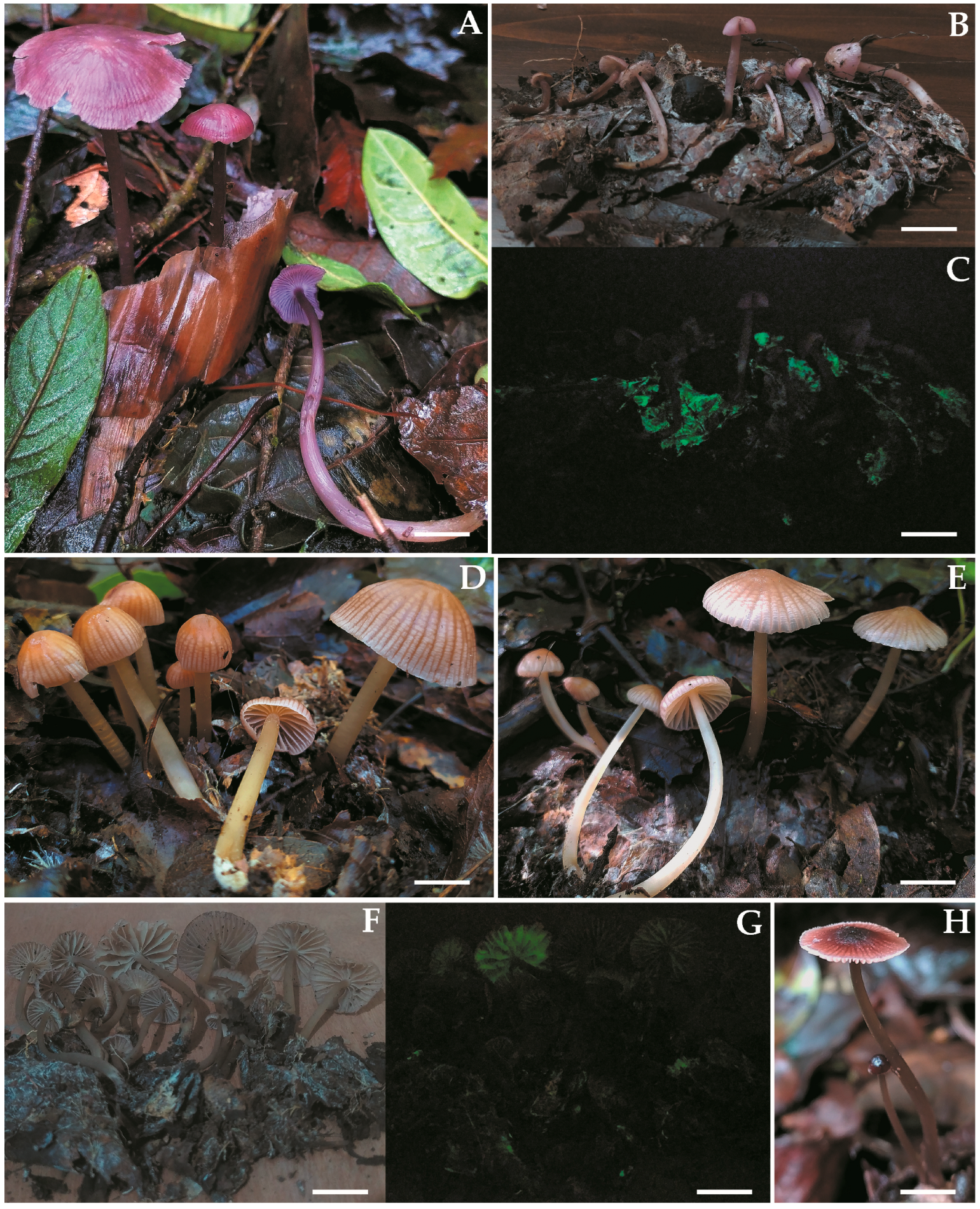
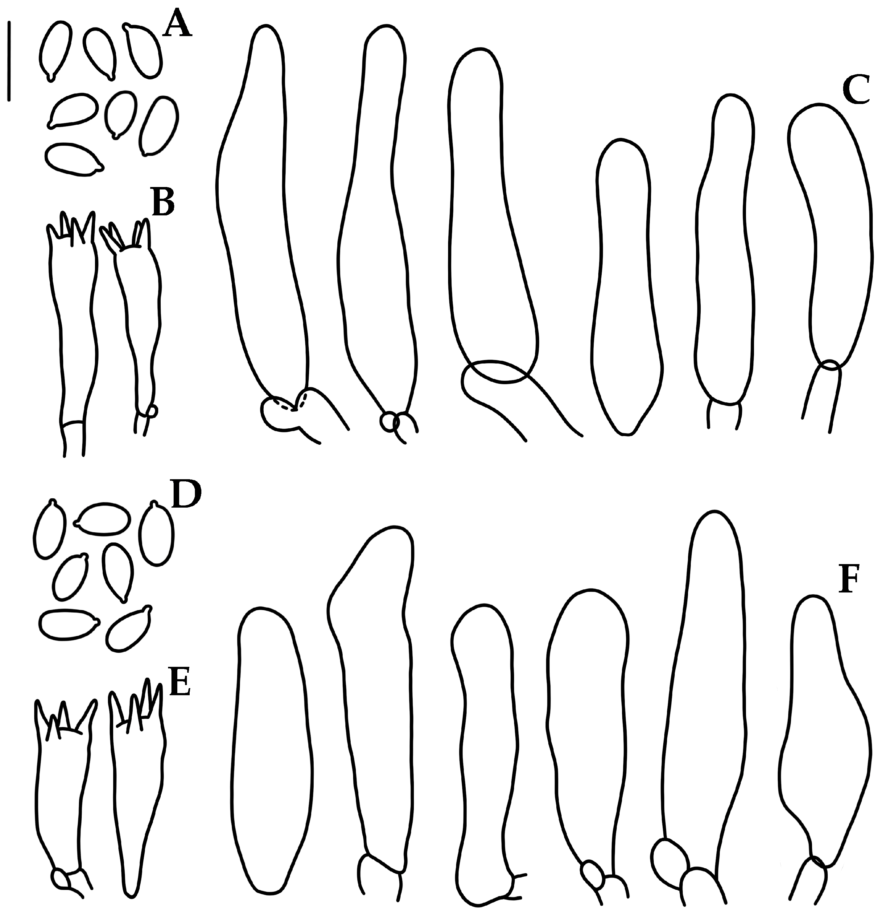
A total of 189 sequences, representing 24 taxa, were used for the phylogenetic reconstruction of Mycena sect. Calodontes. Of these, 32 sequences were generated in this study (12 ITS, 10 rpb1, and 10 Tef-). Detailed information for all sequences can be found in Table 2. The concatenated matrix comprised 70 terminals and 1281 positions, which were distributed as follows: ITS (1–529), rpb1 exon (530–622), rpb1 conserved intron (623–944), Tef exons (945–1250), and Tef- conserved intron (1251–1281). Due to alignment ambiguities, the Tef- not conserved intron and 298 positions from the ITS region were removed. The substitution models for each partition were as follows: TrN+I+G for ITS, JC for rpb1, K80+G for rpb1 intron, TVMef+I+G for Tef- exon, and GTR+G for Tef- intron. Phylogenetic analysis was performed on this concatenated dataset.
Although ML and BI analyses generated slightly different topologies, the placement of the five Mexican taxa on both trees was consistent. The analysis identified 24 statistically supported clades (BS ≥ 70%, PP ≥ 0.90) that separated the different members of the Mycena sect. Calodontes. The ML tree (Figure 1) revealed that the Mycena collections from Mexico did not cluster with any clades of M. pura s.l. or known species of M. sect. Calodontes. Instead, the Mexican collections formed five monophyletic lineages, four of which had high statistical support: M. luceata (BS = 100%, PP = 1), M. lucisnieblae (BS = 100%, PP = 1), M. luxmanantlanensis (BS = 100%, PP = 1), and M. sophiae (BS = 100%, PP = 1). Mycena lucisnieblae was grouped in a statistically supported clade (BS = 74%, PP = 0.99), which included M. shengshanensis Z.W. Liu, Y.P. Ge & Q. Na and M. subulata Z.W. Liu, Y.P. Ge & Q. Na, described in China, as well as M. pearsoniana, known from North America and Europe.
Mycena luxmanantlanensis showed a sister relationship with a specimen from Ecuador, tentatively identified as M. aff. pura (BS = 100%, PP = 1). They formed a clade (BS = 96%, PP = 1) with other specimens from Ecuador, also identified as M. aff. pura. Mycena sophiae formed a clade with M. cahaya from Malaysia, although this relationship lacked statistical support. Furthermore, M. luceata and M. polycystidiata Z.W. Liu, Y.P. Ge, L. Zou & Q. Na from China were found to be sister species, but in an unsupported clade.
For the fifth new Mexican species, M. luciferina, we only had a DNA sequence from the single collected specimen. This sequence was positioned at the base of an unsupported clade that included M. brunneoviolacea A.C. Cooper, Desjardin & B.A. Perry from the Republic of São Tomé and Príncipe, M. rufobrunnea Z.W. Liu, Y.P. Ge & Q. Na from China, and M. seminau A.L.C. Chew & Desjardin from Malaysia.

3.2. Taxonomy

Mycena luceata Cortés-Pérez, Guzm.-Dáv. & Ram.-Cruz, sp. nov. (Figure 2A–D and Figure 3).
MycoBank MB849395.
Etymology. From luceata (Latin), glow up, in reference to the brightly glowing hymenophore.
Holotype. MEXICO. VERACRUZ: Municipality of Banderilla, La Martinica, elev. 1639 m, 19° 35′ 13″ N, 96° 57′ 08″ W, gregarious, growing on leaf litter in cloud forest, 11 September 2021, A. Cortés-Pérez 2116 (holotype IBUG, isotype XAL). GenBank: ITS OR233614, rpb1 OR233746, Tef1-α OR233755.
Diagnosis. Mycena luceata is distinguished by the convex to plane-convex, hygrophanous, violet-brown to grayish-brown pileus; subdistant, intervenose, white to pinkish-white or orange-white lamellae; purplish-pink to pinkish-white stipe; 6–8 × 3.5–5 μm, Q = 1.2–2.1, amyloid basidiospores; 17–36 × 6–12 μm, narrowly to broadly clavate to cylindrical, obtuse, hyaline cheilocystidia, and bioluminescence only in the hymenophore.
Basidiomata medium size, collybioid. Pileus 20–43 mm diam., convex to convex-plane, sometimes with depressed center, margin translucent-striate to sulcate-striate, reflexed, even or eroded in age; surface glabrous, moist, hygrophanous, disc and striations or all violet-brown (10E8, 11E6), ruby (12E8), to grayish-violet (11E4) or pink (11A5), fading to pinkish-white (11A2) or grayish-brown (10D3). Context 1 mm thick, pinkish-white (11A2). Lamellae subdecurrent to adnate, subventricose, subdistant (14–20 reaching the stipe) with 1–3 series of lamellulae, intervenose, white or pinkish-white (11A2) to orange-white (6A2); edge even, pale. Stipe 38–82 × 2.5–5 mm, central or eccentric, cylindrical, with a swollen or narrow base, hollow; surface glabrous, apex purplish-pink (14A3) to purplish-white (14A2), pinkish-white (13A2) towards the base, with the base yellow-white (2A2); with white strigose basal mycelium. Odor not recorded. Bioluminescence in hymenophore, emitting bright green light.
Basidiospores (5.5–)6–8(–8.2) × 3.5–5(–5.5) μm (Xm = 6.6 ± 0.5 × 4.2 ± 0.4 µm, Q = 1.2–2.1, Qm = 1.5 ± 0.1, n = 54), ellipsoid to elongated, thin-walled, hyaline, amyloid. Basidia 28–30 × 5–7 μm, clavate, 4-spored, with sterigmata 3–6 μm long, hyaline, inamyloid. Lamellar edge sterile. Cheilocystidia 17–36 (–37) × 6–12 μm, narrowly to broadly clavate to cylindrical, apex obtuse, thin-walled, hyaline. Pleurocystidia absent. Subhymenium ramose, non-gelatinous, hyaline, non-dextrinoid. Hymenophoral trama regular; hyphae 2–24 μm diam., cylindrical to inflated, thin-walled, non-gelatinous, hyaline, dextrinoid. Pileipellis an ixocutis 6.5–14 μm thick, hyphae 2–6 μm diam., thin-walled, hyaline, non-dextrinoid. Subcutis thick, hyaline, dextrinoid. Pileus trama hyphae 2–24 μm diam., interwoven, cylindrical to inflated, thin-walled, hyaline, dextrinoid. Stipitipellis hyphae 2–6 μm diam., thin-walled, non-gelatinous, hyaline, non-dextrinoid; medullary hyphae of the stipe 3–30 μm diam., thin-walled or wall up to 1 μm thick, hyaline, dextrinoid. Caulocystidia absent. Clamp connections present.
Habitat and distribution—Gregarious, growing on leaf litter in cloud forest, Mexico (Veracruz).
Additional specimens examined—Mexico, Veracruz, Municipality of Banderilla, La Martinica, 19° 35′ 13″ N, 96° 57′ 08″ W, elev. 1639 m a.s.l., 15 September 2021, A. Cortés-Pérez 2115, 2126 (IBUG). Notes—Mycena luceata is distinguished by the violet-brown pileus, white lamellae, basidiospores averaging 6.6 × 4.2 μm, 17–36 × 6–12 μm, clavate to cylindrical cheilocystidia, and by the pileipellis as an ixocutis. A morphologically similar species is M. fenestrata Maas Geest. & de Meijer, described in Brazil, which has a purplish-brown to greyish-violet pileus, 6.7–8.1 × 3.6–4.5 μm basidiospores, and 18–35 × 8–15 μm, clavate, fusiform or subcylindrical cheilocystidia, but they are separate because M. fenestrata presents greyish-violet lamellae, dark purplish-brown stipe, and non-gelatinous pileipellis, and because of the presence on the stipe of 24–27 × 6.5–8 μm clavate terminal cells [23]. Another similar species is M. diosma, which has a basidiome with a dark violet-brown or reddish-purple hygrophanous pileus, but it is distinguished by the fact that M. diosma has dark brownish-violet to dark violet lamellae, larger cheilocystidia, 20–60(–80) × 3.5–20 μm, and pleurocystidia present [13,21]. Mycena luceata is distinguished from Malaysian bioluminescent species by macro- and micromorphological characters [1].
In the phylogenetic reconstruction, M. luceata was recovered as a possible sister to M. polycystidiata from China, but they are different because the Chinese species has a grayish-rose pileus with brownish-orange umbo, 50–78 × 14–31 μm utriform or subclavate cheilocystidia, abundant pleurocystidia, and caulocystidia present [20].
Mycena luciferina Cortés-Pérez, Guzm.-Dáv. & Ram.-Cruz, sp. nov. (Figure 2E–G and Figure 4).
MycoBank MB849396.
Etymology. From lux (Latin), light, -ferre (Latin), bringer: the bringer of light, in reference to the bioluminescent pileus.
Holotype. MEXICO. VERACRUZ: Municipality of Banderilla, 19° 35′ 18″ N, 96° 57′ 22″ W, elev. 1639 m a.s.l., gregarious, growing on leaf litter in cloud forest, 11 September 2021, A. Cortés-Pérez 2114 (holotype IBUG, isotype XAL). GenBank: ITS OR233612, rpb1 OR233744.
Diagnosis. Mycena luciferina is distinguished by the campanulate, reddish-pink, hygrophanous pileus; close, intervenose, white lamellae; white stipe; 6.4–8 × 3.6–4 μm, Q = 1.5–2.2, amyloid basidiospores; 33–64 × 10–17 μm, narrowly to broadly clavate to cylindrical, apex obtuse, hyaline cheilocystidia; with pleurocystidia, rare, similar to cheilocystidia; pileipellis an ixocutis, and bioluminescent pileus.
Basidiomata medium size, mycenoid. Pileus 32–47 mm diam., campanulate, occasionally umbonate; margin translucent-striate to sulcate-striate, even or eroded in age; surface glabrous, moist, hygrophanous, disc reddish-pink (11A7), grayish-red (11C6) to pink (11A5), elsewhere pale pink (11A2–3), with a white margin, fading pinkish-white (11A2). Context 0.5–1 mm thick, pinkish-white (11A2). Lamellae adnate, subventricose, close (26–31 reaching the stipe), with 2–3 series of lamellulae, intervenose, white to yellowish-white (3A2); edge even, concolorous. Stipe 45–82 × 4–5 mm, central or eccentric, cylindrical, with a narrow base, hollow; surface glabrous, white to towards the base yellowish-white (3A2); with white strigose basal mycelium. Odor not recorded. Bioluminescence in pileus, emitting bright green light.
Basidiospores (6–)6.4–8(–8.4)(–8.8) × (3.2–)3.6–4 μm (Xm = 7.4 ± 0.8 × 3.8 ± 0.2 µm, Q = 1.5–2.2, Qm = 1.9 ± 0.1, n = 30), ellipsoid to elongated, thin-walled, hyaline, amyloid. Basidia 15.2–27.2 × 4.5–5.6 μm, clavate, 4-spored, with sterigmata 3–5 μm long, hyaline. Lamellae edge sterile. Cheilocystidia 33–64(–68) × (7–)10–17(–20) μm, narrowly to broadly clavate to cylindrical, apex obtuse, thin-walled, hyaline. Pleurocystidia rare, when present near the edge of the lamella, similar to cheilocystidia. Subhymenium ramose to subcellular, hyaline, non-dextrinoid, non-gelatinous. Hymenophoral trama regular, hyphae 2–20 μm diam., cylindrical to inflated, thin-walled, hyaline, dextrinoid, non-gelatinous. Pileipellis an ixocutis 25–35 μm thick, hyphae 1.5–6.5 μm diam., hyaline, non-dextrinoid. Subcutis with hyphae 2–39 µm diam., inflated or cylindrical, thin-walled, hyaline, dextrinoid. Pileus trama hyphae 2.5–22 μm diam., cylindrical to inflated, thin-walled, interwoven, hyaline, dextrinoid. Stipitipellis with hyphae 2–10 μm diam., thin-walled, non-gelatinous, hyaline, non-dextrinoid; medullary hyphae 2–26 μm diam., thin-walled, hyaline, dextrinoid. Clamp connections present.
Habitat and distribution—Gregarious, growing on leaf litter in cloud forest. Known from Mexico (Veracruz).
Notes—Mycena luciferina is characterized by a campanulate, reddish-pink to pale pink pileus, white intervenose lamellae; basidiospores averaging 7.4 × 3.8 μm, amyloid; cheilocystidia clavate to cylindrical, with a rounded apex, pleurocystidia rare, similar to cheilocystidia, an ixocutis, with simple hyphae, and by its strongly bioluminescent pileus. A morphologically similar species is M. rosea, a European species that has basidiome with a pink pileus but is distinguished by pale pink to lilaceous pink lamellae, larger cheilocystidia, 30–90 × 9–36 μm, and caulocystidia present [13,21]. It is also like some of the species in the Mycena pura complex, especially those with pink pileus; however, they are easily distinguished by having larger cheilocystidia and pleurocystidia [13,21].
BLAST (https://blast.ncbi.nlm.nih.gov/Blast.cgi, accessed on 1 January 2023) sequence similarity searches were performed; comparison of the ITS sequence of the Mexican specimen (ACP2114) showed 96% similarity with sequences determined as Mycena pura from Canada and the USA, and 95% similarity with a sequence determined as M. rosea from Italy. In the phylogenetic tree, M. luciferina was recovered in an unsupported clade formed by M. brunneoviolacea described from Africa [14], M. aff. pura clade X sensu Harder et al. from the USA [18], M. rufobrunnea from China [20], and M. seminau from Malaysia [1]. Although the Mexican specimen is within this clade, it is in an independent lineage, which can be distinguished from the rest of the taxa by its morphological characteristics. Mycena brunneoviolacea is distinguished by its dark violet-brown or dark brown pileus, narrowly lageniform to lageniform cheilocystidia and pleurocystidia, and caulocystidia present [14]. Mycena rufobrunnea is separated by the dark brown to reddish-brown or greyish-brown pileus and utriform cheilocystidia, sometimes clavate, and caulocystidia present [20]. Mycena seminau differs in that it has a dark brown to brown pileus, yellowish-gray to reddish-gray lamellae, and narrower cheilocystidia, 32.8–56.8 × 5.6–9.6 μm [1].
Mycena lucisnieblae Cortés-Pérez, Ram.-Cruz & Guzm.-Dáv., sp. nov. (Figure 5A–C and Figure 6).
MycoBank MB849397.
Etymology. From lucis (Latin), referring to light, -niebla (Spanish, from the Latin nebula), referring to the “bosque de niebla” or cloud forest, in reference to the bioluminescent mycelium that grows in the leaf litter of cloud forest.
Holotype. MEXICO. JALISCO: Municipality of Autlán de Navarro, Estación Científica Las Joyas, Sierra de Manantlán, 19° 35′ 15″ N, 104° 16′ 27″ W, elev. 1926 m a.s.l., gregarious, growing on leaf litter in cloud forest, 18 September 2021, A. Cortés-Pérez 2140 (holotype IBUG, isotype XAL). GenBank: ITS OR233610, rpb1 OR233742, Tef1-α OR233752.
Diagnosis. Mycena lucisnieblae is characterized by the hemispheric-convex to convex, purplish-pink or pink, hygrophanous pileus; arcuate-adnate, intervenose, violet-white to pale pink lamellae; pale violet to pinkish-white stipe; 5.5–7.4 × 4–5 μm, Q = 1.2–2, amyloid basidiospores; 24–41 × 8.5–13 μm, narrowly to broadly clavate, apex obtuse, hyaline cheilocystidia; pleurocystidia absent; pileipellis an ixocutis; without terminal hyphae in the pileipellis or caulocystidia, and bioluminescent mycelium.
Basidiomata small-sized, collybioid. Pileus 7–31 mm diam., hemispheric-convex to convex, some umbonate; margin smooth or translucent-striate to sulcate-striate, even or eroded in age; surface glabrous, moist, hygrophanous, disc and striations grayish-magenta (14D5–7) or purplish-pink (14A4), elsewhere pale pink (13A2), or all pink (13A2–3), fading pinkish-white (13A2). Context 0.2–0.8 mm thick, pinkish-white (13A2). Lamellae arcuate-adnate to adnate, narrow, close (22–29 reaching the stipe), with 2–3 series of lamellulae, intervenose, violet-white (16A2) to pale pink (13A2); edge even, pale. Stipe 26–78 × 1.5–4 mm, central or eccentric, cylindrical, with a swollen or narrow base, hollow; surface glabrous, pale violet (15A3) to violet-white (15A2) or purplish-white (14A2) to pinkish-white (10A2), with the base yellowish-white (3A2); with white strigose basal mycelium. Odor not recorded. Bioluminescence in mycelium, emitting green light, basidiomes non-luminescent.
Basidiospores 5.5–7.4(–8) × (3.8–)4–5 μm (Xm = 6.6 ± 0.5 × 4.4 ± 0.3 µm, Q = 1.2–2, Qm = 1.4 ± 0.1, n = 53), ellipsoid, some elongated, thin-walled, hyaline, amyloid. Basidia 23–33 × 6–7 μm, clavate, 4-spored, with sterigmata 2–4 μm long, hyaline. Lamellae edge sterile. Cheilocystidia 24–41(–44)(–46) × (7.5–)8.5–13 (–14.5) μm, narrowly to broadly clavate, apex obtuse, thin-walled, hyaline. Pleurocystidia absent. Subhymenium subcellular to ramose, non-gelatinous, hyaline, non-dextrinoid. Hymenophoral trama regular; hyphae 2.5–12 μm diam., cylindrical to inflated, thin-walled, non-gelatinous, hyaline, dextrinoid. Pileipellis an ixocutis 13–15 μm thick, hyphae 2–5 μm diam., hyaline, non-dextrinoid. Subcutis with hyphae 3–19 µm diam., inflated or cylindrical, thin-walled or wall up to 0.8 μm thick, hyaline, dextrinoid. Pileus trama hyphae 2–31 μm diam., cylindrical to inflated, thin-walled, interwoven, hyaline, dextrinoid. Stipitipellis hyphae 2–5 μm diam., thin-walled, non-gelatinous, hyaline, non-dextrinoid; medullary hyphae of the stipe 2–21 μm diam., thin-walled or wall up to 0.8 μm thick, hyaline, dextrinoid. Clamp connections present.
Habitat and distribution—Gregarious, growing on leaf litter in cloud forest. Known from Mexico (Jalisco).
Additional specimens examined—Mexico, Jalisco, Municipality of Autlán de Navarro, Estación Científica Las Joyas, Sierra de Manantlán, 19° 35′ 15″ N, 104° 16′ 27″ W, elev. 1926 m a.s.l., 18 September 2021, A. Cortés-Pérez 2139 (IBUG); 19 September 2021, A. Cortés-Pérez 2149 (IBUG); 20 September 2021, A. Cortés-Pérez 2166 (IBUG); 29 September 2022, A. Cortés-Pérez 2352-B (IBUG).
Notes—Mycena lucisnieblae is distinguished by its small basidiome, purplish-pink to pink pileus, violet-white to pale pink intervenose lamellae, pink-white stipe with white-yellow base, basidiospores averaging 6.6 × 4.4 µm, pleurocystidia absent, pileipellis an ixocutis, and by the bioluminescent mycelium that grows on leaf litter of tropical cloud forest trees. In the phylogenetic reconstruction, M. lucisnieblae was recovered in a clade formed by M. pearsoniana, M. shengshanensis, and M. subulata, in which it is distinguished by its morphological characteristics and by the habitat where it grows [17,20,21]. Mycena pearsoniana is a similar morphological species in the pileus with lilaceous or pink tinges; however, it is different by the vinaceous buff lamellae, “vinaceous buff or light cinnamon drab at apex and pale ochraceous buff at base” stipe, 5–6.3 × 3.5–3.8 μm basidiospores, and larger cheilocystidia, 30–80 × 7–12.5 μm, subfusiform, subcylindrical to clavate, with rounded apex or more rarely mucronate [17,21]. Mycena shengshanensis is distinguished by its brown to violet-brown pileus, white lamellae, moderately thick-walled clavate cheilocystidia, and by growing on the litter layer of Larix gmelinii (Rupr.) Kuzen [20]. Mycena subulata, described in China, is morphologically very similar and is the sister species of M. lucisnieblae; it is different in that M. subulata has 43–82 × 4–11 μm, narrowly fusiform with long neck, thick-walled cheilocystidia, clavate with tapering apices caulocystidia, and because it grows on the litter layer in mixed forests of Pinus koraiensis Siebold & Zucc., Larix gmelinii, and Tila sp. [20].
Mycena luxmanantlanensis Cortés-Pérez, Ram.-Cruz & Guzm.-Dáv., sp. nov. (Figure 5D–G and Figure 7).
MycoBank MB849398.
Etymology. From lux (Latin), light, -manantlanensis, referring to the place Manantlán, in reference to the bioluminescent basidiomes that were collected in the Sierra de Manantlán.
Holotype. MEXICO. JALISCO: Municipality of Autlán de Navarro, Estación Científica Las Joyas, Sierra de Manantlán, 19° 35′ 15″ N, 104° 16′ 27″ W, elev. 1926 m a.s.l., gregarious, growing on leaf litter in cloud forest, 20 September 2021, A. Cortés-Pérez 2160 (holotype IBUG, isotype XAL). GenBank: ITS OR233603, rpb1 OR233737, Tef1-α OR233747.
Diagnosis. Mycena luxmanantlanensis is distinguished by the convex, tessellate pileus, brownish-orange to orange, with pink tinges, hygrophanous, then orange-white; distant to subdistant, adnate, intervenose lamellae; yellowish stipe; 6–8 × 4–5.2 μm, Q = 1.3–1.8, amyloid basidiospores; 25–50 × 8–17.5 μm, narrowly to broadly clavate hyaline cheilocystidia, with obtuse or rostrate apex; pileipellis an ixocutis; with bioluminescence in the mycelium and sometimes in the hymenophore.
Basidiomata medium size, collybioid. Pileus 9–55 mm diam., at first hemispheric-convex expanding to convex, some umbonate; translucent-tessellate and sulcate-striate, even to crenate or eroded in age; surface glabrous, moist, hygrophanous, disc and striations brownish-orange (5B8), grayish-orange (6B5), light orange or “melon” (5A6), elsewhere pale orange (5A3), orange-white (5A2), pink (7A2). Context 2–3 mm thick, fleshy, orangish-brown to white. Lamellae adnate, broad, distant to subdistant (13–18 reaching the stipe), with 2–3 series of lamellulae, intervenose, orange-white (6A2) to pinkish-white (7A2); edge even, concolorous. Stipe 44–78 × 2–6 mm, central or eccentric, cylindrical, with a swollen or narrow base, compressed at the base, hollow; surface glabrous, light yellow (3A4) to yellowish-white (3A2); with white (2A1) strigose basal mycelium. Odor not recorded. Bioluminescence in mycelium, sometimes in hymenophore, emitting bright yellowish-green light.
Basidiospores 6–8(–9) × (3.6–)4–5.2 μm (Xm = 6.9 ± 0.6 × 4.4 ± 0.4 µm, Q = 1.3–1.8, Qm = 1.5 ± 0.1, n = 50), ellipsoid to elongated, thin-walled, hyaline, amyloid. Basidia 22–32 × 5–6.5 μm, clavate, 4-spored, with sterigmata 2–5 μm long, hyaline, inamyloid. Lamellar edge sterile. Cheilocystidia (20–)25–50(–52)(–56) × (7–)8–17.5 μm, narrowly to broadly clavate or subfusiform, apex obtuse or rostrate, thin-walled, hyaline. Pleurocystidia absent. Subhymenium ramose to subcellular, non-gelatinous, hyaline, non-dextrinoid. Hymenophoral trama regular, hyphae 2–26.5 μm diam., cylindrical to inflated, thin-walled, non-gelatinous, hyaline, dextrinoid. Pileipellis an ixocutis, 14.5–16 μm thick, hyphae 2–7 μm diam., hyaline, non-dextrinoid. Subcutis with hyphae 2.5–22.5 µm diam., inflated or cylindrical, thin-walled, hyaline, dextrinoid. Pileus trama hyphae 2.5–22.5 μm diam., cylindrical to inflated, interwoven, thin-walled, hyaline, dextrinoid. Stipitipellis hyphae 2–4 μm diam., thin-walled, non-gelatinous, hyaline, non-dextrinoid; medullary hyphae of the stipe 3–31 μm diam., hyaline, dextrinoid. Clamp connections present.
Habitat and distribution—Gregarious or scattered, growing on leaf litter in cloud forest, Mexico (Jalisco).
Additional specimens examined—Mexico, Jalisco, Municipality of Autlán de Navarro, Estación Científica Las Joyas, Sierra de Manantlán, 19° 35′ 15″ N, 104° 16′ 27″ W, elev. 1926 m a.s.l., 20 September 2021, A. Cortés-Pérez 2159 (IBUG).
Notes—Mycena luxmanantlanensis is characterized by its orange pileus, strongly sulcate-striate, orange-white to pink-white, intervenose lamellae, and a yellow stipe, basidiospores averaging 6.9 × 4.4 µm, cheilocystidia clavate, fusiform with the apex obtuse or rostrate. A morphologically similar species is M. luteovariegata B. Harder & Læssøe described in Denmark, but the latter has a yellowish instead of orangish pileus, grayish-pink lamellae, reddish-gray stipe, and pleurocystidia [18]. Mycena luxmanantlanensis differs from the Malaysian bioluminescent species described by Chew et al. [1] in the following: M. cahaya has brown pileus, elongated basidiospores averaging 7.1 × 3.8 µm, and abundant clavate to ventricose cheilocystidia and pleurocystidia; Mycena seminau is different in that it has a dark brown to brown pileus, elongated to cylindrical basidiospores averaging 7.1 × 3.9 µm, and by the narrower cheilocystidia, measuring 32.8–56.8 × 5.6–9.6 µm [1]. Another similar bioluminescent species is M. sinar, which has a brownish-orange to yellowish-brown pileus, but differs in that it has subdecurrent yellowish-gray lamellae, basidiospores averaging 8.3 × 4 µm, and narrower cheilocystidia, 30.4–45.6 × 5.6–9.6 µm [1]. Mycena sinar var. tangkaisinar differs in the brown pileus, reddish-gray lamellae with pink margin, elongated basidiospores averaging 7.2 × 3.6 µm, cylindrical caulocystidia, and a luminescent stipe [1]. In the phylogenetic tree, M. luxmanantlanensis is sister to a taxon from Ecuador determined as M. aff. pura by Harder et al. [18]. These two species are of tropical affinity and in both the pleurocystidia are absent; however, Harder et al. [18] did not provide further morphological data on these Ecuadorian specimens, which could help to better understand the sister relationship between these two taxa.
Mycena sophiae Cortés-Pérez sp. nov. (Figure 5H and Figure 8).
MycoBank MB849399.
Etymology. ‘sophiae’ (Latin), dedicated to Sofía Cortés, niece of the first author.
Holotype. MEXICO. JALISCO: Municipality of Autlán de Navarro, Estación Científica Las Joyas, Sierra de Manantlán, 19° 35′ 15″ N, 104° 16′ 27″ W, elev. 1926 m a.s.l., gregarious, growing on leaf litter in cloud forest, 19 September 2021, A. Cortés-Pérez 2157 (holotype IBUG, isotype XAL). GenBank: ITS OR233606, rpb1 OR233739, Tef1-α OR233749.
Diagnosis. Mycena sophiae has a very small basidiome, with a convex to convex-plane, dark ruby to ruby pileus; adnate with a tooth, subdistant, pinkish-white lamellae; pinkish-white to greyish-orange stipe; 5.8–6.5 × 3–3.8 μm, Q = 1.4–2, amyloid basidiospores; 24–47 × 6.5–11 μm, cylindrical to narrowly clavate, obtuse cheilocystidia; a cutis as pileipellis; with bioluminescent mycelium.
Basidiomata small size, collybioid. Pileus 7–19 mm diam., hemispheric-convex to convex or convex-plane, margin translucent-striate to sulcate-striate, even or eroded in age; surface glabrous, moist, disc and striations dark ruby (12F8) to ruby (12D8), elsewhere pale or pastel red (9A3–4) to pinkish-white (8A2), margin white. Context thin, vinaceous pink to pinkish-white (11A2). Lamellae adnate with a tooth, narrow, subdistant (15–20 reaching the stipe), with 2–3 series of lamellulae, intervenose, pinkish-white (10A2); edge even, concolorous. Stipe 22–55 × 1–1.5 mm, central or eccentric, cylindrical, with a swollen base, hollow; surface glabrous, pinkish-white (11A2) to reddish-brown (8–9E5), with the base pale brownish-orange (7C4); with white strigose basal mycelium. Odor not recorded. Bioluminescence of mycelium observed but not documented, basidiomes non-luminescent.
Basidiospores (5.2–)5.8–6.5(–6.8) × 3–3.8 μm (Xm = 6.0 ±0.3 × 3.3 ±0.2 µm, Q = 1.4–2, Qm = 1.8 ±0.1 n = 63), ellipsoid to elongated, thin-walled, hyaline, amyloid. Basidia 19–24 × 5–6 μm, clavate, 4–spored, with sterigmata 3–4 μm long, hyaline. Lamellar edge sterile. Cheilocystidia 24–47(–52) × 6.5–11 (–13) μm, cylindrical to narrowly clavate, obtuse, thin-walled, hyaline. Pleurocystidia absent. Subhymenium ramose, non-gelatinous, hyaline, non-dextrinoid. Hymenophoral trama regular, hyphae 2–24 μm diam., cylindrical to inflated, thin-walled, non-gelatinous, hyaline, dextrinoid. Pileipellis a cutis, hyphae 2–5 μm diam., hyaline, non-dextrinoid. Subcutis hyphae 2.6–33 µm diam., inflated or cylindrical, thin-walled or wall up to 1 μm thick, hyaline, dextrinoid. Pileus trama hyphae 3–22.6 μm diam., cylindrical to inflated, thin-walled, interwoven, hyaline, dextrinoid. Stipitipellis hyphae 1.5–10 μm diam., thin-walled, non-gelatinous, hyaline, non-dextrinoid; medullary hyphae of the stipe 2–25 μm diam., thin-walled or wall up to 1 μm thick, hyaline, dextrinoid. Clamp connections present.
Habitat and distribution—Gregarious, growing on leaf litter in cloud forest, Mexico (Jalisco).
Additional specimens examined—Mexico, Jalisco, Municipality of Autlán de Navarro, Estación Científica Las Joyas, Sierra de Manantlán 19° 35′ 15″ N, 104° 16′ 27″ W, elev. 1926 m a.s.l., 20 September 2021, A. Cortés-Pérez 2161 (IBUG).
Notes—Mycena sophiae is distinguished by its very small basidiome, ruby pileus, pinkish-white lamellae, relatively small amyloid basidiospores, and cylindrical to narrowly clavate cheilocystidia. Mycena sirayuktha Aravind. & Manim., described in India [12], is a morphologically similar species in its greyish-red pileus and intervenose lamellae, but is different by the pleurocystidia present and gelatinized lageniform, fusiform, clavate, or utriform cheilocystidia. Other similar species are M. clarkeana Grgur. and M. nullawarrensis Grgur., described in Australia, which have a reddish-brown pileus, but both species are different by having pleurocystidia [15]. Mycena cahaya from Malaysia could be a sister group of M. sophiae; they are different because M. cahaya has a brown pileus, basidiospores averaging 7.1 × 3.8 μm, and ventricose to fusoid mucronate cheilocystidia, and pleurocystidia present [1]. Mycena lucisnieblae has small basidiomata resembling those of M. sophiae in size but is distinguished by the fact that M. lucisnieblae has pink or purplish-pink basidiomata.

4. Discussion

This study represents the first occurrence of bioluminescent species belonging to Mycena sect. Calodontes in Mexico. With the inclusion of these five species, the global count of bioluminescent fungi increases to 108, with 15 taxa recorded in that country. The Mexican bioluminescent species of M. sect. Calodontes are exclusively found in cloud forests and can be distinguished from species in other sections by their pileus color, which can be pink, red, purple, orange, or violet, their intervenose lamellae, the presence of oxalate crystals on the basal mycelium, the absence of pleurocystidia (except in M. luciferina), the absence of terminal hyphae in pileipellis and stipitipellis, the pileipellis an ixocutis (except in M. sophiae), and bioluminescence exhibited in the mycelium or some part of the basidiomata, or both.
According to the traditional classification, M. luciferina, characterized by its hyaline cheilocystidia and pleurocystidia, could be classified under M. sect. Calodontes subsect. Purae. Mycena luxmanantlanensis, with its fusiform cheilocystidia with rostrate apex, could be placed in the subsect. Generosae. Lastly, M. luceata, M. lucisnieblae, and M. sophiae could be grouped under subsect. Violacella due to their cheilocystidia with a rounded apex and the absence of pleurocystidia. However, the presence of amyloid basidiospores differentiates them from the species within subsect. Violacella [21,23]. As observed in other studies [1,18,20,25], these subsections are polyphyletic, and the micromorphological characters used in the infrageneric classification exhibit homoplasy. The presence of oxalate crystals on the basal mycelium in the Mexican species was confirmed, consistent with reports by Olariaga et al. and Clémençon for certain species of the M. sect. Calodontes from Europe [25,26]. The information presented here enhances our understanding of the distribution and ecological aspects of the members of M. sect. Calodontes, specifically in the cloud forest of Mexico.
The phylogenetic analysis conducted on M. sect. Calodontes aligns with previous studies [1,18,20,25]. Most of the lineages identified at the species level exhibited strong support, while shallower branches displayed weaker values, consistent with findings in other studies [20,25]. Importantly, this study marks the first inclusion of specimens from M. sect. Calodontes in Mexico, affirming the presence of five distinct clades among the Mexican samples. Additionally, the 11 clades proposed by Harder et al. [16,17,18] for M. pura were indeed recovered; however, they appeared in separate linages from the newly identified species.
We must emphasize that the relationships between certain taxa within M. sect. Calodontes has shifted with the inclusion of Mexican specimens. For instance, Liu et al. [20] recovered M. polycystidiata as a sister species to M. diosma, while in our study a clustering of M. luceata and M. polycystidiata was observed, albeit without support. Similarly, in Liu et al. [20], M. pura clade IX was linked with M. cahaya; however, our phylogeny uncovered an unsupported clade of M. cahaya and M. sophiae. Furthermore, in Harder et al.’s phylogenetic analysis [18], a clade featuring Ecuadorian taxa labeled as M. aff. pura was identified as the sister group of M. pearsoniana. Likewise, Liu et al. [20] found a clade composed of Ecuadorian taxa clustered with M. pearsoniana, M. shengshanensis, and M. subulata from China. In contrast, our analysis placed the Ecuadorian taxa together with M. luxmanantlanensis from Mexico in a well-supported clade. Additionally, another supported clade included M. pearsoniana, M. shengshanensis, and M. subulata along with M. lucisnieblae. Likewise, M. luceata and M. luxmanantlanensis clustered in a large unsupported clade, along with M. lucisnieblae, M. pearsoniana, M. shengshanensis, and M. subulata. This clade represents a mixture of species with temperate affinities, such as M. pearsoniana, and tropical ones, from Ecuador and Mexico, distinct from the M. pura complex.
Mexico is one of the countries with the greatest richness of bioluminescent fungi, with a total of 15 species, most of which are exclusive to the country and with only a few known also from other countries. In that country, M. sect. Calodontes represents the section, so far, with the largest number of species of bioluminescent fungi. It is expected that the knowledge of bioluminescent species will increase as other plant communities and states in the country are explored. On other hand, our phylogenetic reconstruction allowed us to recognize the phylogenetic position of the five new species from Mexico as independent of the M. pura complex. The inclusion of the Mexican specimens in the phylogenetic analyses changed the panorama, generating new relationship hypotheses within M. sect. Calodontes. In this sense, it is expected that, as more taxa are added to the studies, the phylogenetic relationships of this section will continue changing. Finally, this work is the basis for subsequent phylogenetic studies that include Mexican taxa.

Author Contributions

Conceptualization, A.C.-P., L.G.-D. and V.R.-C.; methodology, A.C.-P., L.G.-D., V.R.-C. and E.R.-S.; molecular experiments, A.R.V.-A. and A.C.-P.; formal analysis, A.C.-P. and V.R.-C.; investigation, A.C.-P.; resources, L.G.-D. and A.R.V.-A.; data curation, A.C.-P.; writing—original draft preparation, A.C.-P. and F.R.-G.; writing—review and editing, A.C.-P., L.G.-D., V.R.-C., A.R.V.-A., E.R.-S. and F.R.-G.; supervision, L.G.-D. and V.R.-C.; project administration, L.G.-D.; funding acquisition, L.G.-D., V.R.-C. and A.R.V.-A. All authors have read and agreed to the published version of the manuscript.

Funding

This research was supported by the University of Guadalajara (Programa de Fortalecimiento de la Investigación y el Posgrado 2022–2023 and Pe3 2023 to A.R.V.-A.) and Consejo Nacional de Ciencia y Tecnología (grant number 803773 for doctoral studies to A.C.-P.).

Institutional Review Board Statement

Not applicable.

Informed Consent Statement

Not applicable.

Data Availability Statement

The sequences presented in this study are available at https://www.ncbi.nlm.nih.gov/ accessed on 1 July 2023 (see Table 1 for the accession numbers). The alignments and phylogenetic tree files can be requested directly to the authors. All new taxa were registered in the MycoBank (http://www.mycobank.org/ accessed on 1 July 2023).

Acknowledgments

The authors thank the University of Guadalajara for all the support. V.R.-C. and L.G.-D. recognize CONAHCYT for the academic position at “Investigadoras e Investigadores por México” granted to V.R.-C. We thank Victor Ramón Laureano Güitron, Manager of “Estación Científica Las Joyas”, and the team of guides, Alfredo Aragón, Paulino Chavez, Celso Mendoza, Joel Oleague, and Jaun Loera, as well as Maria Loera and Juana Florentino, who are thanked for all the facilities provided during the fieldwork at the scientific station. Oscar Castro Jauregui is thanked for line drawing.

Conflicts of Interest

The authors declare no conflict of interest.

References

  1. Chew, A.L.C.; Tan, Y.S.; Desjardin, D.E.; Musa, M.Y.; Sabaratnam, V. Four new bioluminescent taxa of Mycena sect. Calodontes from Peninsular Malaysia. Mycologia 2014, 106, 976–988. [Google Scholar] [CrossRef] [PubMed]
  2. Dauner, L.A.P.; Karunarathna, S.C.; Tibpromma, S.; Xu, J.; Mortimer, P.E. Bioluminescent fungus Roridomyces viridiluminus sp. nov. and the first Chinese record of the genus Roridomyces, from Southwestern China. Phytotaxa 2021, 487, 233–250. [Google Scholar] [CrossRef]
  3. Desjardin, D.E.; Oliveira, A.G.; Stevani, C.V. Fungi bioluminescence revisited. Photochem. Photobiol. Sci. 2008, 7, 170–182. [Google Scholar] [CrossRef]
  4. Desjardin, D.E.; Perry, B.A.; Stevani, C.V. New luminescent mycenoid fungi (Basidiomycota, Agaricales) from São Paulo State, Brazil. Mycologia 2016, 108, 1165–1174. [Google Scholar] [CrossRef] [PubMed]
  5. Karunarathna, S.C.; Mortimer, P.E.; Tibpromma, S.; Dutta, A.K.; Paloi, S.; Hu, Y.; Baurah, G.; Axford, S.; Marciniak, C.; Luangharn, T.; et al. Roridomyces phyllostachydis (Agaricales, Mycenaceae), a new bioluminescent fungus from Northeast India. Phytotaxa 2020, 459, 155–167. [Google Scholar] [CrossRef]
  6. Oba, Y.; Hosaka, K. The luminous fungi of Japan. J. Fungi 2023, 9, 615. [Google Scholar] [CrossRef]
  7. Weinstein, P.; Delean, S.; Wood, T.; Austin, A.D. Bioluminescence in the ghost fungus Omphalotus nidiformis does not attract potential spore dispersing insects. IMA Fungus 2016, 7, 229–234. [Google Scholar] [CrossRef]
  8. Matheny, P.B.; Curtis, J.M.; Hofstetter, V.; Aime, M.C.; Moncalvo, J.M.; Ge, Z.W.; Yang, Z.L.; Slot, J.C.; Ammirati, J.F.; Baroni, T.J.; et al. Major clades of Agaricales: A multilocus phylogenetic overview. Mycologia 2006, 98, 982–995. [Google Scholar] [CrossRef]
  9. Nimalrathna, T.S.; Tibpromma, S.; Nakamura, A.; Galappaththi, M.C.A.; Xu, J.; Mortimer, P.E.; Karunarathna, S.C. The case of the missing mushroom: A novel bioluminescent species discovered within Favolaschia in southwestern China. Phytotaxa 2022, 539, 244–256. [Google Scholar] [CrossRef]
  10. Oliveira, J.J.S.; Vargas-Isla, R.; Cabral, T.S.; Cardoso, J.S.; Andriolli, F.S.; Rodrigues, D.P.; Ikeda, T.; Clement, C.R.; Ishikawa, N.K. The Amazonian luminescent Mycena cristinae sp. nov. from Brazil. Mycoscience 2021, 62, 395–405. [Google Scholar] [CrossRef]
  11. Chang, C.C.; Chen, C.Y.; Lin, W.W.; Kao, H.W. Mycena jingyinga, Mycena luguensis, and Mycena venus: Three new species of bioluminescent fungi from Taiwan. Taiwania 2020, 65, 396–406. [Google Scholar] [CrossRef]
  12. Aravindakshan, D.M.; Manimohan, P. Mycenas of Kerala; SporePrint Books: Callicut, India, 2015; 213p. [Google Scholar]
  13. Aronsen, A.; Læssøe, T. The genus Mycena s.l. In The Fungi of Northern Europe; Svampetryk: Copenhagen, Denmark, 2016; Volume 5, 373p. [Google Scholar]
  14. Cooper, A.C.; Desjardin, D.E.; Perry, B.A. The genus Mycena (Basidiomycota, Agaricales, Mycenaceae) and allied genera from Republic of São Tomé and Príncipe, West Africa. Phytotaxa 2018, 383, 1–47. [Google Scholar] [CrossRef]
  15. Grgurinovic, C.A. The genus Mycena in South-Eastern Australia. Fungal Divers. Res. Ser. 2003, 9, 1–329. [Google Scholar]
  16. Harder, C.B.; Læssøe, T.; Kjøller, R.; Frøslev, T.G. A comparison between ITS phylogenetic relationships and morphological species recognition within Mycena sect. Calodontes in Northern Europe. Mycol. Prog. 2010, 9, 395–405. [Google Scholar] [CrossRef]
  17. Harder, C.B.; Lodge, D.J.; Petersen, R.H.; Hughes, K.W.; Cifuentes Blanco, J.; Frøslev, T.G.; Læssøe, T. Amyloidity is not diagnostic for species in the Mycena pearsoniana complex (Mycena sectio Calodontes). Mycol. Prog. 2012, 11, 725–732. [Google Scholar] [CrossRef]
  18. Harder, C.B.; Læssøe, T.; Frøslev, T.G.; Ekelund, F.; Rosendahl, S.; Kjøller, R. A three-gene phylogeny of the Mycena pura complex reveals 11 phylogenetic species and shows ITS to be unreliable for species identification. Fungal Biol. 2013, 117, 764–775. [Google Scholar] [CrossRef]
  19. Liu, Z.; Na, Q.; Cheng, X.; Wu, X.; Ge, Y. Mycena yuezhuoi sp. nov. (Mycenaceae, Agaricales), a purple species from the peninsula areas of China. Phytotaxa 2021, 511, 148–162. [Google Scholar] [CrossRef]
  20. Liu, Z.; Ge, Y.; Zeng, H.; Cheng, X.; Na, Q. Four new species of Mycena sect. Calodontes (Agaricales, Mycenaceae) from northeast China. MycoKeys 2022, 93, 23–56. [Google Scholar] [CrossRef]
  21. Maas Geesteranus, R.A. Mycenas of the Northern Hemisphere. II. Conspectus of the Mycenas of the Northern Hemisphere; North-Holland: Amsterdam, The Netherlands, 1992; 493p. [Google Scholar]
  22. Maas Geesteranus, R.A.; Horak, E. Mycena and related genera from Papua New Guinea and New Caledonia. Taxonomic Monographs of Agaricales. Bibl. Mycol. 1995, 159, 143–229. [Google Scholar]
  23. Maas Geesteranus, R.A.; de Meijer, A.A.R. Mycenae Paranaenses; Koninklijke Nederlandse Akademie van Wetenschappen Verhandelingen, Afd. Natuurkunde, Tweede Reeks: Amsterdam, The Netherlands, 1997; 164p. [Google Scholar]
  24. Métrod, G. Les Mycenes de Madagascar. Prodr. Flore Mycol. Madag. 1949, 3, 1–466. [Google Scholar]
  25. Olariaga, I.; Pérez-de-Gregorio, M.A.; Arrillaga, P. Porpoloma aranzadii is a synonym of Mycena dura further notes in Mycena sect. Calodontes. Cryptogam. Mycol. 2015, 36, 253–264. [Google Scholar] [CrossRef]
  26. Clémençon, H. Les rhizomorphes de Prunulus (Basidiomycètes, Tricholomatales, Mycenaceae). Bull. Soc. Mycol. Fr. 2004, 120, 25–35. [Google Scholar]
  27. Cortés-Pérez, A.; Ramírez-Guillén, F.; Medel, R.; Rockefeller, A. First record of bioluminescence in fungi from Mexico. Mycotaxon 2017, 132, 611–619. [Google Scholar] [CrossRef]
  28. Cortés-Pérez, A.; Desjardin, D.E.; Perry, B.A.; Ramírez-Cruz, V.; Ramírez-Guillén, F.; Villalobos-Arámbula, A.R.; Rockefeller, A. New species and records of bioluminescent Mycena from Mexico. Mycologia 2019, 111, 319–338. [Google Scholar] [CrossRef]
  29. Ramírez-Cruz, V.; Cabarroi-Hernández, M.; Villalobos-Arámbula, A.R.; Castro-Jauregui, O.; Cortés-Pérez, A.; Ramírez-Guillén, F.; Zarco-Velazco, G.; Guzmán-Dávalos, L. Records of lignicolous agaricoid fungi (Agaricales, Basidiomycota) from Mexico. Lilloa 2022, 59, 219–271. [Google Scholar] [CrossRef]
  30. Guzmán, G. Identificación de Los Hongos Comestibles, Venenosos, Alucinantes Y Destructores de la Madera; Limusa: Mexico city, Mexico, 1977; 452p. [Google Scholar]
  31. Cuevas, R.; Núñez, N.; Sánchez, E. Flora arbórea de La Estación Científica Las Joyas Y Áreas Adyacentes, Sierra de Manantlán, México, 1st ed.; Grupo Editorial Universidad de Guadalajara & Centro Universitario de la Costa Sur: Guadalajara, Mexico, 2021; 456p. [Google Scholar]
  32. López-Álvarez, R.L.; Luna-Cavazos, M.; Valdez-Hernández, J.I.; García-Moya, E. Tree structure and diversity of a humid mountain forest in the protected natural area La Martinica, Veracruz, Mexico. Rev. Biol. Trop. 2021, 69, 1189–1203. [Google Scholar] [CrossRef]
  33. Thiers, B.M. Index Herbariorum: A Global Directory of Public Herbaria and Associated Staff; New York Botanical Garden’s Virtual Herbarium: New York, NY, USA; Available online: http://sweetgum.nybg.or/science/ih/ (accessed on 1 July 2023).
  34. Largent, D.; Johnson, D.; Watling, R. How to Identify Mushrooms to Genus III: Microscopic Features; Mad River Press: Eureka, CA, USA, 1977; 166p. [Google Scholar]
  35. Vellinga, E.C. Glossary. In Flora Agaricina Neerlandica Vol. I; Bas, C., Kuyper, T.W., Noordeloos, M.E., Vellinga, E.C., Eds.; AA Balkema: Rotterdam, The Netherlands, 1988; pp. 54–64. [Google Scholar]
  36. Kornerup, A.; Wanscher, J.H. Methuen Handbook of Colour; Methuen Eyre: London, UK, 1978; 252p. [Google Scholar]
  37. Aljanabi, S.M.; Martinez, I. Universal and rapid salt-extraction of high quality genomic DNA for PCR-based techniques. Nucleic Acids Res. 1997, 25, 4692–4693. [Google Scholar] [CrossRef]
  38. Vilgalys, R.; Hester, M. Rapid genetic identification and mapping of enzymatically amplified ribosomal DNA from several Cryptococcus species. J. Bacteriol. 1990, 172, 4238–4246. [Google Scholar] [CrossRef]
  39. White, T.J.; Bruns, T.; Lee, S.; Taylor, J. Amplification and direct sequencing of fungal ribosomal RNA genes for phylogenetics. In PCR Protocols: A Guide to Methods and Applications; Innis, M.A., Gelfand, D.H., Sninsky, J.J., White., T.J., Eds.; Academic Press: New York, NY, USA, 1990; pp. 315–322. [Google Scholar]
  40. Gardes, M.; Bruns, T.D. ITS primers with enhanced specificity for basidiomycetes application to the identification of mycorrhizae and rusts. Mol. Ecol. 1993, 2, 113–118. [Google Scholar] [CrossRef]
  41. Rehner, S.A.; Buckley, E. A Beauveria phylogeny inferred from nuclear ITS and TEF1-α sequences: Evidence for cryptic diversification and links to Cordyceps teleomorphs. Mycologia 2005, 97, 84–88. [Google Scholar] [PubMed]
  42. Chromas vs. 2.6.6 (32 Bit); Technelysium Pty Ltd.: Queensland, Australia, 2018.
  43. Müller, J.; Müller, K.; Neinhuis, C.; Quand, D. PhyDE—Phylogenetic Data Editor; Version 0.9971; PhyDE, 2010; Available online: http://www.phyde.de/index.html (accessed on 1 January 2023).
  44. Edler, D.; Klein, J.; Antonelli, A.; Silvestro, D. RaxmlGUI 2.0: A graphical interface and toolkit for phylogenetic analyses using RAxML. Methods Ecol. Evol. 2021, 12, 373–377. [Google Scholar] [CrossRef]
  45. Stamatakis, A. RAxML-VI-HPC: Maximum likelihood based phylogenetic analyses with thousands of taxa and mixed models. Bioinformatics 2006, 22, 2688–2690. [Google Scholar] [CrossRef] [PubMed]
  46. Darriba, D.; Taboada, G.L.; Doallo, R.; Posada, D. jModelTest 2: More models, new heuristics and parallel computing. Nat. Methods 2012, 9, 772. [Google Scholar] [CrossRef]
  47. Ronquist, F.; Huelsenbeck, J.P. MrBayes 3: Bayesian phylogenetic inference under mixed models. Bioinformatics 2003, 19, 1572–1574. [Google Scholar] [CrossRef] [PubMed]
  48. Rambaut, A. FigTree 1.4.1. 2011. Available online: https://tree.bio.ed.ac.uk/software/figtree (accessed on 1 July 2023).
Figure 1. Phylogenetic tree of Mycena sect. Calodontes from the ML analysis of the ITS + rpb1 + Tef- dataset. Maximum likelihood bootstrap values (≥70%) and posterior probabilities (≥95%) are shown on each branch. The hyphen (-) indicates that the value is absent. Branch lengths are scaled to the expected number of nucleotide substitutions per site. The new species are marked in purple.
Jof 09 00902 g001
Figure 2. Basidiomes of bioluminescent species of Mycena sect. Calodontes from Mexico, in photographs with light and dark exposures. (AD). Mycena luceata [(A). A. Cortés-Pérez 2115; (B). holotype A. Cortés-Pérez 2116; (C,D). A. Cortés-Pérez 2126)] (EG). M. luciferina (holotype A. Cortés-Pérez 2114). Bars: (A,B,E) = 20 mm; (C,D) = 25 mm; (F,G) = 50 mm.
Jof 09 00902 g002
Figure 3. Microscopic features of Mycena luceata. (A,D,G). Basidiospores. (B,E,H). Basidia. (C,F,I). Cheilocystidia. (AC). Holotype A. Cortés-Pérez 2116; (DF). A. Cortés-Pérez 2126; (GI). A. Cortés-Pérez 2115. Bar = 10 µm.
Jof 09 00902 g003
Figure 4. Microscopic features of Mycena luciferina. (A). Basidiospores. (B). Basidia. (C). Cheilocystidia. (AC) Holotype A. Cortés-Pérez 2114. Bar = 10 µm.
Jof 09 00902 g004
Figure 5. Basidiomes of bioluminescent species of Mycena sect. Calodontes from Mexico, in photographs with light and dark exposures. (AC). M. lucisnieblae [(A). A. Cortés-Pérez 2139; (B,C). A. Cortés-Pérez 2149)]. (DG). M. luxmanantlanensis [(D,F,G). holotype A. Cortés-Pérez 2160; (E). A. Cortés-Pérez 2159)]. (H). M. sophiae (holotype A. Cortés-Pérez 2157). Bars: (A,F,G) = 30 mm; (B,C) = 25 mm; (D,E) = 16 mm; (H) = 10 mm.
Jof 09 00902 g005
Figure 6. Microscopic features of Mycena lucisnieblae. (A,D). Basidiospores. (B,E). Basidia. (C,F). Cheilocystidia. (AC) Holotype A. Cortés-Pérez 2140; (DF) A. Cortés Pérez 2149. Bar = 10 µm.
Jof 09 00902 g006
Figure 7. Microscopic features of Mycena luxmanantlanensis. (A,D). Basidiospores. (B,E). Basidia. (C,F). Cheilocystidia. (AC) Holotype A. Cortés-Pérez 2160, (D,E). A. Cortés-Pérez 2159. Bar = 10 µm.
Jof 09 00902 g007
Figure 8. Microscopic features of Mycena sophiae. (A,D). Basidiospores. (B,E). Basidia. (C,F). Cheilocystidia. (AC) Holotype A. Cortés-Pérez 2157. (DF). A. Cortés-Pérez 2161. Bar = 10 µm.
Jof 09 00902 g008
Table 1. Bioluminescent fungi in Mexico recorded in the literature.
TaxonSectionDistributionBioluminescenceReference
Mycena fulgoris Cortés-Pérez & DesjardinRubromarginatae Singer ex Maas Geest.MexicoBasidiome[28]
M. globulispora Maas Geest. & de MeijerSupinae Konrad & Maubl.Brazil, MexicoBasidiome[28]
M. guzmanii Cortés-Pérez, Desjardin & B.A. PerryEuspeirea (Berk. & Curt.) Sacc.MexicoMycelium and basidiome[28]
M. lumina Cortés-Pérez, Desjardin & A. RockefellerRubromarginataeMexicoMycelium and basidiome[28]
M. luxarboricola Desjardin, B. A. Perry & StevaniSupinaeBrazil, MexicoMycelium and basidiome[29]
M. luxfoliicola Cortés-Pérez, Desjardin & Ram.-CruzNigrescentes Maas Geest. & de MeijerMexicoMycelium and basidiome[28]
M. nebula Cortés-Pérez, Desjardin & A. RockefellerSanguinolentae Maas Geest.MexicoBasidiome[28]
M. perlae Cortés-Pérez, Desjardin & A. RockefellerAmparoina T. Bau & Q. NaMexicoBasidiome[28]
M. stylobates (Pers.) P. Kumm.Basipedes (Fr.) Quél.Europe, Mexico, USAMycelium and basidiome[27]
Panellus stipticus (Bull.) P. Karst. Asia, Europe, North AmericaMycelium and basidiome[27]
Table 2. Specimens of Mycena employed in this study. Sequences generated for this work are in bold.
SpeciesSpecimen VoucherCountry GenBank AccessionReferences
ITSrpb1Tef-1α
Mycena aff. puraTL8052EcuadorFN394623KF723687KF723641[16,18]
M. aff. puraTL9433EcuadorFN394622KF723688KF723642[16,18]
M. aff. puraTL9450EcuadorKJ144653KF723689KF723643[16,18]
M. aff. puraTL9678EcuadorFN394621KF723690KF723644[16,18]
M. brunneoviolaceaBAP594, holotypeSão Tomé and PríncipeMH414546[14]
M. brunneaCBH187DenmarkFN394564KF723678KF723632[16,18]
M. brunneaCBH386DenmarkFN394565KF723679KF723633[16,18]
M. cahayaACL134, holotypeMalaysiaKF537248[1]
M. cf. pura ICBH039DenmarkFN394588KF723680KF723634[16,18]
M. cf. pura IICBH105DenmarkFN394581KF723671KF723625[16,18]
M. cf. pura IICBH169DenmarkFN394579KF723672KF723626[16,18]
M. cf. pura IICBH366DenmarkFN394572KF723673KF723627[16,18]
M. cf. pura IICBH404DenmarkFN394566KF723674KF723628[16,18]
M. cf. pura IIICBH019DenmarkFN394605KF723675KF723629[16,18]
M. cf. pura IIICBH022DenmarkFN394574KF723676KF723630[16,18]
M. cf. pura IIIKKSlovakiaFN394606KF723677KF723631[16,18]
M. cf. pura IVCBH410DenmarkFN394595KF723667KF723621[16,18]
M. cf. pura IVJV06979DenmarkFN394585KF723668KF723622[16,18]
M. cf. pura IVTL4571DenmarkFN394583KF723669KF723623[16,18]
M. cf. pura IVTL12786SwedenFN394591KF723670KF723624[16,18]
M. cf. pura VIBAP132USAFN394561KF723660KF723614[16,18]
M. cf. pura VIIIS10/11/200USAFN394611[16,18]
M. cf. pura VIIICBH216DenmarkFN394598KF723662KF723616[16,18]
M. cf. pura VIIICBH402DenmarkFN394599KF723663KF723617[16,18]
M. cf. pura IXCBH166DenmarkFN394607KF723701KF723655[16,18]
M. cf. pura IXCBH358DenmarkFN394608KF723702KF723656[16,18]
M. cf. pura IXCBH367DenmarkKF913022KF723703KF723657[16,18]
M. cf. pura IXCBH371DenmarkKF913023KF723704KF723658[16,18]
M. cf. pura XBAP165AUSAFN394563KF723698KF723652[16,18]
M. diosmaCBH400DenmarkFN394617KF723699KF723653[16,18]
M. diosmaLK1191/2000GermanyFN394619 *KF723700KF723654[16,18]
M. dura10315, holotypeAustriaFN394560KF723694KF723648[16,18]
M. lammiensisTUR165927FinlandFN394552KF723697KF723651[16,18]
M. luceataACP2116, holotypeMexicoOR233614OR233746OR233755This study
M. luceataACP2126MexicoOR233613OR233745OR233754This study
M. luciferinaACP2114, holotypeMexicoOR233612OR233744This study
M. lucisnieblaeACP2140, holotypeMexicoOR233610OR233742OR233752This study
M. lucisnieblaeACP2139MexicoOR233611OR233743OR233753This study
M. lucisnieblaeACP2149MexicoOR233609OR233741OR233751This study
M. lucisnieblaeACP2166MexicoOR233607OR233740This study
M. lucisnieblaeACP2352-BMexicoOR233608OR233756This study
M. luteovariegataCBH226, holotypeDenmarkFN394604KF723664KF723618[16,18]
M. luteovariegataTL5614DenmarkFN394602KF723666KF723620[16,18]
M. luteovariegataDB2005/152DenmarkFN394603[16,18]
M. luxmanantlanensisACP2160, holotypeMexicoOR233603OR233737OR233747This study
M. luxmanantlanensisACP2159MexicoOR233604OR233738OR233748This study
M. pearsonianaCBH068GermanyFN394614KF723691KF723645[16,18]
M. pearsonianaLK880/2002GermanyFN394613KF723693KF723647[16,18]
M. pelianthinaCBH015DenmarkFN394549KF723695KF723649[16,18]
M. pelianthinaCBH016DenmarkFN394547KF723696KF723650[16,18]
M. polycystidiataFFAAS0417, holotypeChinaON427731ON468456ON468469[20]
M. polycystidiataFFAAS0421ChinaON427733ON468458ON468471[20]
M. roseaCBH097DenmarkFN394556KF723681KF723635[16,18]
M. roseaCBH409GermanyFN394551KF723683KF723637[16,18]
M. rufobrunneaFFAAS0414ChinaON427728ON468453ON468466[20]
M. rufobrunneaFFAAS0416, holotypeChinaON427730ON468455ON468468[20]
M. rubromarginataJV362DenmarkFN394624KF723705KF723659[16,18]
M. seminauACL136, holotypeMalaysiaKF537250[1]
M. seminauACL308MalaysiaKF537252[1]
M. shengshanensis, holotypeFFAAS0424ChinaON427739ON468464ON468477[20]
M. shengshanensisFFAAS0425ChinaON427740ON468465ON468478[20]
M. sinarACL092MalaysiaKF537247[1]
M. sinarACL135, holotypeMalaysiaKF537249[1]
M. sinar var. tangkaisinarACL307, holotypeMalaysiaKF537251[1]
M. sophiaeACP2157, holotypeMexicoOR233606OR233739OR233749This study
M. sophiaeACP2161MexicoOR233605OR233757This study
M. subulataFFAAS0419ChinaON427735ON468460ON468473[20]
M. subulataFFAAS0423, holotypeChinaON427737ON468462ON468475[20]
M. yuezhuoiFFAAS0344ChinaMW581490MW868166MW882249[19]
M. yuezhuoiFFAAS0347ChinaMW581493MW868167MW882252[19]
* Correct voucher is LK1191/2002 instead of LK1191/2000 at GenBank.
Disclaimer/Publisher’s Note: The statements, opinions and data contained in all publications are solely those of the individual author(s) and contributor(s) and not of MDPI and/or the editor(s). MDPI and/or the editor(s) disclaim responsibility for any injury to people or property resulting from any ideas, methods, instructions or products referred to in the content.

