Article

Development of Simple Sequence Repeat Markers and Genetic Diversity Evaluation of Mycocentrospora acerina in Yunnan Province, China

1
State Key Laboratory for Conservation and Utilization of Bio-Resources in Yunnan, College of Plant Protection, Yunnan Agricultural University, Kunming 650201, China
2
School of Landscape and Horticulture, Southwest Forestry University, Kunming 650224, China
*
Authors to whom correspondence should be addressed.
These authors contributed equally to this work.
J. Fungi 2023, 9(9), 944; https://doi.org/10.3390/jof9090944
Submission received: 3 August 2023 / Revised: 11 September 2023 / Accepted: 11 September 2023 / Published: 19 September 2023
(This article belongs to the Special Issue Diversity and Molecular Systematics of Plant Endophytic Fungi)

Abstract

:
Round spot is a destructive disease that limits of Panax notoginseng production in China. However, the genetic diversity of its etiological agent Mycocentrospora acerina has yet to be studied. In this work, firstly, we developed 32 M. acerina polymorphic microsatellite markers using MISA and CERVUS 3.0 and selected 14 for further analysis. Then, we studied the genetic diversity of 187 isolates collected from P. notoginseng round spot using simple sequence repeat markers and polyacrylamide gel electrophoresis. The genetic diversity ranged from 0.813 to 0.946, with 264 alleles detected at the 14 microsatellite loci. The expected average heterozygosity was 0.897.

1. Introduction

Sanqi [Panax notoginseng (Burk.) F. H. Chen], a member of the Araliaceae family, is widely distributed throughout the Yunnan Province, China. This species is a valuable traditional Chinese medicinal herb with multiple pharmacological applications [1]. Their ginsenosides and amino acids have a number of positive effects on the circulatory system, cardio-cerebral vascular system, central nervous system, and endocrine system, and reduce inflammation [2,3]. Panax notoginseng is a perennial herb with a 400-year cultivation history, and the increasing awareness of its medicinal properties in recent years has increased its cultivation area [4]. However, large-scale cultivation has brought severe diseases during production, such as root rot caused by Cylindrocarpon destructans var. Crassum [5]; Fusarium oxysporum [6] and F. solani [7]; round spot, caused by Mycocentrospora acerina [8]; and dark speckle, caused by Alternaria panax [9]. During the rainy season, the first symptoms appear on leaves and rapidly spread to the entire plant, causing 20–50% yield losses [10], limiting Sanqi cultivation and production [11].
This disease is characterized by a brown spot with a transparent point in the middle, mainly occurring on the leaves [12]. The causative agent, M. acerina, is a quarantine fungus in China [13]. There are 23 species of fungi belonging to the Mycocentrospora genus (http://www.speciesfungorm.org/, accessed on 12 April 2022), all of which are plant-pathogenic fungi. Moreover, M. acerina infects more than 40 plant species globally, including several economically valuable crops such as Daucus carota L. var. sativa Hoffm [14], Paeonia lactiflora Pall. [15], and Acer rubrum L. [16]. It is worth noting that M. acerina can infect nearly 23 kinds of weeds, including Leptopyrum fumarioides L., Galium spurium L., Matricaria chamomilla L., and Bidens bipinnata L. [17,18,19].
The genotype is affected by the growth and extinction of the host, gene drift, the environment, and reproductive methods. Therefore, the absolute pathogen population is difficult to define. In actual research, all sampled pathogen populations in a limited time and space are usually analyzed as one population [20]. The genetic structure of a plant pathogen population reflects the evolutionary potential and history of the pathogen [21]. Therefore, the ultimate goal of studying pathogen population genetics is to determine the factors that play a major role in the population evolution of pathogenic fungi and understand the rules of interaction between these evolutionary factors. Throughout the history of agriculture, plant pathogens have had important economic and social impacts on humans. Understanding the genetic diversity of plant pathogens will help people better manage the agricultural ecosystem. The pathogenic fungus population and its genetic structure may change with its living environment [22]. The speed of strain evolution is mainly reflected in the number of genetic variations in the pathogen population. Learning the number of these variants will help judge effective maintenance times of disease prevention measures in agriculture. Pathogen populations with complex genetic structures can often adapt faster to host disease resistance and fungicides. Therefore, understanding the genetic structure variations and distributions of phytopathogenic fungi populations will have important guiding significance for disease resistance breeding, the distribution of disease resistance genes, and the use of fungicides in production. The rapid development of molecular biology and genetics has made the accurate detection of genetic variations in plant pathogen populations a reality [23].
Microsatellites or simple sequence repeat (SSR) are often used to evaluate species’ genetic diversity. The term refers to short tandem repeat sequences characterized by nearly identical lengths and base compositions of the repeat unit, with only minor variations observed in a few genes. This method offers simplicity and time efficiency while demonstrating species specificity, excellent stability, and an enhanced ability to accurately reflect species diversity [24,25,26]. Pathogenic fungal populations exhibit variations across different locations; these variations reflect adaptations brought on by differences in control methods and living environments [27]. Additionally, changes in their genetic structure can lead to loss of resistance in host plants [28]. Therefore, the development of molecular markers for M. acerina could offer a more comprehensive genetic basis for M. acerina studies, which would enhance efforts to control round spots in Sanqi. Therefore, understanding variations in genetic structure and distributions of plant pathogenic fungi populations could facilitate appropriate disease control strategies [29,30]. In this study, we analyzed the SSR characteristics in the M. acerina genome and developed SSR primers from M. acerina. Moreover, the effectiveness of the primers in analyzing population genetic diversity structure in M. acerina was evaluated.

2. Materials and Methods

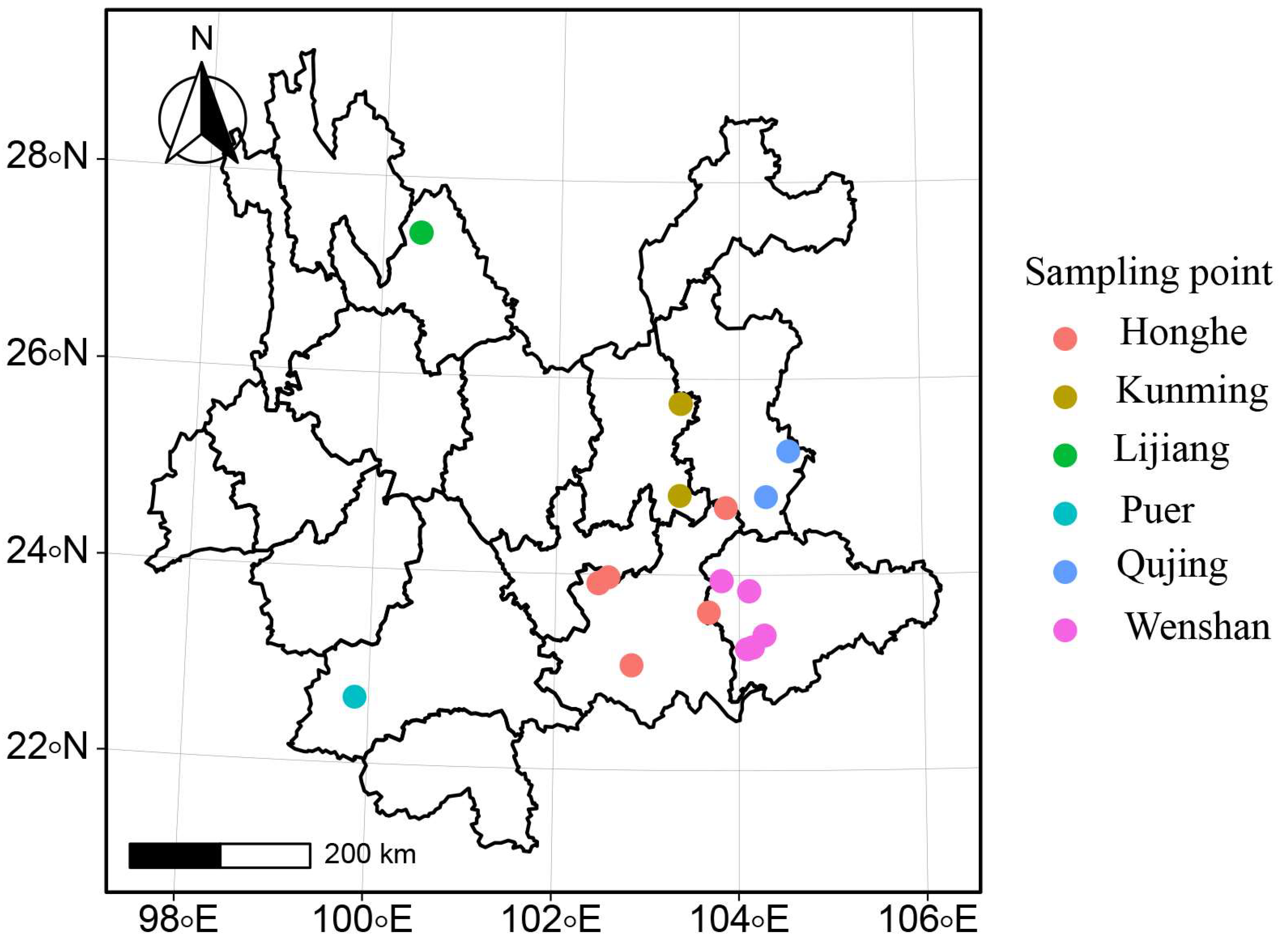
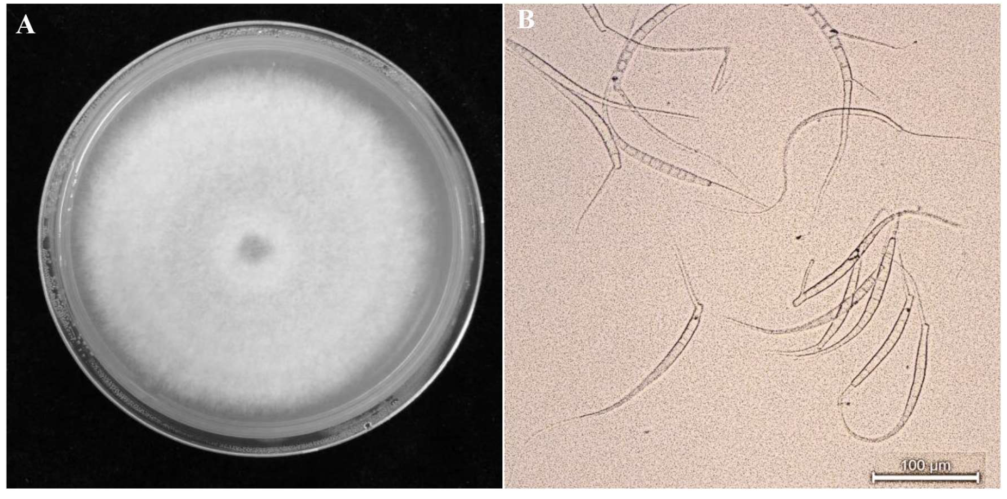
2.1. Strain Isolation and Observation

Leaves from P. notoginseng were collected from the six major production regions in Yunnan Province (i.e., Honghe, Wenshan, Qujing, Kunming, Lijiang, and Puer) (Figure 1, Supplementary Table S1). Plant tissue was washed and disinfected before being transferred onto the Potato Dextrose Agar (PDA) medium [31]. Plates were grown at 20 °C for 4 d, and M. acerina was identified based on morphological characteristics and internal transcribed spacer (ITS) sequence analysis.

2.2. DNA Extraction and Polymerase Chain Reaction Amplification of Internal Transcribed Spacer (ITS1/4) Regions

Genomic DNA was extracted from 0.2 g of mycelium using the Omega Fungi DNA Kit (Kunming Shuoqing Biological Engineering Technology Co. Ltd., Kunming, China) according to the manufacturer’s instructions. Amplification reactions were performed in a 20 μL volume containing 1 μL template DNA, 10 μL mix (DNA polymerase, Buffer, dNTP), 1 μL primer ITS1 (TCCGTAGGTGACCTGCGG), 1 μL primer ITS4 (TCCTCCGCTTATTGATATGC), and 7 μL ddH2O. Polymerase chain reaction (PCR) was performed using a T1 thermocycler (Biometra, Göttingen, Germany), with initial denaturation at 94 °C for 5 min, followed by 35 cycles of 94 °C for 45 s, 60 °C for 45 s, and 72 °C for 90 s, and a final extension at 72 °C for 10 min. Amplification products were separated by electrophoresis on 1% agarose gels in a 0.5× TAE buffer, using a 2000-bp DNA ladder as a DNA molecular weight marker. The PCR products were sequenced at Kunming Shuoqing Biological Engineering Technology Co. Ltd. Molecular Evolutionary Genetics Analysis (MEGA 5.1) was used to construct a phylogenetic tree based on the maximum likelihood method. Bootstrap values were evaluated using 1000 replications [32].

2.3. Simple Sequence Repeat Screening

Microsatellites were screened using MISA (http://pgrc.ipk-gatersleben.de/misa/, accessed on 18 May 2022) based on the whole genome data of M. acerina [33,34]. MISA is a script written in Perl language, which can identify SSRs from genome FASTA files [35]. In the SSR parameter settings, we defined that six, five, four, three, two, and one base were repeated five, five, five, five, six, and ten times.
The genome used in this study was sequenced using Hiseq 2500 from Illumina. After sequencing, two sets of 101 bp long and short paired-end short sequence data were generated. Mycocentrospora acerina produced 20.5 Gb of the original sequence after quality control filtering and removing the linker sequences, low-quality sequences, and low-sequence complexity sequences. The K-mer parameter was 75, and the final assembly result was obtained after automatic assembly and gap filling using SOAP de novo software (BGI, Shenzhen, China). The assembled size of M. acerina was 39 Mb. The N50 index is an evaluation index of the continuity of genome assembly. This value was calculated by sorting the contig sequence length from the largest to the smallest. The larger the N50 value, the better the continuity of the contig generated by the assembly. In this study, the M. acerina genome assembly contig N50 was 151 kb, the scaffold N50 was 567 kb, and the assembly quality was credible.

2.4. Primer Design

Microsatellite primers for the whole genome of M. acerina were designed in the PRIMER 3.0 (https://primer3.org/, accessed on 28 July 2023) website [36] based on the screened SSR results. Primers were synthesized by Shuoqing Biological Engineering Technology Co. Ltd.
For the initial screening, with 24 isolates from different sources, 118 SSR primers were designed and amplified with 20 μL PCR mixtures. Amplification products were separated on 2.5% agarose gels in 0.5× TAE buffer, using a 50-bp DNA ladder as a DNA molecular marker. Finally, the polymorphism of primers was assessed using CERVUS 3.0 [37], and the primers in which PIC exceeded 0.5 were retained for use in genetic diversity analyses [38].

2.5. SSR Analysis of Mycocentrospora acerina Genome

Based on the initial results, 14 primer pairs with high polymorphism in M. acerina populations were selected (MP30, MP36, MP47, MP50, MP51, MP56, MP61, MP62, MP63, MP68, MP84, MP92, MP113, and MP114) for use in amplification reactions. Stable and distinct fragments ranging in size from 100 bp to 800 bp were transformed into a binary character matrix (1 = presence, 0 = absence) [39,40].

2.6. Statistical Analysis

Genetic diversity parameters of each geographic population, including the observed number of alleles, effective number of alleles, Nei’s diversity index, Shannon’s information index, total gene diversity, intrapopulation gene diversity, the coefficient of gene differentiation, and gene flow [41], were calculated using POPGENE 32 (version 1.32) [42]. The NTSYS-pc (version 2.0) software was used to calculate the genetic similarity coefficient [43]. The phylogenetic tree was analyzed using an unweighted pair-group algorithm with arithmetic averages clustering analysis [44].

3. Results

3.1. Isolation and Identification of Strains

All isolates were identified based on their colonies/conidium (Figure 2A,B). Phylogenetic trees constructed based on ITS1/4 sequences showed that the isolates were grouped with M. acerina (Figure 3).

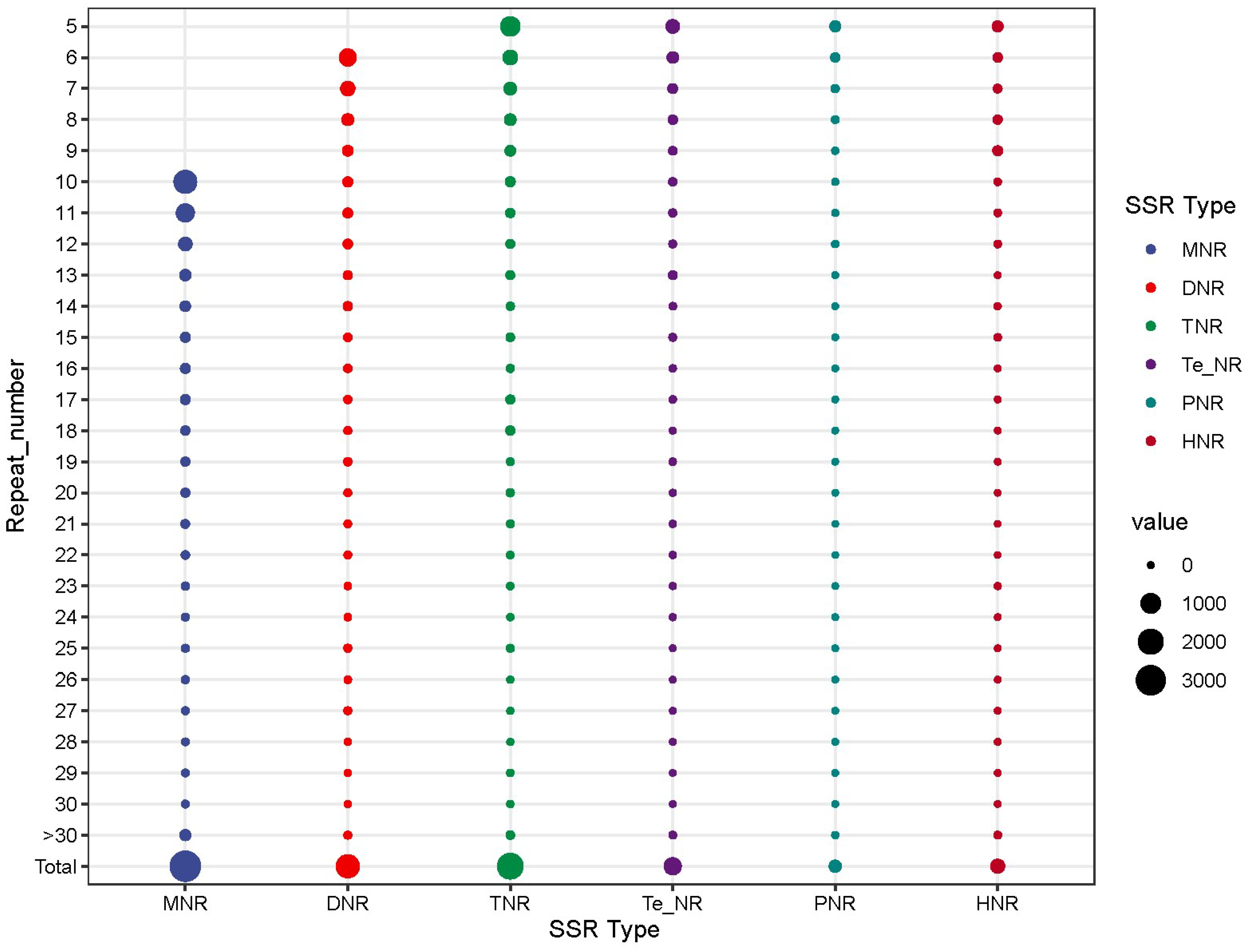
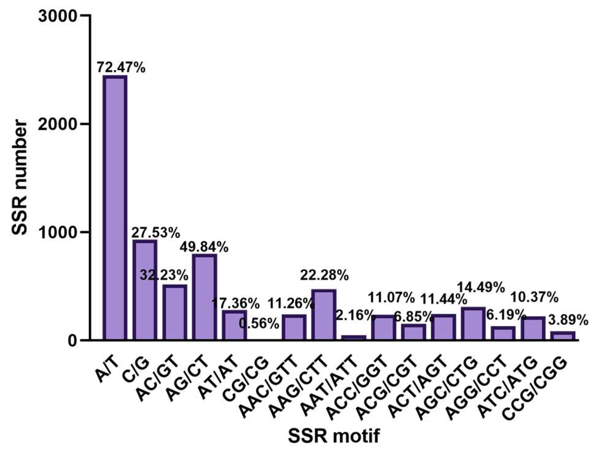
3.2. Genomic SSR Analysis

In the M. acerina genome, 8250 microsatellite sequences with 1 to 6 base repeats were obtained. The average length of simple sequence repeats (SSRs) was 26 bp.
Among the SSRs, there were 3379 mono-nucleotide repeats (40.96%), 1608 di-nucleotide (19.49), 2137 tri-nucleotide (25.90%), 641 tetra-nucleotide repeats (7.77%), 179 penta-nucleotide repeats (2.17%), and 306 hexa-nucleotide (3.71%). The maximum repetition times of each SSRs were 81, 40, 155, 217, 72, and 144 (Figure 4).
In M. acerina, 2449 T or A bases existed in mono-nucleotide SSR (72.47%) and 930 repeats with C or G bases (27.53%). Four types of di-nucleotide repeats were observed: AC (32.23%), AG (49.84%), AT (17.84%), and CG (0.56%). The most frequent types of tri-nucleotide repeats were AAC/GTT (11.26%), AAG/CTT (22.28%), and AAT/ATT (2.16%) (Figure 5).
There were 28 types of tetra-nucleotide repeats, among which ATCC had the highest content (12.48%), and CCCG had the lowest (0.16%). In addition, there were 61 penta-nucleotide repeats, with AACAC (6.7%) and AATCC (5.03%) being the two most common. Hexa-nucleotide repeats were the most abundant (103), with AACCCT being the most common, with 44 motifs (Supplementary Tables S2–S4).

3.3. Polymorphism of SSR Primers

Thirty-two pairs of primers had highly polymorphic loci ([PIC] > 0.5) and were used as SSR markers. According to the SSR primer data (Table 1), the average PIC was 0.649, the average allele number per locus was 5.147, the average proportion of locus types was 1.00, and the average expected heterozygosis (He) was 0.721.

3.4. Genetic Diversity within Populations

A total of 148 polymorphic bands were amplified from 187 populations of M. acerina using 14 SSR primers. The PIC ranged between 0.813 (MP56) and 0.946 (MP114), with an average PIC value of 0.885, all highly polymorphic (Figure S4). The total number is 264 (ranging from 14 on MP61 with the least number and 31 on MP114). The average number of alleles per locus was 18.857. The observed heterozygosis (Ho) of 14 loci was 0, and the expected heterozygosis (He) ranged from 0.831 to 0.951, with an average of 0.897.

3.5. Diversity between Populations

Nei’s genetic diversity (0.0896) and Shannon’s information index (0.1712) were higher in the Honghe population (HH), followed by those in the Puer population (LC) (Nei’s genetic diversity = 0.0893 and Shannon‘s information index = 0.1676). They were the lowest in Lijiang (Nei’s genetic diversity = 0.0842 and Shannon‘s information index = 0.143) (Table 2).
The observed allele number (Na) averaged 2.00, while the effective allele number (Ne) averaged 1.11; Nei’s gene diversity (h) was calculated to be 0.0908, and the average value of the Shannon diversity index was found to be 0.1761. The total genetic diversity (Ht) was estimated at 0.0909, with an intra-population genetic diversity (Hs) of 0.0884 and a genetic differentiation index (Gst) of 0.0277, indicating low inter-population genetic variation at only 2.77%. Notably, the estimated level of gene flow (Nm) stood at a substantial value of 17.5757, suggesting that gene flow has significantly contributed to the observed changes in genes across different regions; however, it is not considered the primary factor influencing the population’s genetic diversity. Among the six populations, Kunming and Honghe exhibited the highest genetic similarity (0.9988) and the smallest genetic distance (0.0012). Furthermore, Lijiang and Lancang displayed the lowest genetic similarity (0.9931) and the most significant genetic distance (0.007) (Table 3). Overall, there was minimal genetic divergence between these populations, with a high coefficient of genetic similarity close to 1, indicating a close genetic relationship among strains within each population and low levels of genetic differentiation between populations.

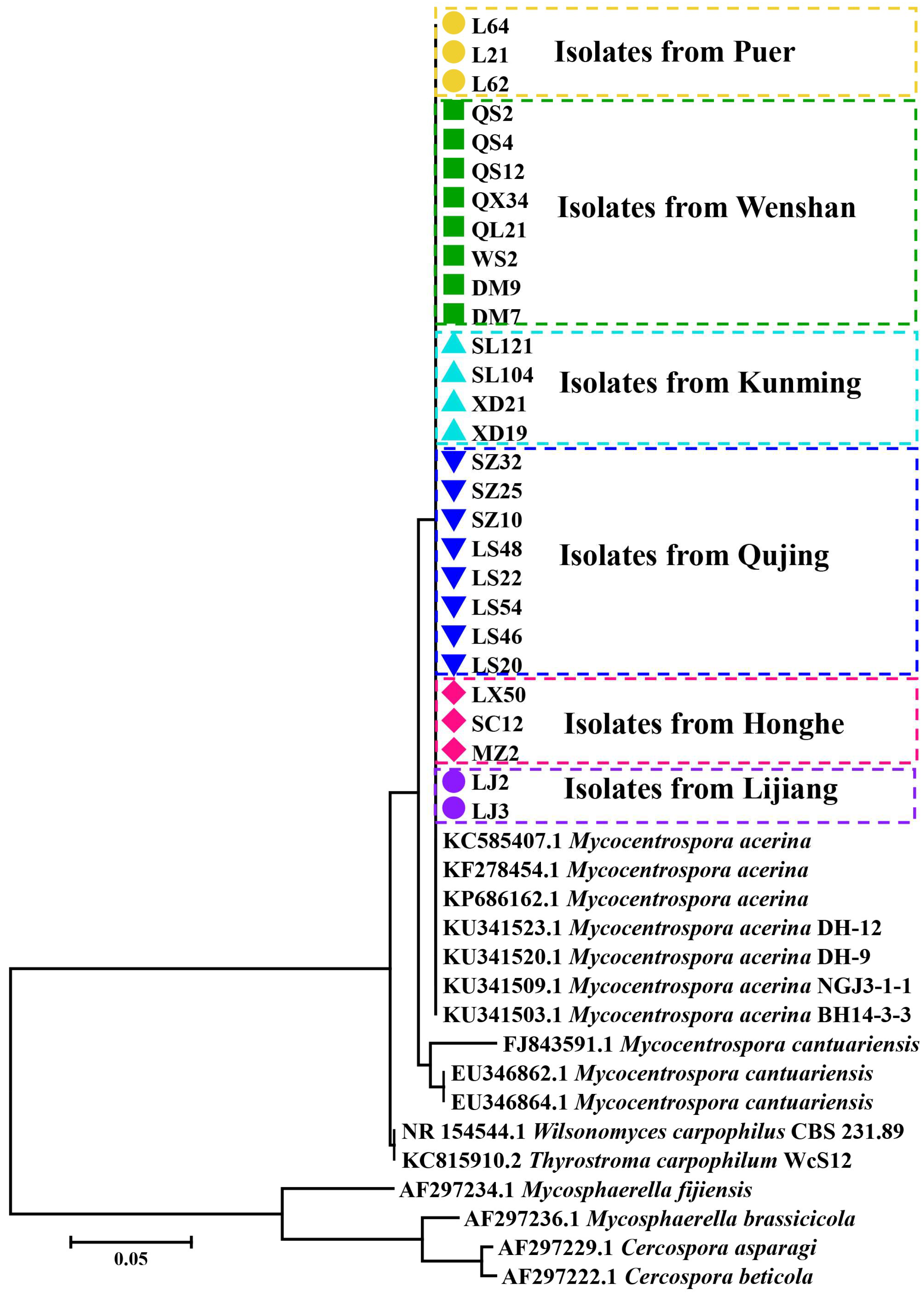
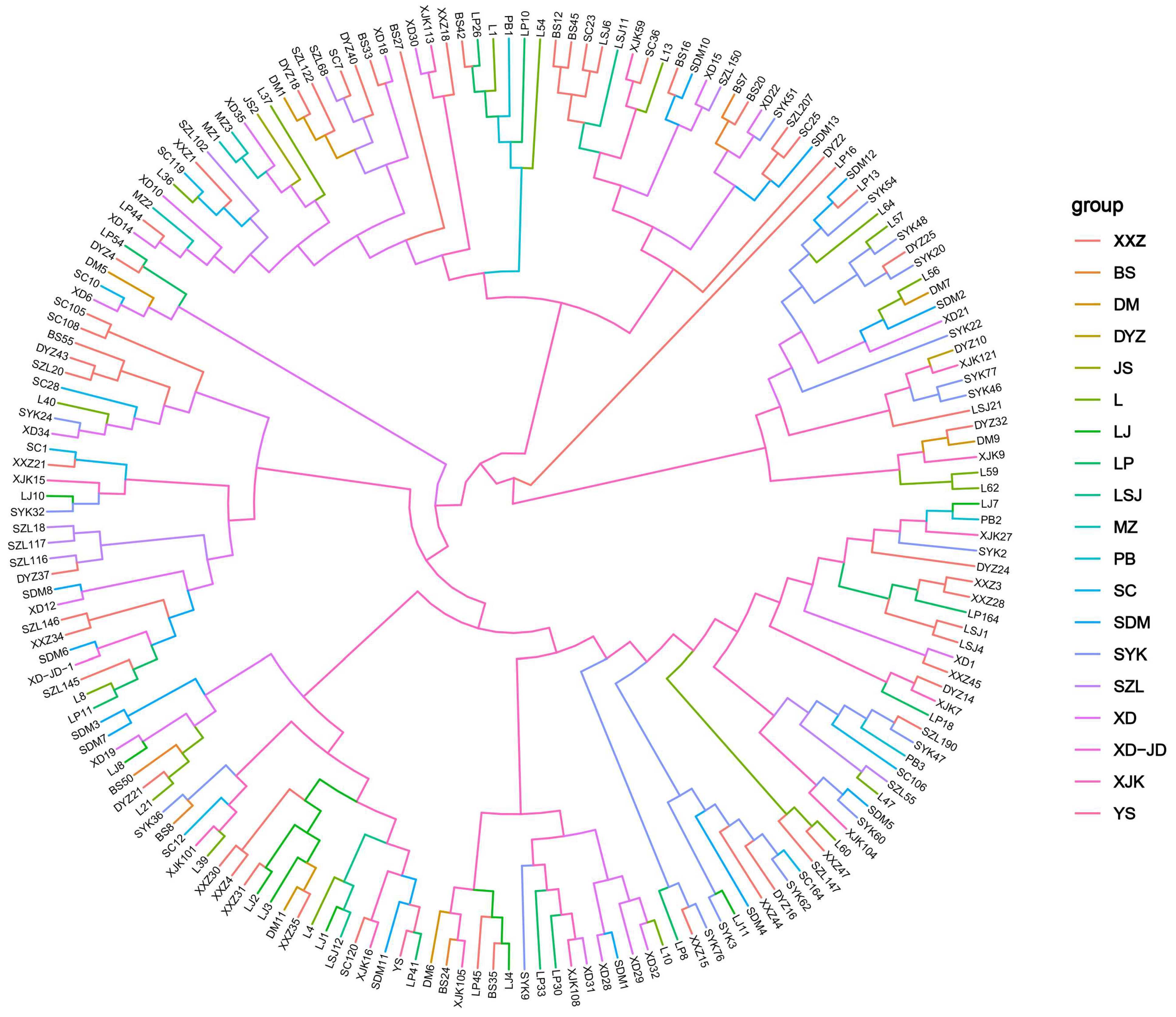
3.6. Cluster Analysis

Cluster analysis revealed that the similarity coefficient ranged between 0.83 and 0.97 (Figure 6).

4. Discussion

4.1. The Biology of M. acerina and Occurrence Regularity of Round Spot of P. notoginseng

In the 20th century, many studies on M. acerina focused mainly on host diversity and transmission methods [13,17]. In contrast, in the 21st century, studies have yet to be conducted on M. acerina. Sébastien Louarn studied the influence of M. acerina on the polyacetylenes and 6-methoxymellein in organic and conventionally cultivated carrots (Daucus carota) during storage [45]. Our laboratory (Key Laboratory of Agricultural Biodiversity and Pest Control of the Ministry of Education) has examined the biological characteristics of M. acerina, and much research has been done on the trait of spread in the field. It was found that M. acerina is a low-temperature-loving fungus. Its conidia will lyse when the temperature exceeds 32 °C. Its optimum growth temperature is 14–22 °C. The latest measured length of M. acerina is (137.36~486.24 μm) × (4.35~16.46 μm) (n = 100), and single conidia can cause infection (Supplementary Figures S1 and S2). M. acerina cause initial infection through chlamydospores stored in the soil and spread in the field through conidia on the surface of infected leaves, causing re-infection. Conidia are mainly spread by rain splash. These results show that the genetic distances between the M. acerina populations are relatively small, and the similarity is high. This may indicate frequent exchange activities between M. acerina from different regions, such as seedlings, cross-regional transportation, and other media dissemination. In addition, P. notoginseng round spot prevention and treatment mainly occur in the rainy season (June to September). Using the technology of facility cultivation to build a rain-proof film in the rainy season can prevent P. notoginseng round spots and reduce the use of chemical pesticides (Supplementary Figure S3). This reduction in the number of chemical pesticides can then reduce the survival pressure of M. acerina and ease the burden of prevention and treatment.

4.2. Features of SSR Loci in M. acerina

A total of 8250 repeats were obtained from the screened SSR sequences, indicating that the number of SSRs was higher in the M. acerina genome than in some eukaryotes [46,47]. The analysis of microsatellite sequences in M. acerina could enhance our understanding of its genome structure, especially the composition of non-coding regions, its mechanisms of pathogenicity, and its heredity at the genome level. Among all SSR types, A and T are abundant, which is consistent with the SSR loci results in most eukaryotic genomes, probably due to the transformation of methylated C residues into T residues [48]. According to Velascor [49], many short repeat sequences indicate a species has a high mutation frequency. In contrast, species with high proportions of long repeat motifs generally have relatively short evolutionary times or low mutation frequencies [49]. Many short repeats of single, dibasic, and tribasic bases were observed in the genome of M. acerina, suggesting this fungus had a relatively high mutation frequency or a relatively short evolutionary time [50].
With advancements in genome sequencing technologies, molecular marker studies have become more cost-effective [51]. Based on genomic data, we obtained 8250 SSRs, which accounted for 0.55% of the whole genome sequences. In the Fusarium graminearum genome, SSR sequences obtained accounted for 0.27% of the whole-genome sequences [52] and 0.21% in the Sphacelotheca reilianm genome [53]. In the present study, more than 100 pairs of primers were designed, of which merely 32 pairs were polymorphic, probably because most of the selected primers existed in the coding regions of the genome, with only a few located in the non-coding regions. Studies have demonstrated that SSRs in coding regions often exhibit low polymorphism. These markers should be designed as much as possible within non-coding regions because coding regions have much greater selection pressure than non-coding regions. Moreover, coding regions are relatively conserved during species evolution, while non-coding regions are more likely to evolve or mutate [54,55].

4.3. Genetic Diversity of M. acerina in Yunnan

In the current study, the PIC of polymorphic loci ranged from 0.53 to 0.8, which was high compared to the PIC in other eukaryotes. For example, PIC ranged from 0.3 to 0.4 in Dactylis glomerata L. [56], from 0 to 0.756 in Magnaporthe oryzae [57], and from 0.305 to 0.726 in Panonychus citri [58]. According to the results based on primer polymorphism, 14 SSR loci were used to analyze the genetic diversity of M. acerina populations. The PIC of screened primers was higher in these loci. However, after population analysis, the genetic diversity of M. acerina does not reflect geographic specificity. The potential reason is that the SSR primer loci are within the coding regions of the genome, which have high degrees of conservation [59]. Judicious selection of primers could improve the accuracy of results.
The genetic diversities of Pyricularia oryzae Cav. and Puccina striiformis f. sp. tritici in Yunnan Province have been reported to be high [60,61]. Therefore, the genetic diversity obtained for M. acerina in the study could be due to its background as a quarantine pest in China [62] or its stable survival in areas with highly homogenous ecological environments for prolonged periods. Some of the primary factors influencing the evolution of the population genetic structures of pathogenic fungi include population size, reproductive mode, and genetic drift [20,63,64,65]. This study included 187 M. acerina strains with a moderate population. The mode of reproduction of M. acerina in the field is asexual reproduction [66], which, to a certain extent, is not conducive to its genetic variation and the evolution of its populations. Pathogenic fungi are small individuals and easily experience genetic drift by natural or artificial means; gene drift is generally considered to hinder the evolution of organisms [67]. In the current study, there were no significant correlations in genetic diversity among strains from different geographical sources. Continuous selection and mutation of pathogenic genes will lead to homozygous individual genes, thus reducing the genetic diversity level of the M. acerina population. These factors partly explain that the isolates from the same region cannot be clustered into a group, and some isolates from different regions have very high genetic similarity coefficients. Another possible explanation is that the host P. notoginseng is only grown in Yunnan and Guangxi, China, and originates from Wenshan in Yunnan. The rest of the sampling points in the paper have been from P. notoginseng planted in the past 5–6 years. Current research shows that, due to the low genetic diversity of sample populations, we can effectively prevent diseases in this area through timely cleaning of diseased leaves, rain-proof cultivation, and alternate use of chemicals [68].

5. Conclusions

In this study, we developed 14 SSR primers of M. acerina with a good polymorphism that can be used in diversity analysis and identification of M. acerina and its closely related species. This result proved that the genetic diversity level of M. acerina was relatively low.

Supplementary Materials

The following supporting information can be downloaded at https://www.mdpi.com/article/10.3390/jof9090944/s1, Supplementary Figure S1: (A) Conidia of M. acerina artificially induced in a petri dish, the size of the conidia is (137.36~486.24 μm) × (4.35~16.46 μm) (n = 100). (B) The diseased spots of P. notoginseng leave four days after inoculation with single conidia. (C) Early lesions of round spot disease of P. notoginseng. Supplementary Figure S2: (A) The diseased spots 72 h after the inoculation of P. notoginseng leaves were sprayed with 5 × 10³ CFU/mL conidia suspension. (B) The lesions are magnified with an optical microscope. (C) The inoculated P. notoginseng leaves were stained with a fluorescent dye (Calcofluor white stain). Conidia can be seen on the lesion under the fluorescence microscope (Leica DM 2000). (D) Conidia produced at the site of a diseased spot, bar = 100 μm. Supplementary Figure S3: (A) P. notoginseng with a serious incidence of round spot disease without shelter from rain. (B) P. notoginseng cultivated in shelter from rain is growing well, and there is almost no occurrence of disease. We first developed M. acerina polymorphic microsatellite markers using CERVUS 3.0. Supplementary Figure S4: Representative result amplification pattern generated in part of Mycocentrospora acerina samples. Lanes M = DL50 marker; lanes 1 to 32 = partial M. acerina isolates. Supplementary Table S1: Mycocentrospora acerina populations examined in the simple sequence repeat analysis. Supplementary Table S2: Tetra-nucleotide simple sequence repeat (SSR) motifs in the whole genome of Mycocentrospora acerina. Supplementary Table S3: Penta-nucleotide simple sequence repeat (SSR) motifs in the whole genome of Mycocentrospora acerina.

Author Contributions

Methodology and formal analysis, H.W. and K.Y.; Writing—original draft, H.W.; Writing—review and editing, H.H. and L.G.; Funding acquisition, X.H. All authors have read and agreed to the published version of the manuscript.

Funding

This work was supported by China Agriculture Research System of MOF and MARA (CARS-21), Major Science and Technology Project of Yunnan and Kunming (202102 AE090042, 202204BI090003, 202205AF150018 and 2021JH002), National and Provincial Excellent Scientist Supporting Programme, and Innovative Research Team of Science and Technology in Yunnan Province (202105AE160016).

Institutional Review Board Statement

Not applicable.

Informed Consent Statement

Not applicable.

Data Availability Statement

Not applicable.

Acknowledgments

We would like to express our sincere appreciation to all teachers, students, and instrument platforms in our research group for their invaluable assistance.

Conflicts of Interest

The authors declare no conflict of interest.

References

  1. Duan, L.; Xiong, X.J.; Hu, J.Y.; Liu, Y.; Li, J.; Wang, J. Panax notoginseng saponins for treating coronary artery disease: A functional and mechanistic overview. Front. Pharmacol. 2017, 8, 702. [Google Scholar] [CrossRef] [PubMed]
  2. Wan, J.Y.; Fan, Y.; Yu, Q.T.; Ge, Y.-Z.; Yan, C.P.; Alolga, R.N.; Li, P.; Ma, Z.H.; Qi, L.W. Integrated evaluation of malonyl ginsenosides, amino acids and polysaccharides in fresh and processed ginseng. J. Pharm. Anal. 2015, 107, 89–97. [Google Scholar] [CrossRef] [PubMed]
  3. Su, L.J.; Ren, Y.C.; Chen, Z.; Ma, H.-F.; Zheng, F.; Li, F.; Zhang, Y.Y.; Gong, S.S.; Kou, J.P. Ginsenoside rb1 improves brain, lung, and intestinal barrier damage in middle cerebral artery occlusion/reperfusion(mcao/r) mice via the pparγ signaling pathway. J. Nat. Med. 2022, 8, 561–571. [Google Scholar]
  4. Xia, P.G.; Zhang, S.C.; Liang, Z.S.; Qi, Z.H. Research history and overview of chemical constituents of Panax notoginseng. Chin. Tradit. Herb. Drugs 2014, 45, 2564–2570. [Google Scholar]
  5. Long, Y.J.; Mao, Z.S.; Zhu, S.S.; Chen, Z.J.; Wei, F.G.; Yin, Z.B.; Zhu, Y.Y.; He, X.H. The pathogen of Panax notoginseng root rust spot. Mycosystema 2015, 34, 177–185. [Google Scholar]
  6. Ma, Y.N.; Chen, C.J.; Li, Q.Q.; Xu, F.R.; Cheng, Y.X.; Dong, X. Monitoring antifungal agents of Artemisia annua against Fusarium oxysporum and Fusarium solani, associated with Panax notoginseng root-rot disease. Molecules 2019, 24, 213. [Google Scholar] [CrossRef]
  7. Tang, B.F.; Li, X.; Pu, L.; Zhao, Q.; Cui, X.; Ge, F.; Liu, D. A pathogenesis-related protein 10 gene pnpr10–3 was involved in molecular interaction between Panax notoginseng and Fusarium solani. Australas. Plant Path. 2019, 48, 447–456. [Google Scholar] [CrossRef]
  8. Lu, N.; Chen, Y.J.; Lu, H.J.; Liu, Y.L. Biological characterization of round spot pathogen on Panax notoginseng. J. Yunnan Agric. Univ. 2015, 2, 193–195. [Google Scholar]
  9. He, C.; Wang, H.L.; Jin, X.; Jin, B.H.; Su, S.; Duan, Y.N.; He, X.H. Identification of Alternaria species associated with black spot disease on Panax ginseng in Yunnan and Guangxi. Acta Phytophys. Sin. 2020, 50, 246–250. [Google Scholar]
  10. Jiang, N.; Qin, L.Y.; Ye, Y.F. Research advances in diseases of Panax notoginseng. J. South. Agric. 2011, 2, 149–153. [Google Scholar]
  11. Li, Y.; Long, S.; Li, X. Occurrence of root rot of Panax notoginseng caused by Fusarium oxysporum in China. Int. J. Agric. Biol. 2018, 20, 2175–2180. [Google Scholar]
  12. Dai, L.; Xu, Y.L.; Long, Y.J.; Du, L.S.; Du, Y.L.; He, X.H. The growth characteristic of mycelia and conidia for Mycocentrospora acerina. J. Yunnan Agric. Univ. (Nat. Sci.) 2017, 32, 27–35. [Google Scholar]
  13. Wall, C.J.; Lewis, B.G. Infection of carrot plants by Mycocentrospora acerina. Trans. Br. Mycol. Soc. 1980, 74, 587–593. [Google Scholar] [CrossRef]
  14. Davies, W.P.; Lewis, B.G.; Day, J.R. Infection of stored carrot roots by Mycocentrospora acerina (hartig) Deighton. Trans. Br. Mycol. Soc. 1977, 77, 139–151. [Google Scholar] [CrossRef]
  15. Garfinkel, A.R.; Chastagner, G.A. Strategies to Address Emerging Fungal Diseases in Peony (Paeonia lactiflora) in the United States; Horticultural Science: Korbeek-Lo, Belgium, 2019; pp. 199–206. [Google Scholar]
  16. Ellis, M.B. Dematiaceous hyphomycetes in Britain. In Transactions of the British Mycological Society; Cambridge University Press: London, UK, 1974. [Google Scholar]
  17. Hermansen, A. Weeds as hosts of Mycocentrospora acerina. Ann. Appl. Biol. 2010, 121, 679–686. [Google Scholar] [CrossRef]
  18. Wang, C.W.; Fu, J.F.; Wang, W.K. Brief report on the study of a new disease- the leaf withering disease of Asarum. J. Shenyang Agric. Univ. 1992, 23, 355–356. [Google Scholar]
  19. Chen, K.; Chen, S.X.; Yu, Z.W. A new disease on sanqi. Plant Prot. 1997, 1, 49. [Google Scholar]
  20. Cumagun, C. Population genetic analysis of plant pathogenic fungi with emphasis on Fusarium species. Philipp. Agric. Sci. 2007, 90, 244–256. [Google Scholar]
  21. Tsedaley, B.A. Review on Disease Detection, Pathogen Identification and Population Genetics in Fungi. J. Biol. Agric. Healthc. 2015, 5, 6–20. [Google Scholar]
  22. Salama, N.; Guillemin, K.; Mcdaniel, T.K.; Sherlock, G.; Tompkins, L.; Falkow, S. Whole-genome microarray reveals genetic diversity among Helicobacter pylori strains. Proc. Natl. Acad. Sci. USA 2000, 97, 14668–14673. [Google Scholar] [CrossRef]
  23. Zhang, Y.Y.; Ren, Z.H.; Liu, P.K.; Zhou, H.; Wang, H.H.; Liu, E.M. Analysis of genetic diversity of Magnaporthe oryzae in rice blast nursery in Taojiang of Hunan by SSR markers. Southwest China J. Agric. Sci. 2018, 31, 725–730. [Google Scholar]
  24. Sreenivasaprasad, S.; Talhinhas, P. Genotypic and phenotypic diversity in Colletotrichum acutatum, a cosmopolitan pathogen causing anthracnose on a wide range of hosts. Mol. Plant Pathol. 2010, 6, 361–378. [Google Scholar] [CrossRef] [PubMed]
  25. Barus, H.; Bayu, E.; Hanafiah, D.S. Identification genetic of soybean mutant (Glycine max L. Merril) based on fatty acid characters using simple sequence repeat (SSR) markers. In Proceedings of the 3rd International Conference Community Research and Service Engagements 2019, North Sumatra, Indonesia, 4 December 2019. [Google Scholar]
  26. Adjebeng-Danquah, J.; Manu-Aduening, J.; Asante, I.K.; Agyare, R.Y.; Gracen, V.; Offei, S.K. Genetic diversity and population structure analysis of Ghanaian and exotic cassava accessions using simple sequence repeat (SSR) markers. Heliyon 2020, 6, e03154. [Google Scholar] [CrossRef]
  27. Crauwels, S.; Zhu, B.; Steensels, J.; Busschaert, P.; Lievens, B. Assessing genetic diversity among Brettanomyces yeasts by DNA fingerprinting and whole-genome sequencing. Appl. Environ. Microbiol. 2014, 80, 4398–4413. [Google Scholar] [CrossRef] [PubMed]
  28. Colling, G.; Matthies, D. The effects of plant population size on the interactions between the endangered plant Scorzonera humilis, a specialised herbivore, and a phytopathogenic fungus. Oikos 2010, 105, 71–78. [Google Scholar] [CrossRef]
  29. Mcgrath, M.T.; Shishkoff, N. Resistance to Triadimefon and Benomyl: Dynamics and Impact on Managing Cucurbit Powdery Mildew. Plant Dis. 2001, 85, 147–154. [Google Scholar] [CrossRef] [PubMed]
  30. Fernández-Ortuño, D.; Pérez-García, A.; López-Ruiz, F.; Romero, D.; Vicente, A.D.; Torés, J.A. Occurrence and distribution of resistance to Qoi fungicides in populations of podosphaera fusca in south central Spain. Eur. J. Plant Pathol. 2006, 115, 215–222. [Google Scholar] [CrossRef]
  31. Wang, W.Y.; Zhao, C.L.; Chen, Z.J.; Wen, G.S.; Wei, F.G.; Long, T.G.; Li, S.W. Studies on the isolation, identification and in vitro growth rates of the three pathogenic fungi from Panax notoginseng cultivated in the Wenshan eparchy. Agric. Sci. Technol. 2015, 16, 1165. [Google Scholar]
  32. Li, H.; Li, J.; Xu, R.Y.; Song, F.S.; Li, L.; Wei, P.C.; Yang, J.B. Isolation of five rice non-endosperm tissue-expressed promoters and evaluation of their activities in transgenic rice. Plant Biotechnol. J. 2018, 16, 1138–1147. [Google Scholar] [CrossRef]
  33. Wang, Z.; Zheng, Q.M.; Cheng, W.T.; Mao, Y.Y.; Cai, Y.Q.; Ma, Y.H. Information analysis and molecular marker development of transcriptome SSR in Pitaya. Guizhou Agric. Sci. 2018, 46, 1–5. [Google Scholar]
  34. Wang, X.; Chen, L.; Zhao, C.L. Mining SSR Molecular Marker Sites with MISA Tool for Different Types of Sequences. Chin. Agric. Sci. Bull. 2016, 32, 150–156. [Google Scholar]
  35. Xu, D.L.; Chen, H.B.; Aci, M.; Pan, Y.H.; Shangguan, Y.N.; Ma, J.; Li, L.; Qian, G.; Wang, Q.X. De Novo assembly, characterization and development of EST-SSRs from Bletilla striata transcriptomes profiled throughout the whole growing period. PLoS ONE 2018, 13, e0205954. [Google Scholar] [CrossRef] [PubMed]
  36. Hou, L.X.; Wei, A.Z.; Wang, L.H.; Liu, Y.L. Analysis of SSR loci and development of molecular markers in Zanthoxylum bungeanum transcriptome. J. Agric. Biotechnol. 2018, 26, 1226–1236. [Google Scholar]
  37. Azam, A.; Zahra, Z.; Zohreh, S.; Alireza, K.; Morteza, K.; Sirous, Z. Characterization and haplotype study of 6 novel STR markers related to the KCNQ1 gene in heterogeneous cardiovascular disorders in the Iranian population. Turk. J. Med. Sci. 2019, 49, 453–457. [Google Scholar]
  38. Zhou, X.J.; Wang, H.L.; Li, F.L.; Zhang, K.; Wang, Y.N.; Ya, H.Y. Development of polymorphic ssr markers in Rhododendron henanense subsp. lingbaoense based on rad-seq. J. Agric. Biotechnol. 2019, 27, 55–62. [Google Scholar]
  39. Zeng, X.Q.; Wang, Y.L.; Xu, Q.J.; Yuan, H.J.; Zaxi, L.B.; Nima, Z.X. Assessment of genetic diversity in Tibetan hulless barley germplasm (Hordeum vulgare L. var. nudum hk.f.) by SSR primers. Tcrop 2013, 33, 260–267. [Google Scholar]
  40. Ei-Komy, H.M.; Saleh, A.A.; Molan, Y.Y. Molecular characterization of early blight disease resistant and susceptible potato cultivars using RAPD and SSR markers. Afr. J. Biotechnol. 2012, 11, 37–45. [Google Scholar] [CrossRef]
  41. Yu, Z.F.; Yan, X.W.; Zhang, Y.H.; Yang, F.; Zhang, G.F. Genetic diversity analysis of different age of a Dalian population of the Manila clam Ruditapes philippinarum by EST-SSR. Acta Ecol. Sin. 2012, 32, 4673–4681. [Google Scholar]
  42. Liu, R.; YANG, J.S. Genetic diversity of some Dendranthema spp. based on RAPD analysis. J. Agric. Univ. Heb. 2010, 33, 60–65+83. [Google Scholar]
  43. Gaikwad, A.B.; Behera, T.K.; Singh, A.K.; Chandel, D.; Karihaloo, J.L.; Staub, J.E. Amplified fragment length polymorphism analysis provides strategies for the improvement of bitter gourd (Momordica charantia L.). Acta Hortic. 2008, 871, 71–78. [Google Scholar] [CrossRef]
  44. Wang, Z.J.; Wu, W.; Hu, B.; Zhang, H.L.; Bai, X.; Zhao, J.J.; Zhang, L.; Yan, X.J. Molecular epidemiology of Aleutian mink disease virus in China. Virus Res. 2014, 184, 14–19. [Google Scholar] [CrossRef] [PubMed]
  45. Louarn, S.; Nawrocki, A.; Edelenbos, M.; Dan, F.J.; Jensen, O.N.; Collinge, D.B.; Jensen, B. The influence of the fungal pathogen Mycocentrospora acerina on the proteome and polyacetylenes and 6-methoxymellein in organic and conventionally cultivated carrots (Daucus carota) during post-harvest storage. J. Proteom. 2012, 75, 962–977. [Google Scholar] [CrossRef]
  46. Luo, H.; Wang, X.; Zhan, G.; Wei, G.; Zhou, X.; Zhao, J.; Huang, L.; Kang, Z. Genome-Wide Analysis of Simple Sequence Repeats and Efficient Development of Polymorphic SSR Markers Based on Whole Genome Re-Sequencing of Multiple Isolates of the Wheat Stripe Rust Fungus. PLoS ONE 2015, 10, e0130362. [Google Scholar] [CrossRef]
  47. Sharapova, N.; McMullen, M.D.; Schultz, L.; Schroeder, S.; Sanchez-Villeda, H. Development and mapping of SSR markers for maize. Plant Mol. Biol. 2002, 48, 463–481. [Google Scholar] [CrossRef] [PubMed]
  48. Toth, G. Microsatellites in Different Eukaryotic Genomes: Survey and Analysis. Genome Res. 2000, 10, 967. [Google Scholar] [CrossRef] [PubMed]
  49. Velasco, R.; Zharkikh, A.; Troggio, M.; Cartwright, D.A.; Viola, R. A high-quality draft consensus sequence of the genome of a Heterozygous grapevine variety. PLoS ONE 2007, 2, 1326. [Google Scholar] [CrossRef]
  50. Sia, E.A.; Kokoska, R.J.; Dominska, M.; Greenwell, P.; Petes, T.D. Microsatellite instability in yeast: Dependence on repeat unit size and DNA mismatch repair genes. Mol. Cell. Biol. 1997, 17, 2851–2858. [Google Scholar] [CrossRef] [PubMed]
  51. Mccouch, S.R.; Teytelman, L.; Xu, Y.; Katarzyna, B.L.; Karen, C.; Mark, W.; Fu, B.Y.; Maghirang, R.; Li, Z.K.; Xing, Y.Z.; et al. Development and mapping of 2240 new SSR markers for rice (Oryza sativa L.) (supplement). DNA Res. 2002, 9, 257–279. [Google Scholar] [CrossRef] [PubMed]
  52. Cho, Y.G.; Ishii, T.; Temnykh, S.; Chen, X.; Lipovich, L.; Mccouch, S.R.; Park, W.D.; Ayres, N.; Cartinhour, S. Diversity of microsatellites derived from genomic libraries and GenBank sequences in rice (Oryza sativaL.). Theor. Appl. Genet. 2000, 100, 713–722. [Google Scholar] [CrossRef]
  53. Lu, X.W.; Brewbaker, J.L. Molecular mapping of QTLs conferring resistance to Sphacelotheca reiliana (kühn) clint. Maize Genet. Coop. Newsl. 1999, 73, 36. [Google Scholar]
  54. Liu, C.; Li, J.; Qin, G. Genome-wide distribution of simple sequence repeats in pomegranate and their application to the analysis of genetic diversity. Tree Genet. Genomes 2020, 16, 1–9. [Google Scholar] [CrossRef]
  55. Wei, H.L.; Edo, K. Evolutionary pressures on simple sequence repeats in prokaryotic coding regions. Nucleic Acids Res. 2011, 6, 6. [Google Scholar]
  56. Xie, W.G.; Zhang, X.Q.; Ma, X.; Peng, Y.; Huang, L.K. Genetic variation and relationship in orchard grass (Dactylis glomerata l.) germplasm detected by SSR markers. Hereditas 2009, 31, 654–662. [Google Scholar] [PubMed]
  57. Zhou, Y.Y.; Wang, L.Q.; Fu, Y.L.; Yang, L.Q. A review of research on biological genetic resources and traditional knowledge. J. Northeast Agric. Univ. 2017, 16, 60–70. [Google Scholar]
  58. Sun, T.; Kong, L.W.; Wang, M.M.; Peng, Y.; Hong, X.Y. Development and characterization of novel EST-microsatellites for the citrus red mite, Panonychus citri (Acari: Tetranychidae). Syst. Appl. Acarol. 2014, 14, 499. [Google Scholar]
  59. Sun, T.T.; Wang, D.W.; Chen, L.; Wang, W.F.; Gong, D.P.; Chen, Y.Q.; Sun, Y.H. Characteristics and distribution of SSR loci in the coding region of potato genome and functional analysis of their encoding proteins. Mol. Plant Breed. 2015, 13, 171–177. [Google Scholar]
  60. Liu, X.F.; Yuan, W.Y.; Liang, D.; Shi, X.W.; Ma, Z.H.; Tian, J. Population genetic structures of Puccinia striiformis f. sp.tritici in Yunnan and Guizhou province. J. Yunnan Agric. Univ. (Nat. Sci.) 2016, 49, 95–100. [Google Scholar]
  61. Iwano, M.; Lee, J.L.; Lee, C.Y. Distribution of pathogenic races and changes in virulence of rice blast fungus, pyricularia oryzae cav. in Yunnan province, China. Jarq 1990, 23, 241–248. [Google Scholar]
  62. Wu, H.; Liu, B.L.; Wang, W.H.; Shi, Y.M.; Wei, S.F.; Zhou, H. Genetic diversity analysis of male sterile lines for three-line hybrid rice mainly applied in Guangxi. J. South. Agric. 2015, 46, 550–554. [Google Scholar]
  63. Mao, Y.X.; Zhang, X.L.; Hu, Z.J.; Zhao, Y. Genetic diversity of Trachycarpus fortunei (Hook.) H. Wendl germplasm resources in Guizhou by SRAP molecular markers analysis. J. South. Agric. 2020, 51, 27–35. [Google Scholar]
  64. Li, Y.F.; Zhang, X.L.; Liao, X.G.; Zhang, D. Evaluation study of multiplex PCR assay for detection diarrheagenic Escherichia coli. Henan J. Prev. Med. 2014, 2, 40. [Google Scholar] [CrossRef]
  65. Ferrucho, R.L.; Ceresini, P.C.; Ramírez-Escobar, U.M.; Mcdonald, B.A.; Domínguez, C.G. The population genetic structure of rhizoctonia solani AG-3PT from potato in the Colombian Andes. Phytopathology 2013, 103, 862–869. [Google Scholar] [CrossRef]
  66. Evenhuis, A.; Verdam, B.; Zadoks, J.C. Splash dispersal of conidia of Mycocentrospora acerina in the field. Plant Pathol. 2010, 46, 459–469. [Google Scholar] [CrossRef]
  67. Mcdermott, J.M.; Mcdonald, B.A. Gene flow in plant pathosystems. Annu. Rev. Phytopathol. 2003, 31, 353–373. [Google Scholar] [CrossRef]
  68. Wang, H.L.; Wang, F.; Jin, B.H.; Zhang, H.; Yang, K.; Wang, W.P.; Yang, M.; Zhu, S.S.; He, X.H. Sensitivity and fitness analysis of round spot of Panax notoginsen Mycocentrospora acerina to azoxystrobin, prochloraz and difenoconazole. Chin. J. Pestic. Sci. 2019, 21, 273–278. [Google Scholar]
Figure 1. The geographical locations of six Mycocentrospora acerina populations in Yunnan.
Jof 09 00944 g001
Figure 2. Mycocentrospora acerina colonies (A), conidia (B).
Jof 09 00944 g002
Figure 3. Phylogenetic tree constructed based on internal transcribed spacer 1 (ITS1) sequences.
Jof 09 00944 g003
Figure 4. Distributions and frequencies of simple sequence repeats (SSRs) in Mycocentrospora acerina. mono-nucleotide repeats, DNR: di-nucleotide repeats; TNR: tri-nucleotide repeats, Te-NR: tetra-nucleotide repeats, PNR: penta-nucleotide repeats, HNR: hexa-nucleotide repeats.
Jof 09 00944 g004
Figure 5. Motifs of mono-, di-, and tri-nucleotide simple sequence repeats (SSRs) in the whole genome of Mycocentrospora acerina.
Jof 09 00944 g005
Figure 6. Cluster analysis of 187 Mycocentrospora acerina strains in Yunnan province based on the genetic similarity coefficient. Different colors mean different sampling locations. SZL (Jianshui), BS (Luxi), JS (Jianshui), MZ (Mengzi), LP (Longpeng), and SC (Shaochong) were the isolates from Honghe. YS (Yanshan), WS (Wenshan), LSJ (Qiubei), SDM (Qiubei), XXZ (Qiubei), DM (Dumeng), and PB (Wenshan) were the fungus from Wenshan. DYZ and SYK were the isolates from Qujing. XD (Xundian), XD-JD (Xundian), and XJK (Shilin) were the fungus from Kunming. LJ was the fungus from Lijiang.
Jof 09 00944 g006
Table 1. Primers of the whole genome of Mycocentrospora acerina.
Locus
(Accession Number)
Repeat MotifPrimer SequenceSize (bp)Expected
Heterozygosity (He)
Polymorphic Information Content (PIC)Annealing Temperature (Tm)
MP40
(OM824348)
(CTA)27F: CACATGCTCAGTCATTTGTGG2430.6810.59956
R: GGTGCAATCGGAAAGAATTG
MP42
(OM824349)
(TC)19F: AAGCGCACTTGCCTATTGAT2250.6090.5358.4
R: GGTGAGTGTTGCTGACGAAA
MP2
(OM824341)
(CAT)14F: CGTCCATCTTCCTCTTCACC2000.710.62256.4
R: GCTCATGTTCGATGGATGTG
MP49
(OM824352)
(GA)25F: GGAAGGAAATCCAGGTGTGA2000.5940.50556
R: CCCACTTCCTGTTTGCTTGT
MP54
(OM824356)
(CAA)19F: GTTGTTGCCAGCAAGAGTGA1990.6230.55258.4
R: AACAACCCTGGCACTACTCG
MP55
(OM824357)
(TTGA)19F: TTCCTCTCCCTCTCCCTCTC2140.6230.55259
R: ATGCTGCAAGTCTGTTGACG
MP56
(OM850345)
(TAG)25F: TGTGTGTGTGTTGTTGTTGTTG2280.870.81259.5
R: TGACAAGCAAGTAGATTTTTACGTTT
MP4
(OM824343)
(GACA)6F: AGGGTAGCTCAAAGCCACTG2730.7250.66558.4
R: CTTTCCAAGCTGAGGGTGAG
MP58
(OM824358)
(GA)25F: TCGTTTTTGGAGCGTTCTTT2090.7390.65954.4
R: TGGACGCACTCCTTCTTTTC
MP52
(OM824355)
(CCA)11F: GCTTCGGTGTCTGGAATCAT1790.6090.5356.4
R: AAACTTCAATGTCGCCAAGG
MP51
(OM824354)
(TTG)19F: CGTCTCTGTTATTGCTGCTTT1590.8260.7655.7
R: CGCACAACCAATGAGAAACA
MP13
(OM824344)
(AGT)12F: CACGTCACGGAGCAAGTAGA2110.6960.62257.4
R: TGATGAGGTCCAACGGAGAT
MP30
(OM824342)
(GT)13F: CATGTGCATTGCTGTGTTGT1700.7250.64461.4
R: CAGCGAGTGAATGGAAGTGA
MP50
(OM824353)
(CTGT)19F: GCTTTACTTTGCCCGTCTGT2000.7680.70155.4
R: TGCATCTCCTCACATCCATC
MP65
(OM824362)
(CA)22F: ACCTCCACACCTGCACCTAC2450.6090.5359.5
R: GCGGGCTTGTAGTCGTAGAG
MP68
(OM824363)
(CAT)7F: GGATATGCCTCACCATTTGC1660.7970.73955.4
R: ATATGGAAGGCCGCAGTGTA
MP83
(OM824364)
(CTT)12
(ATGA)10
F: TGAGCAGGGGCCAAATACTA1560.7790.70254.4
R: TTAAATTCCCATCCCCATCC
MP36
(OM824346)
(CAT)17F: ATCTGTCACCACCATCACCA1930.870.81259
R: AGCTCGCGATCTAAACATCC
MP39
(OM824347)
(AGTG)24F: ATGTGTGTGTGTGCCTGGAT2470.5940.50560
R: TATATGCCCATTCCCATTCC
MP46
(OM824350)
(CACT)10F: TTCCTCTGACGCATCCTCTT2070.6380.53560
R: TGGGCATGTAATGAGTGGTG
MP47
(OM824351)
(CAGG)7F: GATTGTAAGCCGCAGAAGGT2470.7540.6860
R: TCACGACTCCATCACTCCAA
MP20
(OM824345)
(TACA)11F: TGTGTCGCTCACTCACTCAA2390.7540.67159
R: GGAAGGAGTGGAGTTGATGG
MP90
(OM824366)
(TC)18F: TCAAAACCGAAACCCAGAAA1910.5510.50355.4
R: GGGAGAAGAAGGGCAGAGG
MP108
(OM824368)
(TCG)10
(TCA)5
F: TCACTACCCCTACCCCCTTT2370.6810.59957.4
R: CGGTCGGCATAGGGTATTTA
MP92
(OM824367)
(CTA)31F: ACCCCAACACTCAATCATCC2190.710.64354.7
R: TCTGGCAAGAAGAAGAAATGC
MP62
(OM824360)
(CTA)20F: CAGAAAATCCTAGCTACTGCTGCT1740.7250.64456.5
R: TGCAGTCTCTTCACCCTGTTT
MP84
(OM824365)
(AGA)18F: TTCAATCGTGCAAGGTGTGT1670.7390.68658
R: GAGAGGAGCAGGGCATGTAG
MP115
(OM824371)
(AC)7(TC)12F: TCTGCTGCCATGTAGTGCTC2460.7680.69255.4
R: ATGTGATTTTGGGGGAAACA
MP63
(OM824361)
(TC)21F: CAGACTTCCCAGTCACCACA1950.7970.72655.5
R: TTGGCTACTACTGCACCAAAAA
MP113
(OM824369)
(TG)7(AG)10F: CATCTCTCATCTCCCCAGGA2250.8120.74657.4
R: AATCCCATCACACGCTTCTC
MP114
(OM824370)
(CTC)9
(TTC)8
F: GATGTGCAGAGTTTCGGTCA2320.9130.86255.4
R: GGAAGCTGATTCATCCCAGT
MP61
(OM824359)
(CA)36F: TGGTGGCTAGTTGGTTGGAT2120.9280.87856.4
R: GGTCGTCACTGTTGCTTGAA
Table 2. Genetic diversity of the geographic populations of Mycocentrospora acerina.
PopulationNaNeNei’s Genetic DiversityShannon’s Information Index
Qujing1.68921.11080.08880.1658
Honghe1.80411.10890.08960.1712
Kunming1.72971.11030.08910.1674
Puer1.68921.10990.08930.1676
Lijiang1.41221.11870.08420.143
Wenshan1.7771.10990.08930.1693
Table 3. Nei’s genetic identity (upper diagonal) and genetic distance (lower diagonal) of the geographic populations of Mycocentrospora acerina.
popIDHongheLijiangPuerKunmingWenshanQujing
Honghe 0.99380.99760.99880.99820.9977
Lijiang0.0062 0.99310.99370.99590.9946
Puer0.00240.007 0.99750.9980.9978
Kunming0.00120.00640.0025 0.99790.9975
Wenshan0.00180.00410.0020.0021 0.9983
Qujing0.00240.00540.00220.00250.0017
Disclaimer/Publisher’s Note: The statements, opinions and data contained in all publications are solely those of the individual author(s) and contributor(s) and not of MDPI and/or the editor(s). MDPI and/or the editor(s) disclaim responsibility for any injury to people or property resulting from any ideas, methods, instructions or products referred to in the content.

