SlideShare a Scribd company logo
1 of 14
Pathogenic Fungi :
MICROSPORUM
Presented by:
Neha kausar Nasim
sheikh
CONTENT
• Introduction
• Morphological characteristics
• Pathogenesis
• Laboratory diagnosis
• Treatment
INTRODUCTION
• Microsporum is the genus of fungi comes under Dermatophytes
group
• Dermatophytes- it is a group of three types of fungus i.e.
Microsporum, Trichophyton and Epidermophyton that causes skin
diseases in Humans and Animals.
• Microsporum fungi infects only keratinized tissue like skin, hair
and Nails.
• They are restricted to non-viable skin because they are unable to
grow at 37 degree Celsius.
• Enzymes which helps the fungi are Proteases, Elastase,
Keratinases. These are degradative enzymes which degrade
keratinized tissue and serves as nutrients for the fungi.
• Natural habitat of some microsporum spp. Is soil(geophilic) others
primarily affect animals(zoophilic) or human(Anthropophilic).
• The infection caused by Microsporum are known as ringworm or
tinea , Though infections are not caused by worms but it appear as
snake-like on skin, hence named ringworm infection.
MORPHOLOGICAL CHARECTERISTICS
• Microsporum can be identified by their colonial
appearance and microscopic morphology after
growth for 2weeks at 25 degree Celsius on
Sabouraud’s dextrose agar.
• Microsporum species tend to produce distinctive
multicellular machroconidia with echinulate walls.
• Microsporum forms both Macroconidia and
Microconidia on short conidiosphores(hyphae).
MACROCONIDIA MICROCONIDIA
Large asexual
reproductive structure.
Small asexual
reproductive structure.
Hyaline, Multi sepate(2-
15)
Hyaline, single septate
Spindal shaped Club shaped
Rough and Thick walled Smooth and Thin walled
8 to 15 X 40 to 150 µ m 2 to 3 µ m
Microsporum canis
• Colonies :- white cottony surface and
deep yellow color on reverse, Thick
walled.
• On the rice grains, a deep yellow pigment
is produced by M.canis which helps to
differentiate it from M.audouinii
• Macroconidia are abundant. Thick walled
with many septa up to 15 . Macroconidia
are often hooked or curved at ends.
• Microconidia are small and club-shaped.
• In-vitro hair perforation test :- Positive
• M.canis causes tinea capitis and tinea
corporis. Tinea barbae, tinea mannum.
Microsporum audouinii
• Colonies :- dense fur like mat having radiating
edges, Grayish white to salmon pink colored
colonies.
• Pectinate hyphae and rare macroconidia and
microconidia.
• Anthropophilic.
• Hair and skin infected with M.auduinii gives
fluorescence under wood’s UV light.
• In-vitro hair perforation test :- Negative
• It causes tinea capitis and tinea corporis
Microsporum
gypseum
• Colonies :- forms a tan,
powdery colony, reverse colony
often appear ragged around
edges.
• Macroconidia usually have 4-6
septa.
• Microconidia are smaller than
M.canis
• Geophilic as well as Zoophilic
(fur of rodents)
• It causes Tinea capitis & Tinea
corporis
Microsporum ferrugineum
• Colonies:- slow growing , flat, glabrous, wrinkled, leathery
to downy.
• Pale yellow colonies on Lowenstein-Jensen medium
differentiate it from others.
• Septate hyphae usually deformed and no Macroconidia,
Microconidia
• In-vitro hair Perforation test :- Negative
• Causes tinea capitis.
PATHOGENESIS
Tinea Capitis:- Ringworm of scalp and
hair
• Hyphal invasion of skin of scalps
• Spread to the keratinized wall of hair follicle
• Hypha grow downward on nonliving portion
of hair
• As the hair grows upward the hyphae
produce chain of spores that forms a sheath
around hair shaft
• The infection produce dull gray circular itchy
patches.Tinea Corporis:- Ringworm infection of the body(smooth skin)
• Fungal metabolites, Enzymes, Antigens diffuse through the viable
layer of epidermis
• Vesicle formation
• Lesion expand centrifugally and active hyphal growth at periphery.
Tinea Manuum:- Ringworm of the
hands.
• Shows red itchy rash, burning,
cracking and scaling
• May be transmitted sexually or via
scratching and touching.
• It typically starts as a small patch than
gradually becomes larger.
Tinea Barbae:- Ringworm of the
beard and moustache.
• Same as tinea capitis.
• Infection occur as a follicular
inflammation.
• Mostly caused by Trichophyton rarely
caused by Microsporum spp.
SPECIES NATURAL RESERVOIR RINGWORM INFECTION
Microsporum
audouinii
Anthropophilic
Tinea corporis & Tinea
capitis
Microsporum canis
Zoophilic (Cats and
dogs)
Tinea corporis,Tinea
capitis,Tinea mannum,
Tinea barbae
Microsporum
ferrugineum
Anthropophilic
Tinea capitis
Microsporum
gypseum
Geophilic (also isolated
from fur of rodents)
Tinea corporis & Tinea
capitis
1. Nail clipping, skin scrapings, hair placed in a slide than 10-
20%KOH is added with Lactophenol blue stain and examine
the sample with a microscope to determine presence of
hyphae.
2. Wood's lamp examination (ultraviolet light) may be used to
diagnose microsporum spp. that fluoresce.
LABORATORY DIAGNOSIS
Spores of microsporum spp. Impart greenish to
silvery fluorescence when examined under Wood’s
lamp.
M. canis :-
Positive
M. auduinii :-
Negative
3. Fungal culture
medium can be used
for positive
identification of the
species i.e.
Sabouraud’s dextrose
Agar(SDA).
4. In-vitro hair
perforation Test :
Placing an organism
in a petri-dish with
water, yeast extract
and hair sample.
TREATMENT
General Antifungal medications can be used to treat microsporum
(ringworm) infections,
• Topical medications :- clotrimazole, miconazole, econazole
and terbinafine.
• Oral medications :- Ketaconazole, griseofulvin, terbinafine, and
itraconazole.
THANKING YOU

More Related Content

What's hot (20)

Subcutaneous Mycosis
Subcutaneous MycosisSubcutaneous Mycosis
Subcutaneous Mycosis
 
SUBCUTANEOUS AND FUNGAL INFECTIONS
SUBCUTANEOUS AND FUNGAL INFECTIONSSUBCUTANEOUS AND FUNGAL INFECTIONS
SUBCUTANEOUS AND FUNGAL INFECTIONS
 
Superficial mycoses
Superficial mycosesSuperficial mycoses
Superficial mycoses
 
Classification of Mycoses by Dr. Rakesh Prasad Sah
Classification of Mycoses by Dr. Rakesh Prasad SahClassification of Mycoses by Dr. Rakesh Prasad Sah
Classification of Mycoses by Dr. Rakesh Prasad Sah
 
Chromoblastomycosis
ChromoblastomycosisChromoblastomycosis
Chromoblastomycosis
 
Mycology -introduction and lab diagnosis with QC
Mycology -introduction and lab diagnosis with QCMycology -introduction and lab diagnosis with QC
Mycology -introduction and lab diagnosis with QC
 
Subcutaneous mycoses
Subcutaneous mycosesSubcutaneous mycoses
Subcutaneous mycoses
 
Fungal infection in hair
Fungal infection in hairFungal infection in hair
Fungal infection in hair
 
Opportunistic mycoses rhinosporidiosis
Opportunistic mycoses  rhinosporidiosisOpportunistic mycoses  rhinosporidiosis
Opportunistic mycoses rhinosporidiosis
 
Dermatophytes
DermatophytesDermatophytes
Dermatophytes
 
Yersinia & pasteurella
Yersinia & pasteurellaYersinia & pasteurella
Yersinia & pasteurella
 
Superficial mycoses
Superficial mycosesSuperficial mycoses
Superficial mycoses
 
laboratory diagnosis of fungal inections
laboratory diagnosis of fungal inectionslaboratory diagnosis of fungal inections
laboratory diagnosis of fungal inections
 
HISTOPLASMOSIS.pptx
HISTOPLASMOSIS.pptxHISTOPLASMOSIS.pptx
HISTOPLASMOSIS.pptx
 
Dermatomycosis
Dermatomycosis Dermatomycosis
Dermatomycosis
 
Chlamydia
ChlamydiaChlamydia
Chlamydia
 
Mycoplasma pnemoneia.pptx
Mycoplasma pnemoneia.pptxMycoplasma pnemoneia.pptx
Mycoplasma pnemoneia.pptx
 
Mycology - all about fungi
Mycology - all about fungiMycology - all about fungi
Mycology - all about fungi
 
Sporotrichosis
SporotrichosisSporotrichosis
Sporotrichosis
 
Paracoccidioidomycosis
ParacoccidioidomycosisParacoccidioidomycosis
Paracoccidioidomycosis
 

Similar to Microsporum fungi.

100004568.ppt
100004568.ppt100004568.ppt
100004568.pptAvaB3
 
2-Superficial, Cutanous &systemic.ppt
2-Superficial, Cutanous &systemic.ppt2-Superficial, Cutanous &systemic.ppt
2-Superficial, Cutanous &systemic.pptahmedmohammed358733
 
Mycosis-fungal diseases and basic information
Mycosis-fungal diseases and basic informationMycosis-fungal diseases and basic information
Mycosis-fungal diseases and basic informationDr.Aayushi Timbadiya
 
Superficial Mycosis.pptx
Superficial Mycosis.pptxSuperficial Mycosis.pptx
Superficial Mycosis.pptxmicroarunkumar
 
DERMATOPHYTES SUPERFICIAL MYCOSES K R.pptx
DERMATOPHYTES SUPERFICIAL MYCOSES K R.pptxDERMATOPHYTES SUPERFICIAL MYCOSES K R.pptx
DERMATOPHYTES SUPERFICIAL MYCOSES K R.pptxKARTHIK REDDY C A
 
introduction to mycology (1).ppt
introduction to mycology (1).pptintroduction to mycology (1).ppt
introduction to mycology (1).pptFatima Fasih
 
Dermatophytes undergraduates
Dermatophytes undergraduatesDermatophytes undergraduates
Dermatophytes undergraduateselsadekamr
 
fungal disease of medical importance.pptx
fungal disease of medical importance.pptxfungal disease of medical importance.pptx
fungal disease of medical importance.pptxTRUEPRAISEWORSHIPTV
 
DERMATOMYCOSIS Epidermophyton KKR.pptx
DERMATOMYCOSIS Epidermophyton   KKR.pptxDERMATOMYCOSIS Epidermophyton   KKR.pptx
DERMATOMYCOSIS Epidermophyton KKR.pptxKARTHIK REDDY C A
 
pathologyoffungalinfection-190504150346.pptx
pathologyoffungalinfection-190504150346.pptxpathologyoffungalinfection-190504150346.pptx
pathologyoffungalinfection-190504150346.pptxGowthun
 
Dermatophytes.ppt
Dermatophytes.pptDermatophytes.ppt
Dermatophytes.pptKaty738127
 
Dermatophytes , morphology, lifecycle and lab diagnosis
Dermatophytes , morphology, lifecycle and lab diagnosisDermatophytes , morphology, lifecycle and lab diagnosis
Dermatophytes , morphology, lifecycle and lab diagnosisSHIPRA SHRIVASTAVA
 
Mycology revision notes
Mycology  revision notesMycology  revision notes
Mycology revision notesTONY SCARIA
 

Similar to Microsporum fungi. (20)

100004568.ppt
100004568.ppt100004568.ppt
100004568.ppt
 
2-Superficial, Cutanous &systemic.ppt
2-Superficial, Cutanous &systemic.ppt2-Superficial, Cutanous &systemic.ppt
2-Superficial, Cutanous &systemic.ppt
 
Mycosis-fungal diseases and basic information
Mycosis-fungal diseases and basic informationMycosis-fungal diseases and basic information
Mycosis-fungal diseases and basic information
 
Superficial Mycosis.pptx
Superficial Mycosis.pptxSuperficial Mycosis.pptx
Superficial Mycosis.pptx
 
Pathology of fungal infection
Pathology of fungal infectionPathology of fungal infection
Pathology of fungal infection
 
DERMATOPHYTES SUPERFICIAL MYCOSES K R.pptx
DERMATOPHYTES SUPERFICIAL MYCOSES K R.pptxDERMATOPHYTES SUPERFICIAL MYCOSES K R.pptx
DERMATOPHYTES SUPERFICIAL MYCOSES K R.pptx
 
introduction to mycology (1).ppt
introduction to mycology (1).pptintroduction to mycology (1).ppt
introduction to mycology (1).ppt
 
4 superficial mycoses78
4 superficial mycoses784 superficial mycoses78
4 superficial mycoses78
 
Dermatophytes undergraduates
Dermatophytes undergraduatesDermatophytes undergraduates
Dermatophytes undergraduates
 
fungal disease of medical importance.pptx
fungal disease of medical importance.pptxfungal disease of medical importance.pptx
fungal disease of medical importance.pptx
 
Dermatophytes
DermatophytesDermatophytes
Dermatophytes
 
Fungal infections
Fungal infectionsFungal infections
Fungal infections
 
DERMATOMYCOSIS Epidermophyton KKR.pptx
DERMATOMYCOSIS Epidermophyton   KKR.pptxDERMATOMYCOSIS Epidermophyton   KKR.pptx
DERMATOMYCOSIS Epidermophyton KKR.pptx
 
pathologyoffungalinfection-190504150346.pptx
pathologyoffungalinfection-190504150346.pptxpathologyoffungalinfection-190504150346.pptx
pathologyoffungalinfection-190504150346.pptx
 
dermatophytes infections
 dermatophytes infections  dermatophytes infections
dermatophytes infections
 
dermatophytes.pptx
dermatophytes.pptxdermatophytes.pptx
dermatophytes.pptx
 
Dermatophytes.ppt
Dermatophytes.pptDermatophytes.ppt
Dermatophytes.ppt
 
Dermatophytes , morphology, lifecycle and lab diagnosis
Dermatophytes , morphology, lifecycle and lab diagnosisDermatophytes , morphology, lifecycle and lab diagnosis
Dermatophytes , morphology, lifecycle and lab diagnosis
 
Mycology revision notes
Mycology  revision notesMycology  revision notes
Mycology revision notes
 
Skin fungal infection
Skin fungal infectionSkin fungal infection
Skin fungal infection
 

Recently uploaded

An Overview of Mutual Funds Bcom Project.pdf
An Overview of Mutual Funds Bcom Project.pdfAn Overview of Mutual Funds Bcom Project.pdf
An Overview of Mutual Funds Bcom Project.pdfSanaAli374401
 
PROCESS RECORDING FORMAT.docx
PROCESS      RECORDING        FORMAT.docxPROCESS      RECORDING        FORMAT.docx
PROCESS RECORDING FORMAT.docxPoojaSen20
 
Mixin Classes in Odoo 17 How to Extend Models Using Mixin Classes
Mixin Classes in Odoo 17  How to Extend Models Using Mixin ClassesMixin Classes in Odoo 17  How to Extend Models Using Mixin Classes
Mixin Classes in Odoo 17 How to Extend Models Using Mixin ClassesCeline George
 
Application orientated numerical on hev.ppt
Application orientated numerical on hev.pptApplication orientated numerical on hev.ppt
Application orientated numerical on hev.pptRamjanShidvankar
 
Introduction to Nonprofit Accounting: The Basics
Introduction to Nonprofit Accounting: The BasicsIntroduction to Nonprofit Accounting: The Basics
Introduction to Nonprofit Accounting: The BasicsTechSoup
 
Paris 2024 Olympic Geographies - an activity
Paris 2024 Olympic Geographies - an activityParis 2024 Olympic Geographies - an activity
Paris 2024 Olympic Geographies - an activityGeoBlogs
 
Grant Readiness 101 TechSoup and Remy Consulting
Grant Readiness 101 TechSoup and Remy ConsultingGrant Readiness 101 TechSoup and Remy Consulting
Grant Readiness 101 TechSoup and Remy ConsultingTechSoup
 
microwave assisted reaction. General introduction
microwave assisted reaction. General introductionmicrowave assisted reaction. General introduction
microwave assisted reaction. General introductionMaksud Ahmed
 
Unit-IV- Pharma. Marketing Channels.pptx
Unit-IV- Pharma. Marketing Channels.pptxUnit-IV- Pharma. Marketing Channels.pptx
Unit-IV- Pharma. Marketing Channels.pptxVishalSingh1417
 
Web & Social Media Analytics Previous Year Question Paper.pdf
Web & Social Media Analytics Previous Year Question Paper.pdfWeb & Social Media Analytics Previous Year Question Paper.pdf
Web & Social Media Analytics Previous Year Question Paper.pdfJayanti Pande
 
Unit-IV; Professional Sales Representative (PSR).pptx
Unit-IV; Professional Sales Representative (PSR).pptxUnit-IV; Professional Sales Representative (PSR).pptx
Unit-IV; Professional Sales Representative (PSR).pptxVishalSingh1417
 
psychiatric nursing HISTORY COLLECTION .docx
psychiatric  nursing HISTORY  COLLECTION  .docxpsychiatric  nursing HISTORY  COLLECTION  .docx
psychiatric nursing HISTORY COLLECTION .docxPoojaSen20
 
Z Score,T Score, Percential Rank and Box Plot Graph
Z Score,T Score, Percential Rank and Box Plot GraphZ Score,T Score, Percential Rank and Box Plot Graph
Z Score,T Score, Percential Rank and Box Plot GraphThiyagu K
 
Nutritional Needs Presentation - HLTH 104
Nutritional Needs Presentation - HLTH 104Nutritional Needs Presentation - HLTH 104
Nutritional Needs Presentation - HLTH 104misteraugie
 
ICT Role in 21st Century Education & its Challenges.pptx
ICT Role in 21st Century Education & its Challenges.pptxICT Role in 21st Century Education & its Challenges.pptx
ICT Role in 21st Century Education & its Challenges.pptxAreebaZafar22
 
APM Welcome, APM North West Network Conference, Synergies Across Sectors
APM Welcome, APM North West Network Conference, Synergies Across SectorsAPM Welcome, APM North West Network Conference, Synergies Across Sectors
APM Welcome, APM North West Network Conference, Synergies Across SectorsAssociation for Project Management
 

Recently uploaded (20)

An Overview of Mutual Funds Bcom Project.pdf
An Overview of Mutual Funds Bcom Project.pdfAn Overview of Mutual Funds Bcom Project.pdf
An Overview of Mutual Funds Bcom Project.pdf
 
PROCESS RECORDING FORMAT.docx
PROCESS      RECORDING        FORMAT.docxPROCESS      RECORDING        FORMAT.docx
PROCESS RECORDING FORMAT.docx
 
Mixin Classes in Odoo 17 How to Extend Models Using Mixin Classes
Mixin Classes in Odoo 17  How to Extend Models Using Mixin ClassesMixin Classes in Odoo 17  How to Extend Models Using Mixin Classes
Mixin Classes in Odoo 17 How to Extend Models Using Mixin Classes
 
Application orientated numerical on hev.ppt
Application orientated numerical on hev.pptApplication orientated numerical on hev.ppt
Application orientated numerical on hev.ppt
 
Código Creativo y Arte de Software | Unidad 1
Código Creativo y Arte de Software | Unidad 1Código Creativo y Arte de Software | Unidad 1
Código Creativo y Arte de Software | Unidad 1
 
Mattingly "AI & Prompt Design: Structured Data, Assistants, & RAG"
Mattingly "AI & Prompt Design: Structured Data, Assistants, & RAG"Mattingly "AI & Prompt Design: Structured Data, Assistants, & RAG"
Mattingly "AI & Prompt Design: Structured Data, Assistants, & RAG"
 
Introduction to Nonprofit Accounting: The Basics
Introduction to Nonprofit Accounting: The BasicsIntroduction to Nonprofit Accounting: The Basics
Introduction to Nonprofit Accounting: The Basics
 
Paris 2024 Olympic Geographies - an activity
Paris 2024 Olympic Geographies - an activityParis 2024 Olympic Geographies - an activity
Paris 2024 Olympic Geographies - an activity
 
Grant Readiness 101 TechSoup and Remy Consulting
Grant Readiness 101 TechSoup and Remy ConsultingGrant Readiness 101 TechSoup and Remy Consulting
Grant Readiness 101 TechSoup and Remy Consulting
 
microwave assisted reaction. General introduction
microwave assisted reaction. General introductionmicrowave assisted reaction. General introduction
microwave assisted reaction. General introduction
 
Unit-IV- Pharma. Marketing Channels.pptx
Unit-IV- Pharma. Marketing Channels.pptxUnit-IV- Pharma. Marketing Channels.pptx
Unit-IV- Pharma. Marketing Channels.pptx
 
Web & Social Media Analytics Previous Year Question Paper.pdf
Web & Social Media Analytics Previous Year Question Paper.pdfWeb & Social Media Analytics Previous Year Question Paper.pdf
Web & Social Media Analytics Previous Year Question Paper.pdf
 
INDIA QUIZ 2024 RLAC DELHI UNIVERSITY.pptx
INDIA QUIZ 2024 RLAC DELHI UNIVERSITY.pptxINDIA QUIZ 2024 RLAC DELHI UNIVERSITY.pptx
INDIA QUIZ 2024 RLAC DELHI UNIVERSITY.pptx
 
Unit-IV; Professional Sales Representative (PSR).pptx
Unit-IV; Professional Sales Representative (PSR).pptxUnit-IV; Professional Sales Representative (PSR).pptx
Unit-IV; Professional Sales Representative (PSR).pptx
 
psychiatric nursing HISTORY COLLECTION .docx
psychiatric  nursing HISTORY  COLLECTION  .docxpsychiatric  nursing HISTORY  COLLECTION  .docx
psychiatric nursing HISTORY COLLECTION .docx
 
Z Score,T Score, Percential Rank and Box Plot Graph
Z Score,T Score, Percential Rank and Box Plot GraphZ Score,T Score, Percential Rank and Box Plot Graph
Z Score,T Score, Percential Rank and Box Plot Graph
 
Nutritional Needs Presentation - HLTH 104
Nutritional Needs Presentation - HLTH 104Nutritional Needs Presentation - HLTH 104
Nutritional Needs Presentation - HLTH 104
 
Mehran University Newsletter Vol-X, Issue-I, 2024
Mehran University Newsletter Vol-X, Issue-I, 2024Mehran University Newsletter Vol-X, Issue-I, 2024
Mehran University Newsletter Vol-X, Issue-I, 2024
 
ICT Role in 21st Century Education & its Challenges.pptx
ICT Role in 21st Century Education & its Challenges.pptxICT Role in 21st Century Education & its Challenges.pptx
ICT Role in 21st Century Education & its Challenges.pptx
 
APM Welcome, APM North West Network Conference, Synergies Across Sectors
APM Welcome, APM North West Network Conference, Synergies Across SectorsAPM Welcome, APM North West Network Conference, Synergies Across Sectors
APM Welcome, APM North West Network Conference, Synergies Across Sectors
 

Microsporum fungi.

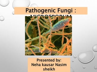
  • 1. Pathogenic Fungi : MICROSPORUM Presented by: Neha kausar Nasim sheikh
  • 2. CONTENT • Introduction • Morphological characteristics • Pathogenesis • Laboratory diagnosis • Treatment
  • 3. INTRODUCTION • Microsporum is the genus of fungi comes under Dermatophytes group • Dermatophytes- it is a group of three types of fungus i.e. Microsporum, Trichophyton and Epidermophyton that causes skin diseases in Humans and Animals. • Microsporum fungi infects only keratinized tissue like skin, hair and Nails. • They are restricted to non-viable skin because they are unable to grow at 37 degree Celsius. • Enzymes which helps the fungi are Proteases, Elastase, Keratinases. These are degradative enzymes which degrade keratinized tissue and serves as nutrients for the fungi. • Natural habitat of some microsporum spp. Is soil(geophilic) others primarily affect animals(zoophilic) or human(Anthropophilic). • The infection caused by Microsporum are known as ringworm or tinea , Though infections are not caused by worms but it appear as snake-like on skin, hence named ringworm infection.
  • 4. MORPHOLOGICAL CHARECTERISTICS • Microsporum can be identified by their colonial appearance and microscopic morphology after growth for 2weeks at 25 degree Celsius on Sabouraud’s dextrose agar. • Microsporum species tend to produce distinctive multicellular machroconidia with echinulate walls. • Microsporum forms both Macroconidia and Microconidia on short conidiosphores(hyphae). MACROCONIDIA MICROCONIDIA Large asexual reproductive structure. Small asexual reproductive structure. Hyaline, Multi sepate(2- 15) Hyaline, single septate Spindal shaped Club shaped Rough and Thick walled Smooth and Thin walled 8 to 15 X 40 to 150 µ m 2 to 3 µ m
  • 5. Microsporum canis • Colonies :- white cottony surface and deep yellow color on reverse, Thick walled. • On the rice grains, a deep yellow pigment is produced by M.canis which helps to differentiate it from M.audouinii • Macroconidia are abundant. Thick walled with many septa up to 15 . Macroconidia are often hooked or curved at ends. • Microconidia are small and club-shaped. • In-vitro hair perforation test :- Positive • M.canis causes tinea capitis and tinea corporis. Tinea barbae, tinea mannum.
  • 6. Microsporum audouinii • Colonies :- dense fur like mat having radiating edges, Grayish white to salmon pink colored colonies. • Pectinate hyphae and rare macroconidia and microconidia. • Anthropophilic. • Hair and skin infected with M.auduinii gives fluorescence under wood’s UV light. • In-vitro hair perforation test :- Negative • It causes tinea capitis and tinea corporis
  • 7. Microsporum gypseum • Colonies :- forms a tan, powdery colony, reverse colony often appear ragged around edges. • Macroconidia usually have 4-6 septa. • Microconidia are smaller than M.canis • Geophilic as well as Zoophilic (fur of rodents) • It causes Tinea capitis & Tinea corporis
  • 8. Microsporum ferrugineum • Colonies:- slow growing , flat, glabrous, wrinkled, leathery to downy. • Pale yellow colonies on Lowenstein-Jensen medium differentiate it from others. • Septate hyphae usually deformed and no Macroconidia, Microconidia • In-vitro hair Perforation test :- Negative • Causes tinea capitis.
  • 9. PATHOGENESIS Tinea Capitis:- Ringworm of scalp and hair • Hyphal invasion of skin of scalps • Spread to the keratinized wall of hair follicle • Hypha grow downward on nonliving portion of hair • As the hair grows upward the hyphae produce chain of spores that forms a sheath around hair shaft • The infection produce dull gray circular itchy patches.Tinea Corporis:- Ringworm infection of the body(smooth skin) • Fungal metabolites, Enzymes, Antigens diffuse through the viable layer of epidermis • Vesicle formation • Lesion expand centrifugally and active hyphal growth at periphery.
  • 10. Tinea Manuum:- Ringworm of the hands. • Shows red itchy rash, burning, cracking and scaling • May be transmitted sexually or via scratching and touching. • It typically starts as a small patch than gradually becomes larger. Tinea Barbae:- Ringworm of the beard and moustache. • Same as tinea capitis. • Infection occur as a follicular inflammation. • Mostly caused by Trichophyton rarely caused by Microsporum spp.
  • 11. SPECIES NATURAL RESERVOIR RINGWORM INFECTION Microsporum audouinii Anthropophilic Tinea corporis & Tinea capitis Microsporum canis Zoophilic (Cats and dogs) Tinea corporis,Tinea capitis,Tinea mannum, Tinea barbae Microsporum ferrugineum Anthropophilic Tinea capitis Microsporum gypseum Geophilic (also isolated from fur of rodents) Tinea corporis & Tinea capitis
  • 12. 1. Nail clipping, skin scrapings, hair placed in a slide than 10- 20%KOH is added with Lactophenol blue stain and examine the sample with a microscope to determine presence of hyphae. 2. Wood's lamp examination (ultraviolet light) may be used to diagnose microsporum spp. that fluoresce. LABORATORY DIAGNOSIS Spores of microsporum spp. Impart greenish to silvery fluorescence when examined under Wood’s lamp.
  • 13. M. canis :- Positive M. auduinii :- Negative 3. Fungal culture medium can be used for positive identification of the species i.e. Sabouraud’s dextrose Agar(SDA). 4. In-vitro hair perforation Test : Placing an organism in a petri-dish with water, yeast extract and hair sample. TREATMENT General Antifungal medications can be used to treat microsporum (ringworm) infections, • Topical medications :- clotrimazole, miconazole, econazole and terbinafine. • Oral medications :- Ketaconazole, griseofulvin, terbinafine, and itraconazole.