SlideShare a Scribd company logo
1 of 30
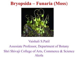
Bryopsida – Funaria (Moss)
Vaishali S.Patil
Assosiate Professor, Department of Botany
Shri Shivaji College of Arts, Commerce & Science
Akola
Division: Bryophyta
Class: Bryopsida
Subclass: Funariidae
Order: Funariales
Family: Funariaceae
Genus: Funaria
Habit-
210 species.
•Grows in dense patches or cushions
in moist shady and cool places during
the rainy seasons. These are primitive
multicellular, autotrophic, amphibious
plants. It can also be found on moist
walls and the crevices of rocks and places where recent fires have taken
place
External structure-
•The plant body is green, soft, and upright, about half an inch
tall (3–5 cm).
• It shows a radial symmetry with a differentiation of an axis
or stem, leaves or phylloids are multicellular colorless
branched rhizoids with oblique septa. The main axis of the
plant, which is upright, bears a set of spirally sessile leaves
having a clearly distinguishable midrib.
•The obovate leaves form a bulb-like cluster at the top of the
shortish, long stem. The leaves are long with an acute apex
and weakly serrate margins. The midrib ends just below, or
extends just beyond the leaf apex. They reproduce by spore
formation.
•They have no vascular system. Plant body consists of a long,
twisting stalk (seta) with an asymmetrical,grooved capsule
(spore-bearing part of the plant i.e.sporophyte) at the tip.
•At the apex of the main plant axis,the antheridium is borne.
This is the male part of the shoot. A lateral branch from the
main plant axisbears the female shoot archegonium at
its meristem.
Internal Structure:
1. Axis or stem
The transverse section (T. S.) of axis can be differentiated into
three distinct regions:
(i) Epidermis
(ii) Cortex
(iii) Central conducting strand or central cylinder.
(i) Epidermis:
It is the outer most single layered protective covering
consisting of small tangentially elongated chlorophyll bearing
cells. Cuticle and stomata are absent.
(ii) Cortex:
It is present between the epidermis and conducting tissue. It is
made up to parenchymatous cells. Younger part of the cortex
contains chloroplasts but in the older part they are lacking. At maturity
few outer layers of cortex become thick walled and are reddish brown in
colour but those of the inner layers become thin walled.
(iii) Central Conducting Strand:
It is made up of long, narrow thin walled dead cells which lack
protoplasm. These cells are now commonly called as hydroids.
Conducting strand besides providing a certain amount of mechanical
support, functions in the upward conduction of water and solutes.
.
2. Leaf:
Transverse section (T. S.) of ‘leaf’ shows a well-defined
midrib with two lateral wings. Except the midrib region,
the ‘leaf’ is composed of single layer of parenchymatous
polygonal cells. The cells contain many large and prominent
chloroplasts. The central part of the mid rib has narrow
conducting strand of thick walled cells which help in
conduction
Reproduction -
i) Vegetative Reproduction:
1. By multiplication of primary protonema:
In Funaria, spores on germination form a branched, filamentous,
multicellular structure. It’s called primary protonema. In it certain
colourless separation cells are formed by intercalary divisions. These cells
die out and break up the protonema into single cell or many celled
fragments. These fragments grow into new protonemata which bear buds.
Each bud develops into a leafy gametophore.
2. By secondary protonema:
When protonema is developed by other than the germination of spore, it is
called secondary protonema. It may be developed from any detached
living part of the gametophyte such as ‘stem’, ‘leaves’, antheridium,
archegonium paraphysis, sterile cells of capsule, seta or when the rhizoids
are exposed to sun light in moist atmosphere . It is similar to primary
protonema and develops into leafy gametophore.
3. By Gemmae:
During unfavorable conditions, the terminal cells of the protonemal
branches divide by transverse, longitudinal divisions and form green
multicellular bodies of 10-30 cells. These are called gemmae. At maturity
gemmae become slightly reddish brown in colour. On the return of
favourable conditions gemmae germinate and form new plants.
4. By Bulbils:
When such gemmae like structures are produced on rhizoids inside the
substratum, these are called bulbils. These are devoid of chloroplasts but
capable of developing into leafy individuals under favourable conditions.
5. Apospory:
Development of gametophyte from sporophyte without the formation of
spores is known as apospory. Any vegetative cell of the sporophyte may
form green protonemal filaments which bear lateral buds. These buds
later develop into leafy gametophores.
The gametophores thus formed are diploid. Sexual reproduction in such
gametophores results in the formation of tetraploid (4n) zygote. The
sporophytes from tetraploid are sterile because they are not capable of
bearing spores.
ii) Sexual Reproduction -
Sexual reproduction is oogamous. Male reproductive structure is known
as antheridium and female as archegonium. Funaria is monoecious
(having male and female sex organs on the same thallus) and autoicous
(antheridia and archegonia develop on separate branches of the same
thallus). Sex organs are borne on leafy gametophores in terminal clusters.
The main shoot of the leafy gametophore bears antheridia and act as
male branch. Female branch develops as a lateral outgrowth from the
base of the male branch and bears archegonia. It grows higher than the
male branch. Funaria is protandrous (antheridia mature before the
archegonia). It ensures the cross fertilization.
Male Branch or Antheridiophore: Longitudinal section (L. S.) of male
branch shows that its apex is expanded and convex shaped. It bears large
number of reddish brown or orange antheridia in different stages of
development. Projected antheridia are surrounded by a rosette of
spreading leaves called perigonial leaves.
The antheridial cluster with surrounding perigonial leaves is called
perigonium. The antheridia are intermingled with large number of sterile
hair like club shaped structures called paraphyses . Paraphyses store
water, protect developing antheridia, help in photosynthesis and
dehiscence of antheridia.
Structure of an Antheridium:
The antheridium is club shaped. It can be differentiated into two
parts:
(a) Short multicellular stalk
(b) Body of antheridium.
Body of antheridium has sterile, single layered jacket of polyhedral
flattened cells. When young the cells of the jacket contain chloroplasts
which turn orange or reddish brown at maturity. Jacket encloses a large
number of androcytes (antherozoid mother cells).
At maturity the distal end of the antheridium
bears one or two thick walled, colourless
cells called operculum.
The opercular cells become mucilaginous,
absorb water and swell, break
connections with the neighbouring cells and
form a narrow pore. Androcytes ooze out
in the form of a viscous fluid through this
pore.
Female Branch or Archegoniphore:
The female branch arises from the base of the male branch. Longitudinal
section (L. S.) of female branch shows that many archegonia
intermingled with paraphyses occurs at its apex. The terminal cell of
paraphyses is not swollen. The cluster of archegonia is enclosed by a
group of green foliage ‘leaves’ called perichaetial leaves. The
archegonial cluster with the surrounding perichaetial leaves is called
perichaetium.
Structure of an Archegonium:
A mature archegonium is flask shaped structure. It remains attached to
the female branch by a massive stalk. It consists upper elongated slender
neck and basal globular portion called venter .
The neck is slightly tubular, twisted, single layered and consists of six
vertical rows of neck cells, which enclose an axial row of ten or more
neck canal cells. The venter wall is two layered and encloses venter canal
cell and egg cell. Venter canal cell is situated just below the neck canal
cells.
Fertilization in Funaria:
Water is essential for fertilization. The opercular cells of the antheridium
rupture and releases mass of antherozoids. When archegonium reaches at
maturity, the neck canal cells and venter canal cell disintegrate to form a
mucilaginous mass. It absorbs water, swells up and comes out of the
archegonial mouth by pushing the cover cells apart. This mucilaginous
mass consists chemical substances (mainly sugars).
The cover cells of the neck separate widely from each other and form a
passage leading to the egg. Rosette like perigonial leaves serve as splash
cup from which rain drops disperse antheroziods to some distance (rain
drops falling on the archegonial cluster situated at lower level).
Many antherozoids enter the archegonial neck because of chemical
response but only one of them fuses with the egg to form the zygote.
Union of male and female nuclei is complete within 10 hours.
Fertilization ends the gametophytic phase.
Sporophytic Phase:
Zygote is the first cell of the sporophytic phase. Development of
sporophyte takes place within the venter of the archegonium.
Structure of Sporophyte:
The sporophyte is semi-parasitic in nature, the mature sporophyte can be
differentiated into three distinct parts—foot, seta and capsule.
(i) Foot:
It is the basal portion of the sporogonium. It is small dagger like conical
structure embedded in the apex of female branch. It functions as
anchoring and absorbing organ.
(ii) Seta:
It is long, slender, stalk like hygroscopic structure. It bears the capsule at
its tip. It raises the capsule above the apex of leafy gametophore.
(iii) Capsule:
It is the terminal part of the sporophyte and is developed at the apex of
the seta. It is green in colour when young but on maturity it becomes
bright orange coloured. It is covered by a cap like structure called
calyptra. (gametophytic tissue develops from the upper part of the
archegonium).
Internal Structure of the Capsule:
Longitudinal Section (L.S.) of the capsule shows that it can be
differentiated into three distinct regions-apophasis, theca and operculum.
(a) Apophysis:
It is the basal sterile part of the capsule. It is bounded by the single
layered epidermis which is interrupted by stomata. The sotmata have
single ring like guard cells. Below the epidermis is spongy parenchyma.
The central part of the apophysis is made up of elongated thin walled
cells forming a conducting strand. It is called neck of the capsule. It is
the photosynthetic region and connects seta with capsule.
(b) Theca:
It is the middle, slightly bent spore bearing region of the capsule. It lies
between the apophysis and operculum.
Longitudinal section (L. S.) passing through the theca shows the
following regions:(i) Epidermis:
It is the outer most layer. It is single layered with or without stomata.
(ii) Hypodermis:
It is present below the epidermis. It consists two to three layers of
compactly arranged colourless cells.
(iii) Spongy parenchyma:
It consists two to three layers of loosely arranged chlorophyllous cells. It
is present inner to hypodermis. These cells are capable to manufacture
their own food but dependent on gametophyte for water and mineral
nutrients. Therefore, the sporophyte of Funaria is partially dependent on
gametophyte.
(iv) Air spaces:
These are present just below the spongy parenchyma and outside the
spore sacs. Air spaces are traversed by green cells (chlorenchymatous
cells) called trabecular (elongated parenchymatous cells).
(v) Spore sac:
These are present below the air spaces on either side of the columella. It
is ‘U’ shaped and broken at the base. (It separates its both arms).
It has an outer wall (3-4 cells thick) and an inner wall (single cell in
thickness). Between the outer wall and inner wall is the cavity of the
spore sac. When young, the cavity of the spore sac is filled with many
spore mother cells. At maturity the spore mother cells divide by meiotic
divisions and form many haploid spores.
(vi) Columella:
It is the central part of the theca region. It is made up of compactly
arranged colourless parenchymatous cells. It is wide above and narrow
below, connecting the central strand of apophysis. It helps in conduction
of water and mineral nutrients.
(c) Operculum:
It is the upper region of the capsule. It is dome shaped and consists four
to five layers of cells. The outermost layer is thick walled and called
epidermis. Operculum is differentiated from theca by a well-marked
constriction. Just below the constriction there is a diaphragm (rim).
It is composed of two to three layers of radially elongated pitted cells.
Immediately above the rim is annulus which consists of 5-6
superimposed layers of cells. Its upper cells are thick but two lowermost
layers of cells are thin. Annulus separates the theca from the operculum.
Below the operculum lies the peristome . It is attached below to the edge
of the diaphragm. The peristome consists of two rings of radially
arranged peristomial teeth. In each ring there are sixteen teeth.
The teeth are not cellular but they are simply the strips of the cuticle. The
teeth of the outer ring are conspicuous, red with thick transverse bands
while the teeth of the inner ring are small, delicate, colourless and
without transverse bands. Inner to peristome teeth lies a mass of thin
walled parenchymatous cells.
Dehiscence of the Capsule:
Funaria is a stegocarpous moss (dehisce along a pre-determined line)
Dehiscence of the capsule is achieved by ‘breaking off’ of annulus. As
the capsule matures it becomes inverted due to epinasty. The thin walled
cells of the annulus break away, the operculum is thrown off and the
peristome teeth are exposed.
The outer peristomial teeth (exostome) are hygroscopic. The inner
peristomial teeth (endostome) do not show any hygroscopic movements
but act as a sieve allowing only a few spores to disperse at a time. The
lengthening and shortening of the outer peristomial teeth help in the
dispersal of spores.
In high humidity the exostome absorb water, increase in length and curve
inwards. In dry weather, the exostome teeth lose water, bend outwards
with jerky movements. It allows the dispersal of spores from the capsule
in instalments. At maturity the seta also shows jerky movements.
Twisting and swinging of seta in dry weather further aids in the dispersal
of spores.
Structure and Germination of Spore:
Spore is the first cell of the gametophytic phase. Each spore is spherical ,
12-20 µ in diameter and surrounded by two wall layers . The outer wall
is thick, smooth, brown and known as exosporium, while the inner wall
is thin, hyaline and called endosporium. Spore wall encloses single
nucleus, chloroplasts and many oil globules.
Under favourable conditions (sufficient moisture) spores germinate.
Exosporium ruptures and endosporium comes out in the form of one or
two germ tubes. Each germ tube is multicellular, green with oblique
septa. The germ tube grows in length, divides by septa to form green
algal filament like structure called primary protonema.
Primary Protonema:
It is the juvenile (young) stage of the gametophyte formed by the
germination of spore. It forms two different types of branches.
Most of the branches grow horizontally on the moist surface of the soil
and are known as chloronemal branches (positive phototrophic, thick and
rich in chloroplast) while some branches grow down in the soil and are
called rhizoidal branches (non-green, thin and possess oblique septa).
These branches can develop chlorophyll if expose to light.
Rhizoidal branches function as anchoring and absorbing organs while
chloronemal branches develop minute green buds behind the cross walls
which develop into leafy gametophores. From one primary protonema
many moss plants develop, so the moss is gregarious in habit. Primary
protonema is short lived.
According to Sirnoval (1947) development of protonema under
laboratory conditions can be differentiated into two stages—chloronemal
stage and caulonemal stage. Chloronemal stage is characterised by
irregular branching, right angle colourless cross walls, and many evenly
distributed discoid chloroplast.
It is positive phototropic but never produce buds. Nearly after 20 days
chloronemal stage matures into caulonemal stage. This stage is
characterised by regular branching brown cell walls, oblique cross walls
and fewer chloroplasts. It is negative phototropic and produce buds
which later develop into leafy gametophores. Rhizoids arise from the
base of a bud.
Bryopsida – funaria
Bryopsida – funaria
Bryopsida – funaria

More Related Content

What's hot (20)

Anthoceros
AnthocerosAnthoceros
Anthoceros
 
Selaginella
SelaginellaSelaginella
Selaginella
 
Funaria ( bryophytes)
Funaria ( bryophytes)Funaria ( bryophytes)
Funaria ( bryophytes)
 
Anthoceros ppt
Anthoceros pptAnthoceros ppt
Anthoceros ppt
 
General characters of pteridophytes
General characters of pteridophytesGeneral characters of pteridophytes
General characters of pteridophytes
 
Marchantia
MarchantiaMarchantia
Marchantia
 
Funaria
FunariaFunaria
Funaria
 
Riccia
RicciaRiccia
Riccia
 
Pteriduphytes 2
Pteriduphytes 2Pteriduphytes 2
Pteriduphytes 2
 
Classification of brayophyta
Classification of brayophytaClassification of brayophyta
Classification of brayophyta
 
Life cycle of_sphagnum
Life cycle of_sphagnumLife cycle of_sphagnum
Life cycle of_sphagnum
 
HETEROSPORY and SEED HABIT.pptx
HETEROSPORY and SEED HABIT.pptxHETEROSPORY and SEED HABIT.pptx
HETEROSPORY and SEED HABIT.pptx
 
Vaucheria
VaucheriaVaucheria
Vaucheria
 
Sphenophyllum
SphenophyllumSphenophyllum
Sphenophyllum
 
Life cycle of pinus
Life cycle of pinusLife cycle of pinus
Life cycle of pinus
 
Psilotum
PsilotumPsilotum
Psilotum
 
Reproduction and life cycle of Psilotum
Reproduction and life cycle of PsilotumReproduction and life cycle of Psilotum
Reproduction and life cycle of Psilotum
 
Psilotum
PsilotumPsilotum
Psilotum
 
Classification of pteridophytes
Classification of pteridophytesClassification of pteridophytes
Classification of pteridophytes
 
General Characters of Gymnosperms.pptx
General Characters of Gymnosperms.pptxGeneral Characters of Gymnosperms.pptx
General Characters of Gymnosperms.pptx
 

Similar to Bryopsida – funaria

Funaria botany
Funaria botanyFunaria botany
Funaria botanyMANSI
 
Structural variations in reproductive structures of division Bryophyta
Structural variations in reproductive structures of division BryophytaStructural variations in reproductive structures of division Bryophyta
Structural variations in reproductive structures of division BryophytaDarshi Thamali Paranagama
 
Morphology, Structure and Reproduction of Equisetum.
Morphology, Structure and Reproduction of Equisetum. Morphology, Structure and Reproduction of Equisetum.
Morphology, Structure and Reproduction of Equisetum. Dhaneesh Ram
 
MARSILEA notes in detail for II year Botany.ppt
MARSILEA  notes in detail for II year Botany.pptMARSILEA  notes in detail for II year Botany.ppt
MARSILEA notes in detail for II year Botany.pptaigil2
 
Riccia Bryophytes presentation
Riccia             Bryophytes         presentationRiccia             Bryophytes         presentation
Riccia Bryophytes presentationAnzaDar3
 
Life-cycle-of-Sphagnum.pdf
Life-cycle-of-Sphagnum.pdfLife-cycle-of-Sphagnum.pdf
Life-cycle-of-Sphagnum.pdfVainyaVerma
 
Adiantum.pptx(intoduction,species,reproduction)
Adiantum.pptx(intoduction,species,reproduction)Adiantum.pptx(intoduction,species,reproduction)
Adiantum.pptx(intoduction,species,reproduction)lalimaevane17
 

Similar to Bryopsida – funaria (20)

Life cycle of_funaria-jb
Life cycle of_funaria-jbLife cycle of_funaria-jb
Life cycle of_funaria-jb
 
Plant Kingdom III
Plant Kingdom IIIPlant Kingdom III
Plant Kingdom III
 
Funaria botany
Funaria botanyFunaria botany
Funaria botany
 
Structural variations in reproductive structures of division Bryophyta
Structural variations in reproductive structures of division BryophytaStructural variations in reproductive structures of division Bryophyta
Structural variations in reproductive structures of division Bryophyta
 
Pteridophytes
PteridophytesPteridophytes
Pteridophytes
 
Marchantia jb
Marchantia jbMarchantia jb
Marchantia jb
 
Morphology, Structure and Reproduction of Equisetum.
Morphology, Structure and Reproduction of Equisetum. Morphology, Structure and Reproduction of Equisetum.
Morphology, Structure and Reproduction of Equisetum.
 
Marchantia
MarchantiaMarchantia
Marchantia
 
Plant kingdom II
Plant kingdom IIPlant kingdom II
Plant kingdom II
 
Anthoceros
AnthocerosAnthoceros
Anthoceros
 
Type polytrichum
Type polytrichumType polytrichum
Type polytrichum
 
MARSILEA notes in detail for II year Botany.ppt
MARSILEA  notes in detail for II year Botany.pptMARSILEA  notes in detail for II year Botany.ppt
MARSILEA notes in detail for II year Botany.ppt
 
Adiantum .pptx
Adiantum .pptxAdiantum .pptx
Adiantum .pptx
 
Riccia Bryophytes presentation
Riccia             Bryophytes         presentationRiccia             Bryophytes         presentation
Riccia Bryophytes presentation
 
Marchantia important points
Marchantia important pointsMarchantia important points
Marchantia important points
 
Life-cycle-of-Sphagnum.pdf
Life-cycle-of-Sphagnum.pdfLife-cycle-of-Sphagnum.pdf
Life-cycle-of-Sphagnum.pdf
 
Furneria
FurneriaFurneria
Furneria
 
Anthoceros.ppt
Anthoceros.pptAnthoceros.ppt
Anthoceros.ppt
 
Funaria
FunariaFunaria
Funaria
 
Adiantum.pptx(intoduction,species,reproduction)
Adiantum.pptx(intoduction,species,reproduction)Adiantum.pptx(intoduction,species,reproduction)
Adiantum.pptx(intoduction,species,reproduction)
 

More from vaishalidandge3

Host parasite relationship ,interaction & mechanism of
Host parasite relationship ,interaction & mechanism ofHost parasite relationship ,interaction & mechanism of
Host parasite relationship ,interaction & mechanism ofvaishalidandge3
 
Economic importance of fungi
Economic importance of fungiEconomic importance of fungi
Economic importance of fungivaishalidandge3
 
Signs and symptoms of plant disease
Signs and symptoms of plant diseaseSigns and symptoms of plant disease
Signs and symptoms of plant diseasevaishalidandge3
 
General account of post harvest diseases of vegetables
General account of post harvest diseases of vegetablesGeneral account of post harvest diseases of vegetables
General account of post harvest diseases of vegetablesvaishalidandge3
 
Classification of fungi proposed by Ainsworth (1971)
Classification of fungi proposed by Ainsworth (1971)Classification of fungi proposed by Ainsworth (1971)
Classification of fungi proposed by Ainsworth (1971)vaishalidandge3
 
Important fungal disease on vegetables-Brinjal
Important fungal disease on vegetables-BrinjalImportant fungal disease on vegetables-Brinjal
Important fungal disease on vegetables-Brinjalvaishalidandge3
 
History,classification & importance of plant pathology
History,classification & importance of plant pathologyHistory,classification & importance of plant pathology
History,classification & importance of plant pathologyvaishalidandge3
 
Koch's postulate –principals & method
Koch's postulate –principals & methodKoch's postulate –principals & method
Koch's postulate –principals & methodvaishalidandge3
 
Industrial fungal metabolites enzymes pectinases
Industrial fungal metabolites enzymes  pectinasesIndustrial fungal metabolites enzymes  pectinases
Industrial fungal metabolites enzymes pectinasesvaishalidandge3
 
Industrial fungal metabolites enzymes lipases
Industrial fungal metabolites enzymes  lipasesIndustrial fungal metabolites enzymes  lipases
Industrial fungal metabolites enzymes lipasesvaishalidandge3
 
Industrial fungal metabolites enzymes proteases
Industrial fungal metabolites enzymes proteasesIndustrial fungal metabolites enzymes proteases
Industrial fungal metabolites enzymes proteasesvaishalidandge3
 
Fungal diseases of vegetables tomato
Fungal diseases of vegetables tomatoFungal diseases of vegetables tomato
Fungal diseases of vegetables tomatovaishalidandge3
 
Fungal diseases of vegetables potato
Fungal diseases of vegetables potatoFungal diseases of vegetables potato
Fungal diseases of vegetables potatovaishalidandge3
 
Fungal diseases of vegetables bhindi
Fungal diseases of vegetables bhindiFungal diseases of vegetables bhindi
Fungal diseases of vegetables bhindivaishalidandge3
 
Fungal diseases of vegetables-Cabbage
Fungal diseases of vegetables-CabbageFungal diseases of vegetables-Cabbage
Fungal diseases of vegetables-Cabbagevaishalidandge3
 

More from vaishalidandge3 (20)

Host parasite relationship ,interaction & mechanism of
Host parasite relationship ,interaction & mechanism ofHost parasite relationship ,interaction & mechanism of
Host parasite relationship ,interaction & mechanism of
 
Zygomycotina
ZygomycotinaZygomycotina
Zygomycotina
 
Plasmodiophoromycetes
PlasmodiophoromycetesPlasmodiophoromycetes
Plasmodiophoromycetes
 
Oomycetes
OomycetesOomycetes
Oomycetes
 
Mastigomycotina
MastigomycotinaMastigomycotina
Mastigomycotina
 
Myxomycotina
MyxomycotinaMyxomycotina
Myxomycotina
 
Economic importance of fungi
Economic importance of fungiEconomic importance of fungi
Economic importance of fungi
 
Signs and symptoms of plant disease
Signs and symptoms of plant diseaseSigns and symptoms of plant disease
Signs and symptoms of plant disease
 
General account of post harvest diseases of vegetables
General account of post harvest diseases of vegetablesGeneral account of post harvest diseases of vegetables
General account of post harvest diseases of vegetables
 
Classification of fungi proposed by Ainsworth (1971)
Classification of fungi proposed by Ainsworth (1971)Classification of fungi proposed by Ainsworth (1971)
Classification of fungi proposed by Ainsworth (1971)
 
Important fungal disease on vegetables-Brinjal
Important fungal disease on vegetables-BrinjalImportant fungal disease on vegetables-Brinjal
Important fungal disease on vegetables-Brinjal
 
History,classification & importance of plant pathology
History,classification & importance of plant pathologyHistory,classification & importance of plant pathology
History,classification & importance of plant pathology
 
Koch's postulate –principals & method
Koch's postulate –principals & methodKoch's postulate –principals & method
Koch's postulate –principals & method
 
Industrial fungal metabolites enzymes pectinases
Industrial fungal metabolites enzymes  pectinasesIndustrial fungal metabolites enzymes  pectinases
Industrial fungal metabolites enzymes pectinases
 
Industrial fungal metabolites enzymes lipases
Industrial fungal metabolites enzymes  lipasesIndustrial fungal metabolites enzymes  lipases
Industrial fungal metabolites enzymes lipases
 
Industrial fungal metabolites enzymes proteases
Industrial fungal metabolites enzymes proteasesIndustrial fungal metabolites enzymes proteases
Industrial fungal metabolites enzymes proteases
 
Fungal diseases of vegetables tomato
Fungal diseases of vegetables tomatoFungal diseases of vegetables tomato
Fungal diseases of vegetables tomato
 
Fungal diseases of vegetables potato
Fungal diseases of vegetables potatoFungal diseases of vegetables potato
Fungal diseases of vegetables potato
 
Fungal diseases of vegetables bhindi
Fungal diseases of vegetables bhindiFungal diseases of vegetables bhindi
Fungal diseases of vegetables bhindi
 
Fungal diseases of vegetables-Cabbage
Fungal diseases of vegetables-CabbageFungal diseases of vegetables-Cabbage
Fungal diseases of vegetables-Cabbage
 

Recently uploaded

Presentation Vikram Lander by Vedansh Gupta.pptx
Presentation Vikram Lander by Vedansh Gupta.pptxPresentation Vikram Lander by Vedansh Gupta.pptx
Presentation Vikram Lander by Vedansh Gupta.pptxgindu3009
 
Orientation, design and principles of polyhouse
Orientation, design and principles of polyhouseOrientation, design and principles of polyhouse
Orientation, design and principles of polyhousejana861314
 
Biopesticide (2).pptx .This slides helps to know the different types of biop...
Biopesticide (2).pptx  .This slides helps to know the different types of biop...Biopesticide (2).pptx  .This slides helps to know the different types of biop...
Biopesticide (2).pptx .This slides helps to know the different types of biop...RohitNehra6
 
Raman spectroscopy.pptx M Pharm, M Sc, Advanced Spectral Analysis
Raman spectroscopy.pptx M Pharm, M Sc, Advanced Spectral AnalysisRaman spectroscopy.pptx M Pharm, M Sc, Advanced Spectral Analysis
Raman spectroscopy.pptx M Pharm, M Sc, Advanced Spectral AnalysisDiwakar Mishra
 
GFP in rDNA Technology (Biotechnology).pptx
GFP in rDNA Technology (Biotechnology).pptxGFP in rDNA Technology (Biotechnology).pptx
GFP in rDNA Technology (Biotechnology).pptxAleenaTreesaSaji
 
STERILITY TESTING OF PHARMACEUTICALS ppt by DR.C.P.PRINCE
STERILITY TESTING OF PHARMACEUTICALS ppt by DR.C.P.PRINCESTERILITY TESTING OF PHARMACEUTICALS ppt by DR.C.P.PRINCE
STERILITY TESTING OF PHARMACEUTICALS ppt by DR.C.P.PRINCEPRINCE C P
 
Hubble Asteroid Hunter III. Physical properties of newly found asteroids
Hubble Asteroid Hunter III. Physical properties of newly found asteroidsHubble Asteroid Hunter III. Physical properties of newly found asteroids
Hubble Asteroid Hunter III. Physical properties of newly found asteroidsSérgio Sacani
 
A relative description on Sonoporation.pdf
A relative description on Sonoporation.pdfA relative description on Sonoporation.pdf
A relative description on Sonoporation.pdfnehabiju2046
 
Biological Classification BioHack (3).pdf
Biological Classification BioHack (3).pdfBiological Classification BioHack (3).pdf
Biological Classification BioHack (3).pdfmuntazimhurra
 
Zoology 4th semester series (krishna).pdf
Zoology 4th semester series (krishna).pdfZoology 4th semester series (krishna).pdf
Zoology 4th semester series (krishna).pdfSumit Kumar yadav
 
Recombination DNA Technology (Nucleic Acid Hybridization )
Recombination DNA Technology (Nucleic Acid Hybridization )Recombination DNA Technology (Nucleic Acid Hybridization )
Recombination DNA Technology (Nucleic Acid Hybridization )aarthirajkumar25
 
Unlocking the Potential: Deep dive into ocean of Ceramic Magnets.pptx
Unlocking  the Potential: Deep dive into ocean of Ceramic Magnets.pptxUnlocking  the Potential: Deep dive into ocean of Ceramic Magnets.pptx
Unlocking the Potential: Deep dive into ocean of Ceramic Magnets.pptxanandsmhk
 
Boyles law module in the grade 10 science
Boyles law module in the grade 10 scienceBoyles law module in the grade 10 science
Boyles law module in the grade 10 sciencefloriejanemacaya1
 
Discovery of an Accretion Streamer and a Slow Wide-angle Outflow around FUOri...
Discovery of an Accretion Streamer and a Slow Wide-angle Outflow around FUOri...Discovery of an Accretion Streamer and a Slow Wide-angle Outflow around FUOri...
Discovery of an Accretion Streamer and a Slow Wide-angle Outflow around FUOri...Sérgio Sacani
 
Animal Communication- Auditory and Visual.pptx
Animal Communication- Auditory and Visual.pptxAnimal Communication- Auditory and Visual.pptx
Animal Communication- Auditory and Visual.pptxUmerFayaz5
 
Isotopic evidence of long-lived volcanism on Io
Isotopic evidence of long-lived volcanism on IoIsotopic evidence of long-lived volcanism on Io
Isotopic evidence of long-lived volcanism on IoSérgio Sacani
 
G9 Science Q4- Week 1-2 Projectile Motion.ppt
G9 Science Q4- Week 1-2 Projectile Motion.pptG9 Science Q4- Week 1-2 Projectile Motion.ppt
G9 Science Q4- Week 1-2 Projectile Motion.pptMAESTRELLAMesa2
 
Artificial Intelligence In Microbiology by Dr. Prince C P
Artificial Intelligence In Microbiology by Dr. Prince C PArtificial Intelligence In Microbiology by Dr. Prince C P
Artificial Intelligence In Microbiology by Dr. Prince C PPRINCE C P
 
Chemistry 4th semester series (krishna).pdf
Chemistry 4th semester series (krishna).pdfChemistry 4th semester series (krishna).pdf
Chemistry 4th semester series (krishna).pdfSumit Kumar yadav
 

Recently uploaded (20)

Presentation Vikram Lander by Vedansh Gupta.pptx
Presentation Vikram Lander by Vedansh Gupta.pptxPresentation Vikram Lander by Vedansh Gupta.pptx
Presentation Vikram Lander by Vedansh Gupta.pptx
 
Orientation, design and principles of polyhouse
Orientation, design and principles of polyhouseOrientation, design and principles of polyhouse
Orientation, design and principles of polyhouse
 
Biopesticide (2).pptx .This slides helps to know the different types of biop...
Biopesticide (2).pptx  .This slides helps to know the different types of biop...Biopesticide (2).pptx  .This slides helps to know the different types of biop...
Biopesticide (2).pptx .This slides helps to know the different types of biop...
 
Raman spectroscopy.pptx M Pharm, M Sc, Advanced Spectral Analysis
Raman spectroscopy.pptx M Pharm, M Sc, Advanced Spectral AnalysisRaman spectroscopy.pptx M Pharm, M Sc, Advanced Spectral Analysis
Raman spectroscopy.pptx M Pharm, M Sc, Advanced Spectral Analysis
 
GFP in rDNA Technology (Biotechnology).pptx
GFP in rDNA Technology (Biotechnology).pptxGFP in rDNA Technology (Biotechnology).pptx
GFP in rDNA Technology (Biotechnology).pptx
 
STERILITY TESTING OF PHARMACEUTICALS ppt by DR.C.P.PRINCE
STERILITY TESTING OF PHARMACEUTICALS ppt by DR.C.P.PRINCESTERILITY TESTING OF PHARMACEUTICALS ppt by DR.C.P.PRINCE
STERILITY TESTING OF PHARMACEUTICALS ppt by DR.C.P.PRINCE
 
Hubble Asteroid Hunter III. Physical properties of newly found asteroids
Hubble Asteroid Hunter III. Physical properties of newly found asteroidsHubble Asteroid Hunter III. Physical properties of newly found asteroids
Hubble Asteroid Hunter III. Physical properties of newly found asteroids
 
A relative description on Sonoporation.pdf
A relative description on Sonoporation.pdfA relative description on Sonoporation.pdf
A relative description on Sonoporation.pdf
 
Biological Classification BioHack (3).pdf
Biological Classification BioHack (3).pdfBiological Classification BioHack (3).pdf
Biological Classification BioHack (3).pdf
 
Zoology 4th semester series (krishna).pdf
Zoology 4th semester series (krishna).pdfZoology 4th semester series (krishna).pdf
Zoology 4th semester series (krishna).pdf
 
Recombination DNA Technology (Nucleic Acid Hybridization )
Recombination DNA Technology (Nucleic Acid Hybridization )Recombination DNA Technology (Nucleic Acid Hybridization )
Recombination DNA Technology (Nucleic Acid Hybridization )
 
Unlocking the Potential: Deep dive into ocean of Ceramic Magnets.pptx
Unlocking  the Potential: Deep dive into ocean of Ceramic Magnets.pptxUnlocking  the Potential: Deep dive into ocean of Ceramic Magnets.pptx
Unlocking the Potential: Deep dive into ocean of Ceramic Magnets.pptx
 
Boyles law module in the grade 10 science
Boyles law module in the grade 10 scienceBoyles law module in the grade 10 science
Boyles law module in the grade 10 science
 
Discovery of an Accretion Streamer and a Slow Wide-angle Outflow around FUOri...
Discovery of an Accretion Streamer and a Slow Wide-angle Outflow around FUOri...Discovery of an Accretion Streamer and a Slow Wide-angle Outflow around FUOri...
Discovery of an Accretion Streamer and a Slow Wide-angle Outflow around FUOri...
 
Animal Communication- Auditory and Visual.pptx
Animal Communication- Auditory and Visual.pptxAnimal Communication- Auditory and Visual.pptx
Animal Communication- Auditory and Visual.pptx
 
Isotopic evidence of long-lived volcanism on Io
Isotopic evidence of long-lived volcanism on IoIsotopic evidence of long-lived volcanism on Io
Isotopic evidence of long-lived volcanism on Io
 
CELL -Structural and Functional unit of life.pdf
CELL -Structural and Functional unit of life.pdfCELL -Structural and Functional unit of life.pdf
CELL -Structural and Functional unit of life.pdf
 
G9 Science Q4- Week 1-2 Projectile Motion.ppt
G9 Science Q4- Week 1-2 Projectile Motion.pptG9 Science Q4- Week 1-2 Projectile Motion.ppt
G9 Science Q4- Week 1-2 Projectile Motion.ppt
 
Artificial Intelligence In Microbiology by Dr. Prince C P
Artificial Intelligence In Microbiology by Dr. Prince C PArtificial Intelligence In Microbiology by Dr. Prince C P
Artificial Intelligence In Microbiology by Dr. Prince C P
 
Chemistry 4th semester series (krishna).pdf
Chemistry 4th semester series (krishna).pdfChemistry 4th semester series (krishna).pdf
Chemistry 4th semester series (krishna).pdf
 

Bryopsida – funaria

  • 1. Bryopsida – Funaria (Moss) Vaishali S.Patil Assosiate Professor, Department of Botany Shri Shivaji College of Arts, Commerce & Science Akola
  • 2. Division: Bryophyta Class: Bryopsida Subclass: Funariidae Order: Funariales Family: Funariaceae Genus: Funaria Habit- 210 species. •Grows in dense patches or cushions in moist shady and cool places during the rainy seasons. These are primitive multicellular, autotrophic, amphibious plants. It can also be found on moist walls and the crevices of rocks and places where recent fires have taken place
  • 3. External structure- •The plant body is green, soft, and upright, about half an inch tall (3–5 cm). • It shows a radial symmetry with a differentiation of an axis or stem, leaves or phylloids are multicellular colorless branched rhizoids with oblique septa. The main axis of the plant, which is upright, bears a set of spirally sessile leaves having a clearly distinguishable midrib. •The obovate leaves form a bulb-like cluster at the top of the shortish, long stem. The leaves are long with an acute apex and weakly serrate margins. The midrib ends just below, or extends just beyond the leaf apex. They reproduce by spore formation. •They have no vascular system. Plant body consists of a long, twisting stalk (seta) with an asymmetrical,grooved capsule (spore-bearing part of the plant i.e.sporophyte) at the tip.
  • 4. •At the apex of the main plant axis,the antheridium is borne. This is the male part of the shoot. A lateral branch from the main plant axisbears the female shoot archegonium at its meristem.
  • 5. Internal Structure: 1. Axis or stem The transverse section (T. S.) of axis can be differentiated into three distinct regions: (i) Epidermis (ii) Cortex (iii) Central conducting strand or central cylinder. (i) Epidermis: It is the outer most single layered protective covering consisting of small tangentially elongated chlorophyll bearing cells. Cuticle and stomata are absent. (ii) Cortex: It is present between the epidermis and conducting tissue. It is made up to parenchymatous cells. Younger part of the cortex
  • 6. contains chloroplasts but in the older part they are lacking. At maturity few outer layers of cortex become thick walled and are reddish brown in colour but those of the inner layers become thin walled. (iii) Central Conducting Strand: It is made up of long, narrow thin walled dead cells which lack protoplasm. These cells are now commonly called as hydroids. Conducting strand besides providing a certain amount of mechanical support, functions in the upward conduction of water and solutes.
  • 7. . 2. Leaf: Transverse section (T. S.) of ‘leaf’ shows a well-defined midrib with two lateral wings. Except the midrib region, the ‘leaf’ is composed of single layer of parenchymatous polygonal cells. The cells contain many large and prominent chloroplasts. The central part of the mid rib has narrow conducting strand of thick walled cells which help in conduction
  • 8. Reproduction - i) Vegetative Reproduction: 1. By multiplication of primary protonema: In Funaria, spores on germination form a branched, filamentous, multicellular structure. It’s called primary protonema. In it certain colourless separation cells are formed by intercalary divisions. These cells die out and break up the protonema into single cell or many celled fragments. These fragments grow into new protonemata which bear buds. Each bud develops into a leafy gametophore. 2. By secondary protonema: When protonema is developed by other than the germination of spore, it is called secondary protonema. It may be developed from any detached living part of the gametophyte such as ‘stem’, ‘leaves’, antheridium, archegonium paraphysis, sterile cells of capsule, seta or when the rhizoids are exposed to sun light in moist atmosphere . It is similar to primary protonema and develops into leafy gametophore.
  • 9.
  • 10. 3. By Gemmae: During unfavorable conditions, the terminal cells of the protonemal branches divide by transverse, longitudinal divisions and form green multicellular bodies of 10-30 cells. These are called gemmae. At maturity gemmae become slightly reddish brown in colour. On the return of favourable conditions gemmae germinate and form new plants. 4. By Bulbils: When such gemmae like structures are produced on rhizoids inside the substratum, these are called bulbils. These are devoid of chloroplasts but capable of developing into leafy individuals under favourable conditions.
  • 11. 5. Apospory: Development of gametophyte from sporophyte without the formation of spores is known as apospory. Any vegetative cell of the sporophyte may form green protonemal filaments which bear lateral buds. These buds later develop into leafy gametophores. The gametophores thus formed are diploid. Sexual reproduction in such gametophores results in the formation of tetraploid (4n) zygote. The sporophytes from tetraploid are sterile because they are not capable of bearing spores. ii) Sexual Reproduction - Sexual reproduction is oogamous. Male reproductive structure is known as antheridium and female as archegonium. Funaria is monoecious (having male and female sex organs on the same thallus) and autoicous (antheridia and archegonia develop on separate branches of the same thallus). Sex organs are borne on leafy gametophores in terminal clusters. The main shoot of the leafy gametophore bears antheridia and act as male branch. Female branch develops as a lateral outgrowth from the base of the male branch and bears archegonia. It grows higher than the
  • 12. male branch. Funaria is protandrous (antheridia mature before the archegonia). It ensures the cross fertilization. Male Branch or Antheridiophore: Longitudinal section (L. S.) of male branch shows that its apex is expanded and convex shaped. It bears large number of reddish brown or orange antheridia in different stages of development. Projected antheridia are surrounded by a rosette of spreading leaves called perigonial leaves. The antheridial cluster with surrounding perigonial leaves is called perigonium. The antheridia are intermingled with large number of sterile hair like club shaped structures called paraphyses . Paraphyses store water, protect developing antheridia, help in photosynthesis and dehiscence of antheridia.
  • 13.
  • 14. Structure of an Antheridium: The antheridium is club shaped. It can be differentiated into two parts: (a) Short multicellular stalk (b) Body of antheridium. Body of antheridium has sterile, single layered jacket of polyhedral flattened cells. When young the cells of the jacket contain chloroplasts which turn orange or reddish brown at maturity. Jacket encloses a large number of androcytes (antherozoid mother cells). At maturity the distal end of the antheridium bears one or two thick walled, colourless cells called operculum. The opercular cells become mucilaginous, absorb water and swell, break connections with the neighbouring cells and form a narrow pore. Androcytes ooze out in the form of a viscous fluid through this pore.
  • 15. Female Branch or Archegoniphore: The female branch arises from the base of the male branch. Longitudinal section (L. S.) of female branch shows that many archegonia intermingled with paraphyses occurs at its apex. The terminal cell of paraphyses is not swollen. The cluster of archegonia is enclosed by a group of green foliage ‘leaves’ called perichaetial leaves. The archegonial cluster with the surrounding perichaetial leaves is called perichaetium. Structure of an Archegonium: A mature archegonium is flask shaped structure. It remains attached to the female branch by a massive stalk. It consists upper elongated slender neck and basal globular portion called venter . The neck is slightly tubular, twisted, single layered and consists of six vertical rows of neck cells, which enclose an axial row of ten or more neck canal cells. The venter wall is two layered and encloses venter canal cell and egg cell. Venter canal cell is situated just below the neck canal cells.
  • 16.
  • 17. Fertilization in Funaria: Water is essential for fertilization. The opercular cells of the antheridium rupture and releases mass of antherozoids. When archegonium reaches at maturity, the neck canal cells and venter canal cell disintegrate to form a mucilaginous mass. It absorbs water, swells up and comes out of the archegonial mouth by pushing the cover cells apart. This mucilaginous mass consists chemical substances (mainly sugars). The cover cells of the neck separate widely from each other and form a passage leading to the egg. Rosette like perigonial leaves serve as splash cup from which rain drops disperse antheroziods to some distance (rain drops falling on the archegonial cluster situated at lower level). Many antherozoids enter the archegonial neck because of chemical response but only one of them fuses with the egg to form the zygote. Union of male and female nuclei is complete within 10 hours. Fertilization ends the gametophytic phase.
  • 18. Sporophytic Phase: Zygote is the first cell of the sporophytic phase. Development of sporophyte takes place within the venter of the archegonium. Structure of Sporophyte: The sporophyte is semi-parasitic in nature, the mature sporophyte can be differentiated into three distinct parts—foot, seta and capsule. (i) Foot: It is the basal portion of the sporogonium. It is small dagger like conical structure embedded in the apex of female branch. It functions as anchoring and absorbing organ. (ii) Seta: It is long, slender, stalk like hygroscopic structure. It bears the capsule at its tip. It raises the capsule above the apex of leafy gametophore.
  • 19. (iii) Capsule: It is the terminal part of the sporophyte and is developed at the apex of the seta. It is green in colour when young but on maturity it becomes bright orange coloured. It is covered by a cap like structure called calyptra. (gametophytic tissue develops from the upper part of the archegonium). Internal Structure of the Capsule: Longitudinal Section (L.S.) of the capsule shows that it can be differentiated into three distinct regions-apophasis, theca and operculum. (a) Apophysis: It is the basal sterile part of the capsule. It is bounded by the single layered epidermis which is interrupted by stomata. The sotmata have single ring like guard cells. Below the epidermis is spongy parenchyma. The central part of the apophysis is made up of elongated thin walled cells forming a conducting strand. It is called neck of the capsule. It is the photosynthetic region and connects seta with capsule. (b) Theca: It is the middle, slightly bent spore bearing region of the capsule. It lies
  • 20. between the apophysis and operculum. Longitudinal section (L. S.) passing through the theca shows the following regions:(i) Epidermis: It is the outer most layer. It is single layered with or without stomata. (ii) Hypodermis: It is present below the epidermis. It consists two to three layers of compactly arranged colourless cells. (iii) Spongy parenchyma: It consists two to three layers of loosely arranged chlorophyllous cells. It is present inner to hypodermis. These cells are capable to manufacture their own food but dependent on gametophyte for water and mineral nutrients. Therefore, the sporophyte of Funaria is partially dependent on gametophyte. (iv) Air spaces: These are present just below the spongy parenchyma and outside the spore sacs. Air spaces are traversed by green cells (chlorenchymatous cells) called trabecular (elongated parenchymatous cells).
  • 21. (v) Spore sac: These are present below the air spaces on either side of the columella. It is ‘U’ shaped and broken at the base. (It separates its both arms). It has an outer wall (3-4 cells thick) and an inner wall (single cell in thickness). Between the outer wall and inner wall is the cavity of the spore sac. When young, the cavity of the spore sac is filled with many spore mother cells. At maturity the spore mother cells divide by meiotic divisions and form many haploid spores. (vi) Columella: It is the central part of the theca region. It is made up of compactly arranged colourless parenchymatous cells. It is wide above and narrow below, connecting the central strand of apophysis. It helps in conduction of water and mineral nutrients. (c) Operculum: It is the upper region of the capsule. It is dome shaped and consists four to five layers of cells. The outermost layer is thick walled and called epidermis. Operculum is differentiated from theca by a well-marked constriction. Just below the constriction there is a diaphragm (rim).
  • 22. It is composed of two to three layers of radially elongated pitted cells. Immediately above the rim is annulus which consists of 5-6 superimposed layers of cells. Its upper cells are thick but two lowermost layers of cells are thin. Annulus separates the theca from the operculum. Below the operculum lies the peristome . It is attached below to the edge of the diaphragm. The peristome consists of two rings of radially arranged peristomial teeth. In each ring there are sixteen teeth. The teeth are not cellular but they are simply the strips of the cuticle. The teeth of the outer ring are conspicuous, red with thick transverse bands while the teeth of the inner ring are small, delicate, colourless and without transverse bands. Inner to peristome teeth lies a mass of thin walled parenchymatous cells.
  • 23.
  • 24. Dehiscence of the Capsule: Funaria is a stegocarpous moss (dehisce along a pre-determined line) Dehiscence of the capsule is achieved by ‘breaking off’ of annulus. As the capsule matures it becomes inverted due to epinasty. The thin walled cells of the annulus break away, the operculum is thrown off and the peristome teeth are exposed. The outer peristomial teeth (exostome) are hygroscopic. The inner peristomial teeth (endostome) do not show any hygroscopic movements but act as a sieve allowing only a few spores to disperse at a time. The lengthening and shortening of the outer peristomial teeth help in the dispersal of spores. In high humidity the exostome absorb water, increase in length and curve inwards. In dry weather, the exostome teeth lose water, bend outwards with jerky movements. It allows the dispersal of spores from the capsule in instalments. At maturity the seta also shows jerky movements. Twisting and swinging of seta in dry weather further aids in the dispersal of spores.
  • 25. Structure and Germination of Spore: Spore is the first cell of the gametophytic phase. Each spore is spherical , 12-20 µ in diameter and surrounded by two wall layers . The outer wall is thick, smooth, brown and known as exosporium, while the inner wall is thin, hyaline and called endosporium. Spore wall encloses single nucleus, chloroplasts and many oil globules. Under favourable conditions (sufficient moisture) spores germinate. Exosporium ruptures and endosporium comes out in the form of one or two germ tubes. Each germ tube is multicellular, green with oblique septa. The germ tube grows in length, divides by septa to form green algal filament like structure called primary protonema.
  • 26. Primary Protonema: It is the juvenile (young) stage of the gametophyte formed by the germination of spore. It forms two different types of branches. Most of the branches grow horizontally on the moist surface of the soil and are known as chloronemal branches (positive phototrophic, thick and rich in chloroplast) while some branches grow down in the soil and are called rhizoidal branches (non-green, thin and possess oblique septa). These branches can develop chlorophyll if expose to light. Rhizoidal branches function as anchoring and absorbing organs while chloronemal branches develop minute green buds behind the cross walls which develop into leafy gametophores. From one primary protonema many moss plants develop, so the moss is gregarious in habit. Primary protonema is short lived. According to Sirnoval (1947) development of protonema under laboratory conditions can be differentiated into two stages—chloronemal stage and caulonemal stage. Chloronemal stage is characterised by irregular branching, right angle colourless cross walls, and many evenly distributed discoid chloroplast.
  • 27. It is positive phototropic but never produce buds. Nearly after 20 days chloronemal stage matures into caulonemal stage. This stage is characterised by regular branching brown cell walls, oblique cross walls and fewer chloroplasts. It is negative phototropic and produce buds which later develop into leafy gametophores. Rhizoids arise from the base of a bud.