MTU.edu: Chapter 2-7 Bryopsida
MTU.edu: Chapter 2-7 Bryopsida
MTU.edu: Chapter 2-7 Bryopsida

You also want an ePaper? Increase the reach of your titles
YUMPU automatically turns print PDFs into web optimized ePapers that Google loves.
CHAPTER 2-7<br />
BRYOPSIDA<br />
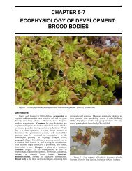
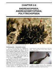
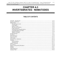
Figure 1. Aulacomnium androgynum with asexual gemmae on a modified stem tip. Photo by Michael Lüth.<br />
<strong>Bryopsida</strong> Definition<br />
By far the largest class of Bryophyta (sensu stricto)<br />
(84% of families) (Goffinet et al. 2001) and ~98% of the<br />
species, this class is unquestionably the most diverse.<br />
Their evolution by both advancement and r<strong>edu</strong>ction makes<br />
circumscription difficult, with nearly every character<br />
having exceptions. It appears that the only unique and<br />
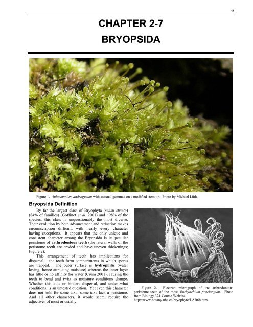
consistent character among the <strong>Bryopsida</strong> is its peculiar<br />
peristome of arthrodontous teeth (the lateral walls of the<br />
peristome teeth are eroded and have uneven thickenings;<br />
Figure 2).<br />
This arrangement of teeth has implications for<br />
dispersal – the teeth form compartments in which spores<br />
are trapped. The outer surface is hydrophilic (water<br />
loving, hence attracting moisture) whereas the inner layer<br />
has little or no affinity for water (Crum 2001), causing the<br />
teeth to bend and twist as moisture conditions change.<br />
Whether this aids or hinders dispersal, and under what<br />
conditions, is an untested question. Yet even this character<br />
does not hold for some taxa; some taxa lack a peristome.<br />
And all other characters, it would seem, require the<br />
adjectives of most or usually.<br />
Figure 2. Electron micrograph of the arthrodontous<br />
peristome teeth of the moss Eurhynchium praelongum. Photo<br />
from Biology 321 Course Website,<br />
http://www.botany.ubc.ca/bryophyte/LAB6b.htm.<br />
45
46 <strong>Chapter</strong> 2-7: <strong>Bryopsida</strong><br />
Spore Production and Protonemata<br />
As in all bryophytes, the spores are produced within<br />
the capsule by meiosis. In the <strong>Bryopsida</strong>, once germinated<br />
(Figure 3), they produce a filamentous protonema (first<br />
thread) that does not develop into a thalloid body (Figure<br />
4). This germination process can be rapid (1-3 days in<br />
Funaria hygrometrica) or lengthy, involving a long<br />
dormancy period.<br />
Figure 3. Germinating spore of Fontinalis squamosa. Photo<br />
by Janice Glime.<br />
Figure 4. Protonemata among leafy plants of Plagiomnium.<br />
Photo by Janice Glime.<br />
Many mosses differentiate their protonemata into<br />
chloronema and caulonema (Figure 5, Figure 6). The<br />
chloronema, meaning light green thread or chlorophyll<br />
thread, is the first part of the protonema to form when the<br />
spore germinates. The caulonema, meaning stem thread, is<br />
the portion that develops later, but not in all mosses, and<br />
that gives rise to the upright gametophores, or leafy plants.<br />
The caulonema differs from the younger parts of the<br />
protonema, the chloronema, in having longer cells with<br />
slanting cross walls, usually brownish cell walls, and fewer,<br />
less evenly distributed, smaller spindle-shaped chloroplasts.<br />
The chloronema exhibits irregular branching, whereas the<br />
caulonema exhibits regular branching.<br />
Figure 5. Protonema of moss such as Funaria hygrometrica<br />
with differentiated caulonema and chloronema. Drawing by Noris<br />
Salazar Allen.<br />
Figure 6. Protonema of Funaria hygrometrica showing<br />
chloronema (short cells with perpendicular walls and dense<br />
chloroplasts) and caulonema (long cells with diagonal cross walls<br />
and more dispersed chloroplasts). Photo by Janice Glime.<br />
Gametophore Buds<br />
As the protonema continues to develop and produce<br />
buds (Figure 7, Figure 8), the mosses and liverworts again<br />
differ. In liverworts, the bud is produced by the apical cell,<br />
hence ending further growth of the protonema and<br />
accounting for its single gametophore. In mosses, on the<br />
other hand, the bud originates from a cell behind the apical<br />
cell, hence permitting the apical cell to continue to divide<br />
and the protonema to continue to grow. The result is that<br />
moss protonemata produce many buds and upright plants<br />
(Figure 9). This provides the possibility for somatic<br />
mutations to arise, affording genetic variation among the<br />
leafy plants.<br />
Figure 7. Protonema and young developing bud of the moss<br />
Funaria hygrometrica. Photo by Martin Bopp.<br />
As the bud develops, rhizoids (Figure 25) form,<br />
functioning largely in anchorage, but at least in some<br />
mosses, also functioning in moving water and nutrients<br />
from substrate to moss. This may be especially important<br />
as the atmosphere dries and the rhizoids help to maintain a<br />
humid substrate.
Figure 8. Moss protonema with developed bud. Photo by<br />
Janice Glime.<br />
Figure 9. Leafy buds on the protonemata of Funaria<br />
hygrometrica forming a doughnut shape. Each of these circles of<br />
buds is the result of one spore. The hole in the middle is the area<br />
where the protonemata is in the chloronema stage and does not<br />
produce buds. Photo by Janice Glime.<br />
Gametophores<br />
The bud develops into the upright (or horizontal)<br />
gametophore. These plants are leafy haploid (1n) plants;<br />
thus, they are the dominant gametophyte generation of<br />
the life cycle. The stem may have a central strand (Figure<br />
10), or lack it (Figure 11); this strand may or may not have<br />
hydroids.<br />
Their leaves, more accurately known as phyllids (but<br />
rarely called that), are usually in more than three rows,<br />
but there are exceptions with two or three rows. Typically<br />
they are one cell thick, but there are modifications on this<br />
scheme that are expressed in some mosses by leaves folded<br />
over on themselves, creating a pocket in the genus<br />
Fissidens (Figure 12), or alternating hyaline (colorless) and<br />
photosynthetic layers as in Leucobryum, or just multiple<br />
layers of tissue, sometimes in patches. Some may have<br />
borders which likewise can be one or more layers thick.<br />
These leaves often have a multi-layered costa (Figure 13)<br />
in the center, or double, or even triple costa. The costa<br />
itself (Figure 25) consists of long, narrow cells that offer<br />
support and seem to function in moving water more quickly<br />
than their wider and often shorter neighboring cells.<br />
<strong>Chapter</strong> 2-7: <strong>Bryopsida</strong> 47<br />
Figure 10. Stem cross section of Rhizogonium illustrating<br />
central strand of hydroids. Photo by Isawa Kawai.<br />
Figure 11. Cross section of stem of the brook moss<br />
Fontinalis dalecarlica showing absence of central strand and<br />
conducting tissues. Photo by Janice Glime.<br />
Figure 12. Pockets in leaf of Fissidens arnoldii. Photo by<br />
Michael Lüth.<br />
Figure 13. Cross section of <strong>Bryopsida</strong> leaf showing one cell<br />
thick lamina (blade) portion and thickened costa. Photo by Janice<br />
Glime.
48 <strong>Chapter</strong> 2-7: <strong>Bryopsida</strong><br />
Location of Sex Organs<br />
Based on the branching patterns and location of sexual<br />
organs, the <strong>Bryopsida</strong> have traditionally been divided into<br />
two major groups, although there are good arguments for<br />
additional groupings. The acrocarpous mosses (Figure<br />
14) are generally those upright mosses with terminal<br />
sporangia. They usually are unbranched or sparsely<br />
branched. Pleurocarpous mosses (Figure 15), by contrast,<br />
produce their sporangia on short, specialized lateral<br />
branches or buds and typically are prostrate, forming freely<br />
branched mats. The truly pleurocarpous mosses appear to<br />
represent a single monophyletic clade (Buck & Goffinet<br />
2000; Buck et al. 2000a, b; Cox et al. 2000) and may be an<br />
adaptation to forming mats of continuous growth in mesic<br />
conditions (Vitt 1984). Those mosses that bear<br />
sporophytes terminally on short, lateral branches form a<br />
special category of pleurocarpous mosses termed<br />
cladocarpous. The branching patterns and positions of<br />
sporangia determine not only the growth form, but also<br />
influence success of fertilization, availability of water, and<br />
ability to spread horizontally across a substrate.<br />
Figure 14. Barbula unguiculata, an acrocarpous moss.<br />
Setae originate at the apex of the previous year's growth. Photo<br />
by Michael Lüth.<br />
Figure 15. Pleuroziopsis ruthenica, a pleurocarpous moss.<br />
Photo by Janice Glime.<br />
The upright or sprawling stems of the gametophyte<br />
produce antheridia (sperm-containers) and archegonia<br />
(egg-containers). In mosses, antheridia and archegonia<br />
may be located at the end of the main stem, at the ends of<br />
lateral branches, or along the main stem, either at the ends<br />
of very short branches or nearly sessile (Figure 26). Often<br />
the chloroplasts of the antheridial jacket cells are converted<br />
into chromoplasts as the antheridia mature, causing the<br />
characteristic red-orange color (Bold et al. 1987; see Figure<br />
16).<br />
Sperm Dispersal<br />
The dispersal of the sperm from the antheridium is an<br />
interesting phenomenon. In Mnium hornum, within about<br />
four minutes of placing water into an antheridial cup,<br />
dehiscence will occur (Muggoch & Walton 1942). The<br />
spermatocytes (cells in which sperm have differentiated)<br />
emerge in a banana-shaped package into the water<br />
surrounding the antheridium, usually within 4-10 minutes.<br />
Then, when (or if) that package connects with the water-air<br />
interface, the sperm spread apart rapidly to form a surface<br />
layer of regularly spaced sperm. This suggests that some<br />
substance with a low surface tension might be present in<br />
the sperm package because the mass spreads much like an<br />
oil spill. The behavior suggests that there is a small<br />
amount of fat present in the sperm mass. Muggoch and<br />
Walton considered this to be a widespread phenomenon,<br />
perhaps true of all mosses, and that it was important in<br />
permitting insects to carry sperm to female plants.<br />
However, there seem to be few observations of such insect<br />
dispersal except in Polytrichum (Class Polytrichopsida) and<br />
Bryum (or Rosulabryum) capillare (<strong>Bryopsida</strong>; Figure 16).<br />
Figure 16. Bryum capillare showing antheridial head of<br />
male plants. Photo by Michael Lüth.<br />
It appears that Bryum capillare may indeed be<br />
fertilized, at least some of the time, by animals. When<br />
covered by a fine net to discourage winged insects and<br />
other creatures, females were not fertilized, but when the<br />
net was removed, fertilization occurred 2 m(!) from the<br />
nearest males (Gayat 1897). However, it is difficult to rule<br />
out the possibility of raindrops in this case, or even<br />
squirrels, for that matter. Raindrops are likely to trap the<br />
mucilage with its sperm load in the tiny capillary spaces of<br />
the net. The success of fertilization would depend on the<br />
success of these drops getting bounced from one plant to<br />
another, and that bounce would surely be inhibited by such<br />
a filter to diminish the impact and retain the mucilage.<br />
Observations on Bryum argenteum are more<br />
conclusive. Cronberg et al. (2006), in an experiment in
which male and female plants were separated by 0, 2, and 4<br />
cm, demonstrated that help from such agents as<br />
invertebrates are essential. These treatment distances were<br />
combined either with no animals, or with mites (Acarina:<br />
Scutovertex minutus) or springtails (Collembola: Isotoma<br />
caerulea, Figure 17) (Cronberg et al. 2006; Milius 2006).<br />
After three months, those females in contact with male<br />
plants (0 cm) produced sporophytes. Those without this<br />
contact (2 or 4 cm) and without either animal group<br />
produced no sporophytes. But those housed with<br />
springtails or with mites produced numerous sporophytes,<br />
with springtails being the more effective conveyor.<br />
Springtails are more mobile than mites, and in this<br />
experiment, more sporophytes were produced at greater<br />
distances when springtails were available as dispersal<br />
agents.<br />
Figure 17. Isotoma caerulea, a springtail that is instrumental<br />
in fertilizing Bryum argenteum. Photo by Katrina Hedlund.<br />
But how do these springtails find the mosses? Flowers<br />
provide odors and colors to attract their pollinators. It<br />
appears that these mosses also have a way to attract their<br />
dispersal agents. When springtails and mites were given<br />
choices of plants with mature gametangia vs those that<br />
were sterile, fertile plants were chosen over non-fertile ones<br />
about five times as often (Beckman 2006) in the cases of<br />
both males and females and by both organisms. Cronberg<br />
et al. (2006) suggest that fertile plants may attract the<br />
invertebrates with sucrose (Pfeffer 1884), starch, fatty<br />
acids, and/or mucilage (Harvey-Gibson & Miller-Brown<br />
1927; Paolillo 1979; Renzaglia & Garbary 2001).<br />
Anderson (2002) managed to catch the dispersal of<br />
Plagiomnium affine on video to see the effectiveness of the<br />
splash platform of that moss. Although many drops will<br />
miss the tiny platform completely, a few manage full hits.<br />
Impact causes a "crown" of water to form, like dropping a<br />
rock into a lake. The capillary spaces between the<br />
antheridia and adjoining paraphyses (sing. paraphysis:<br />
sterile filaments located among reproductive organs; Figure<br />
18, Figure 26, Figure 27) fill with water. The impact of the<br />
drop causes the swollen antheridia to burst, releasing the<br />
swimming sperm. For the splash to be effective in making<br />
the crown, the diameter of the drop should be 1 mm or less,<br />
a common size in most rain showers. The rim of the crown<br />
has small droplets that are propelled away by the action.<br />
Since these droplets include water from within the splash<br />
platform, they also contain the sperm and thus propel them<br />
away from the plant. These droplets can travel 100 mm or<br />
more and manage to fertilize most of the females within 80<br />
<strong>Chapter</strong> 2-7: <strong>Bryopsida</strong> 49<br />
mm. The dioicous liverwort Marchantia has a splash<br />
platform that performs a similar function.<br />
Splash cups and platforms seem to be rare in<br />
monoicous taxa (exceptions include species of<br />
Brachymenium and Rosulabryum per John Spence),<br />
suggesting fertilization is accomplished with close<br />
neighbors. For most <strong>Bryopsida</strong>, however, there is no<br />
antheridial splash cup or platform, so seemingly sperm<br />
must swim all the way. However, other things can create<br />
splash. Jonathan Shaw (pers. comm.) has considered that<br />
F. hygrometrica has wide-spreading bracts surrounding the<br />
antheridia and the flexible nature of these bracts permits<br />
them to bend back and create an effective cup from which<br />
sperm in that species might be splashed. Angela Newton<br />
(pers. comm.) has suggested that platform surfaces among<br />
the more dendroid and shelf-forming taxa could be viewed<br />
as water-trapping mechanisms that would promote surface<br />
flow and dripping to the next level down as a mode of<br />
transporting sperm between individual plants or parts of<br />
plants. One complication in this arrangement is that the<br />
complex texture would act to trap water drops rather than<br />
encouraging them to splash out and away. However, in<br />
some of the plants with large smooth leaves, these leaves<br />
might act as springboards, but she considered that in such a<br />
case the water drops would be unlikely to carry sperm,<br />
although they might carry the smaller kinds of vegetative<br />
propagules. Nevertheless, sperm that had gotten as far as a<br />
leaf might benefit from this splash as well.<br />
Figure 18. Mature antheridia and paraphyses of the moss<br />
Rhizomnium. Photo by Janice Glime.<br />
Whereas flowering plants frequently rely on animals,<br />
especially insects, to transport their male gametophytes,<br />
and ultimately the sperm, to the female reproductive organ,<br />
this seems rarely to be the case in bryophytes.<br />
Surprisingly, it appears that the only documented case of<br />
such animal transport of sperm is in Polytrichum commune<br />
(Polytrichopsida), which has well-developed splash cups<br />
for the purpose of sperm dispersal. Nevertheless, it was in<br />
this species that Harvey-Gibson and Miller-Brown (1927)<br />
found motile sperm on the bodies of small arthropods<br />
(flies, leafhoppers, mites, spiders, and springtails) on both<br />
male and female reproductive inflorescences. Schofield<br />
(1985) suggests that mucilage produced in both the<br />
perigonia (modified leaves enclosing male reproductive<br />
structures) and perichaetia (modified leaves enclosing
50 <strong>Chapter</strong> 2-7: <strong>Bryopsida</strong><br />
female reproductive structures) sometimes attracts<br />
invertebrates.<br />
One might expect that many antheridia burst as they<br />
and their surrounding paraphyses swell from a desiccated<br />
state to a hydrated state during early minutes of a<br />
precipitation event. Could it be that the same external<br />
capillary forces that carry water rapidly to other parts of the<br />
plant could move sperm, thus r<strong>edu</strong>cing the energy<br />
requirements for getting these tiny cells to their<br />
destinations? Or are these forces to be reckoned with,<br />
forcing the sperm to swim against a current?<br />
If animal dispersal is so rare, then how, in this vast<br />
world, does an unintelligent sperm find an archegonium<br />
(Figure 19) and an egg? Fortunately for the moss, the<br />
archegonium at this time has dissolved the neck canal cells<br />
(entry canal through neck to egg in base of archegonium;<br />
Figure 26) leading down to the egg, and the resulting liquid<br />
provides a chemical attractant for the sperm. Meanwhile,<br />
the egg exudes mucilage into the cavity of the venter (Lal<br />
et al. 1982). When the canal opens, the liquid exudes from<br />
the opening of the neck, creating a chemical gradient. The<br />
sperm follows the concentration gradient toward the<br />
archegonium and finally swims down the neck canal of the<br />
archegonium to the egg. The exact nature of this liquid is<br />
unknown, but it seems that sugars and sometimes boron are<br />
necessary. It seems also likely that something specific,<br />
perhaps a protein, might guide the sperm to the correct<br />
species. Otherwise, it would seem that in spring, when so<br />
many species are producing sexual structures, some of<br />
these sperm would find their way into the wrong<br />
archegonium – or perhaps they do!<br />
Figure 19. Archegonia of the moss Fontinalis duriaei.<br />
Photo by Janice Glime<br />
Embryo Development<br />
When a sperm reaches and fertilizes an egg, the<br />
resulting diploid (having two sets of chromosomes; 2n)<br />
zygote begins dividing by mitosis to form an embryo that<br />
starts to stretch the archegonium (Figure 20). But the<br />
archegonium cannot stretch indefinitely, and as the embryo<br />
gets larger, the archegonium finally tears. Here, mosses<br />
and liverworts differ. In most mosses, part of the<br />
archegonium remains perched on top of the developing<br />
embryo (young sporophyte). This separated piece of<br />
archegonium is the cap you often see on top of the capsule<br />
and is now called a calyptra (Figure 26). So the calyptra is<br />
a 1n covering over the 2n capsule.<br />
The emerging embryo grows into the sporophyte of the<br />
moss. The mature sporophyte has a capsule and stalk<br />
(seta), with a foot embedded into the gametophyte tissue<br />
(Figure 21). Meiosis occurs in the mature capsule,<br />
producing haploid (1n) spores, as in all plants. Note that<br />
this is a major difference from meiosis in animals, which<br />
results in gametes. These spores are dispersed from the<br />
capsule by wind (or in a few cases – Splachnaceae – by<br />
insects) and grow into new gametophytes.<br />
Figure 20. Development of the calyptra of a moss. a. egg<br />
in archegonium, with neck canal cells not yet disintegrated. b.<br />
archegonium after fertilization and early development of embryo,<br />
showing elongation of the archegonium as embryo grows. c.<br />
elongated seta with calyptra perched on top of it before capsule<br />
has developed. d. mature capsule with calyptra and fully<br />
elongated seta. c & d indicate the remains of the venter of the<br />
archegonium at the base of the sporophyte. Drawings by Janice<br />
Glime.<br />
The calyptra that covers the capsule of mosses most<br />
likely plays multiple roles. We know that in many species,<br />
normal development ceases if the calyptra is removed<br />
(Paolillo 1968; French & Paolillo 1976a, b). One could<br />
assume that it provides protection from UV light and other<br />
environmental influences, as well as changing the internal<br />
environment, and that these influences are important in<br />
shaping the further development of the capsule, as will be<br />
discussed in another chapter.<br />
Capsule Development<br />
In mosses, once the calyptra has been shed, the<br />
operculum (lid) of the capsule is exposed. This operculum<br />
must come off before the spores can be dispersed. The<br />
dehiscence of the operculum is usually facilitated by<br />
drying of the capsule that causes it to shrink and compress<br />
the contents. But a few are cleistocarpous (indehiscent;<br />
lacking a regular means of opening), thus lacking an<br />
operculum.
Figure 21. Aloina rigida with stalk and capsule and with<br />
foot imbedded in gametophyte tissue. Photo by Michael Lüth.<br />
Just under the lid of most moss capsules you will find<br />
the peristome teeth (in mosses, fringe of teeth around<br />
opening of capsule, involved in spore dispersal; Figure 22,<br />
Figure 23). These are usually hygroscopic (responding to<br />
humidity changes) and may flex back and forth in response<br />
to moisture changes to aid in gradual dispersal. In most<br />
cases, these function best as the capsule is drying, but in<br />
some taxa moisture actually facilitates dispersal. Perhaps<br />
their best role is in preventing the spores from all exiting<br />
the capsule at the same time, as happens in the liverworts<br />
and Sphagnum and most likely also in the mosses with<br />
valvate capsules. The sporophyte capsule usually has a<br />
columella (Figure 22) that is columnar like those in<br />
Polytrichopsida, providing structure.<br />
Unlike the valvate capsules of liverworts and some<br />
moss classes, the sporophytes of the <strong>Bryopsida</strong> are<br />
photosynthetic. The same pigments often occur in both<br />
generations: chlorophylls a and b, carotene, lutein,<br />
violaxanthin, and zeaxanthin (Freeland 1957). Even the<br />
ratio of chlorophyll a to b is approximately the same –<br />
about 2.5:1 (Rastorfer 1962). Nevertheless, the<br />
gametophyte contains a higher chlorophyll concentration<br />
than does the sporophyte and the ratio of photosynthesis to<br />
respiration is likewise higher in the gametophyte. Despite<br />
its photosynthetic abilities, the sporophyte still depends on<br />
the gametophyte for some of its carbohydrates (Krupa<br />
1969).<br />
These stages of the life cycle are summarized in Figure<br />
24.<br />
<strong>Chapter</strong> 2-7: <strong>Bryopsida</strong> 51<br />
Figure 22. Section of Mnium capsule. This capsule actually<br />
hangs down, so teeth are on the bottom of the picture. Photo by<br />
Janice Glime.<br />
Figure 23. Arthrodontous peristome teeth. Left: peristome of Bartramia pomiformis. Photo by Zen Iwatsuki. Right: spores and<br />
peristome teeth of Trichostomum showing cell thickenings. Photo by Janice Glime.
52 <strong>Chapter</strong> 2-7: <strong>Bryopsida</strong><br />
Figure 24. Life cycle of the moss Funaria hygrometrica. Drawn by Shelly Meston.
. <strong>Chapter</strong> 2-7: <strong>Bryopsida</strong> 53<br />
Figure 25. Vegetative characters (gametophyte) of Class <strong>Bryopsida</strong>. Upper Left: Plagiomnium medium stem and leaves. Photo<br />
by Michael Lüth. Upper right: Plagiomnium stem cross section showing central strand of hydroids. Note smaller darkened areas in<br />
stem cortex that are leaf traces. Photo by Janice Glime. Middle Left: Leaf of Rhizomnium illustrating a border, small, roundish cells,<br />
and a distinct costa. Tip of leaf lacking a costa, illustrating elongate cells and undifferentiated apical leaf cells. Photo by Zen Iwatsuki.<br />
Middle Right: Portion of Plagiomnium leaf showing border. Photo by Janice Glime. Lower Left: Fontinalis stem, leaves, and tuft of<br />
rhizoids. Photo by Janice Glime. Lower Right: Microscopic view of rhizoids showing single cell thickness and diagonal cross walls.<br />
Photo by Janice Glime.
54 <strong>Chapter</strong> 2: The Life Cycle: Surviving Change<br />
Figure 26. Sexual reproduction of mosses. Upper row shows male reproductive parts. Splash platforms (left) of Mnium hornum<br />
in which antheridia may be located, or they can be among ordinary leaves (center); among the antheridia are paraphyses (center and<br />
right) that help in retaining water and in forcing sperm out of the antheridia at maturity. Lower row shows female reproductive parts.<br />
Perichaetial leaves and young sporophytes of Plagiomnium cuspidatum (left), archegonia from leaf bases of Pleurozium schreberi<br />
(center), and a section of archegonia (right) with sperm in the neck canal. Plant photos by Michael Lüth; photomicrographs by Janice<br />
Glime.
<strong>Chapter</strong> 2-7: <strong>Bryopsida</strong> 55<br />
Figure 27. Life cycle of a moss such as Mnium (<strong>Bryopsida</strong>). G represents Gametophyte; S represents Sporophyte. Drawings by<br />
Allison Slavick, Noris Salazar Allen, and Janice Glime.
56 <strong>Chapter</strong> 2: The Life Cycle: Surviving Change<br />
Summary<br />
The <strong>Bryopsida</strong> is the largest and most diverse class<br />
of Bryophyta. In <strong>Bryopsida</strong>, as in Polytrichopsida, an<br />
operculum usually covers peristome teeth that often<br />
aid dispersal. <strong>Bryopsida</strong> have arthrodontous<br />
peristome teeth, separating them from the<br />
Polytrichopsida, which have nematodontous teeth.<br />
All other classes of Bryobiotina lack peristomes.<br />
The life cycle of <strong>Bryopsida</strong> involves a protonema<br />
that is usually threadlike and develops from the<br />
germinating spore, developing numerous buds and<br />
gametophores. Gametophores produce archegonia<br />
and/or antheridia and the embryo develops within the<br />
archegonium.<br />
Sporophytes remain attached to the gametophyte<br />
and produce spores by meiosis. As in all Bryophyta,<br />
<strong>Bryopsida</strong> produce spores from the sporophyte only<br />
once.<br />
Vegetative reproduction is common among<br />
bryophytes. Bryophyta can reproduce by fragments as<br />
well as specialized asexual structures and thus add a<br />
new dimension to life cycle strategies.<br />
Acknowledgments<br />
I appreciate the comments and suggestions of Karla<br />
Werner, who offered a beginner's perspective. Noris<br />
Salazar Allen offered constructive criticisms on the<br />
taxonomic descriptions and helped with the proof reading<br />
and life cycle diagrams.<br />
Literature Cited<br />
Anderson, L. E. 2000. Great discoveries in bryology and<br />
lichenology. Charles E. Allen and sex chromosomes.<br />
Bryologist 103: 442-448.<br />
Beckman, M. 2006. The birds, the bees, and the mites.<br />
ScienceNOW Daily News 901: 1 Accessed on 3 September<br />
at<br />
http://sciencenow.sciencemag.org/cgi/content/fujll/2006/901/<br />
1>.<br />
Bold, H. C., Alexopoulos, C. J., and Delevoryas, T. 1987.<br />
Morphology of Plants and Fungi. Harper & Row,<br />
Publishers, Inc., New York, NY. 912 pp.<br />
Buck, W. R. and Goffinet, B. 2000. Morphology and<br />
classification of mosses. In: Shaw, J. A. and Goffinet, B.<br />
(eds.). Bryophyte Biology. Cambridge University Press, pp.<br />
71-123.<br />
Buck, W. R., Goffinet, B., and Shaw, A. J. 2000a. Novel<br />
relationships in pleurocarpous mosses as revealed by cpDNA<br />
sequences. Bryologist 103: 774-789.<br />
Buck, W. R., Goffinet, R. B., and Shaw, A. J. 2000b. Testing<br />
morphological concepts of orders of pleurocarpous mosses<br />
(Bryophyta) using phylogenetic reconstructions based on<br />
trnL-trnF and rps4 sequences. Molec. Phylogen. Evol. 16:<br />
180-198.<br />
Cox, C. J., Goffinet, B., Newton, A. E., Shaw, A. J., and<br />
Hedderson, T. A. J. 2000. Phylogenetic relationships among<br />
the diplolepideous-alternate mosses (Bryidae) inferred from<br />
nuclear and chloroplast DNA sequences. Bryologist 103:<br />
224-241.<br />
Cronberg, N., Natcheva, R., and Hedlund, K. 2006.<br />
Microarthropods mediate sperm transfer in mosses. Science<br />
313: 1225.<br />
Crum, H. 2001. Structural Diversity of Bryophytes. University<br />
of Michigan Herbarium, Ann Arbor, 379 pp.<br />
Freeland, R. O. 1957. Plastid pigments of gametophytes and<br />
sporophytes of Musci. Plant Physiol. 32: 64-66.<br />
French, J. C. and Paolillo, D. J. Jr. 1976a. Effect of the calyptra<br />
on intercalary meristematic activity in the sporophyte of<br />
Funaria (Musci). Amer. J. Bot. 63: 492-498.<br />
French, J. C. and Paolillo, D. J. Jr. 1976b. Effect of light and<br />
other factors on capsule expansion in Funaria hygrometrica.<br />
Bryologist 79: 457-465.<br />
Gayat, L. A. 1897. Recherches sur le developpement de<br />
l'archegore chez les Muscinees. Ann. Sci. Nat. Ser. 8, 3:161-<br />
285. In: Clayton-Greene, K. A., Green, T. G. A., and<br />
Staples, B. 1977. Studies of Dawsonia superba. 1.<br />
Antherozoid dispersal. Bryologist 80: 439-444.<br />
Goffinet, B., Cox, C. J., Shaw, A. J., and Hedderson, T. A. J.<br />
2001. The Bryophyta (mosses): Systematic and<br />
evolutionary inferences from an rps4 gene (cpDNA)<br />
phylogeny. Ann. Bot. 87: 191-208.<br />
Harvey-Gibson, R. J. and Miller-Brown, D. 1927. Fertilization<br />
of Bryophyta. Ann. Bot. 41: 190-191.<br />
Krupa, J. 1969. Photosynthetic activity and productivity of the<br />
sporophyte of Funaria hygrometrica during ontogenesis.<br />
Acta Soc. Bot. Poloniae 38: 207-215.<br />
Lal, M., Kaur, G., and Chauhan, E. 1982. Ultrastructural studies<br />
on archegonial development in the moss Physcomitrium<br />
cyathicarpum. New Phytol. 92: 441-452.<br />
Milius, S. 2006. Moss express. Insects and mites tote mosses'<br />
sperm. Science News 170: 148.<br />
Muggoch, H. and Walton, J. 1942. On the dehiscence of the<br />
antheridium and the part played by surface tension in the<br />
dispersal of spermatocytes in Bryophyta. Proc. Roy. Soc.<br />
London Sec. B Biol. Sci. 130: 448-461.<br />
Paolillo, D. 1968. The effect of the calyptra on capsule<br />
symmetry in Polytrichum juniperinum Hedw. Bryologist 71:<br />
327-334.<br />
Paolillo, D. J. Jr. 1979. On the lipids of the sperm masses of<br />
three mosses. Bryologist 82: 93-96.<br />
Pfeffer, W. 1884. Untersuchungen aus dem botanischen Institut<br />
in Tübingen 1: 363.<br />
Rastorfer, J. R. 1962. Photosynthesis and respiration in moss<br />
sporophytes and gametophytes. Phyton 19: 169-177.<br />
Renzaglia, K. S. and Garbary, D. J. 2001. Motile gametes of land<br />
plants: Diversity, development, and evolution. CRC Crit.<br />
Rev. Plant Sci. 20(2): 107-213.<br />
Schofield, W. B. 1985. Introduction to Bryology. Macmillan<br />
Publishing Co., New York, 431 pp.<br />
Vitt, D. H. 1984. Classification of the <strong>Bryopsida</strong>. In: Schuster,<br />
R. M. (ed.). New Manual of Bryology, Vol. 2. The Hattori<br />
Botanical Laboratory, Nichinan, Japan, pp. 696-759.