Morphological And Molecular Identification of Phaeoacremonium ...
Morphological And Molecular Identification of Phaeoacremonium ...
Morphological And Molecular Identification of Phaeoacremonium ...

Create successful ePaper yourself
Turn your PDF publications into a flip-book with our unique Google optimized e-Paper software.
Journal <strong>of</strong> Basrah Researches ((Sciences)) Volume 37. Number 4. E ((2011))<br />
Available online at: http://www.basra-science-journal.org<br />
ISSN18172695<br />
<strong>Morphological</strong> <strong>And</strong> <strong>Molecular</strong> <strong>Identification</strong> <strong>of</strong> <strong>Phaeoacremonium</strong><br />
aleophilum Associated with Grapevines Decline Phenomenon in Duhok<br />
Governorate<br />
1 Raed A. Haleem , 2 Samir K. Abdullah and 3 Jaladat M.S. Jubraeel<br />
1 Department <strong>of</strong> Plant Protection, College <strong>of</strong> Agriculture, University <strong>of</strong> Duhok, Iraq<br />
2 Department <strong>of</strong> Biology, College <strong>of</strong> Science, Zakho University, Iraq , samer_abdalh@yahoo.com<br />
3 Scientific Research Centre, University <strong>of</strong> Duhok, Iraq<br />
Abstract<br />
Decline symptoms on grapevine included plants that failed to thrive normal with reducing<br />
shoot growth and chloratic interveinal areas that latter became necrotic. In a cross section <strong>of</strong><br />
grapevine arms, the internal wood tissue were frequently dark brown to black with a wedgeshaped<br />
necrotic sectors. <strong>Phaeoacremonium</strong> aleophilum was isolated from infected tissues <strong>of</strong><br />
declined plants in pure culture and identified on the basis <strong>of</strong> its morphological and cultural<br />
characteristics. For accurate identification <strong>of</strong> P. aleophilum the PCR technique was employed.<br />
Ten isolates were selected from different locations. These isolates were subjected to specific<br />
PCR assay. The specific primers for P. aleophilum were used to amplify the ITS region <strong>of</strong><br />
nuclear ribosomal DNA (rDNA) containing ITS1, ITS2 and the intervening 5.8 rRNA genes.<br />
PCR results obtained from <strong>Phaeoacremonium</strong> isolates indicated that only three isolates were<br />
related to P. aleophilum. The remaining isolates may represent different species <strong>of</strong><br />
<strong>Phaeoacremonium</strong>. P. aleophilum is reported for the first time in Iraq.<br />
Keywords: grapevines decline, Phaeoacremnium aleophilum, molecular detection.<br />
Introduction<br />
The genus <strong>Phaeoacremonium</strong> is<br />
intermediate between Acremonium Link,<br />
Fr. and Phialophora Medlar.<br />
<strong>Phaeoacremonium</strong> parasiticum, under its<br />
original name Phialophora parasitica<br />
Ajello, Georg & C.J.K. Wang is the type<br />
species for the genus. <strong>Phaeoacremonium</strong><br />
can be distinguished from Phialophora by<br />
its aculeate phialides and inconspicuous,<br />
non-flaring collarettes, and from<br />
Acremonium by its pigmented vegetative<br />
hyphae (Crous et al., 1996). Togninia minima<br />
(Diaporthales: Ascomycota) was recently<br />
confirmed as the sexual stage <strong>of</strong><br />
1<br />
<strong>Phaeoacremonium</strong> aleophilum. T. minima is<br />
characterized by having dark globose longbeaked<br />
and non-stromatic perithecia<br />
(Rooney-Latham et al., 2005a,b).<br />
<strong>Phaeoacremonium</strong> species have wide<br />
host range and world wide distribution ,<br />
however, majority <strong>of</strong> them are found on<br />
Vitis vinifera, others were reported from<br />
Olea europaea, Fraxinus spp., Prunus spp.,<br />
Salix spp., and Quercus spp. Few species are<br />
parasitic on human (Auger et<br />
al.2005,Eskalen et al.2005, Mostert et al.<br />
2005b).
Haleem, Abdullah & Jubraeel: <strong>Morphological</strong> <strong>And</strong> <strong>Molecular</strong> <strong>Identification</strong> <strong>of</strong> <strong>Phaeoacremonium</strong> aleophilum…<br />
Six species <strong>of</strong> <strong>Phaeoacremonium</strong> were<br />
originally identified based on morphology<br />
and cultural characters (Crous et al., 1996).<br />
It soon became apparent that the taxon once<br />
referred to as ‘Cephalosporium’ species or<br />
P. Chlamydosporum represented a new<br />
genus, Phaeomoniella Crous & W. Gams,<br />
which resided within the Chaetothyriales<br />
(Crous and Gams, 2000). <strong>Morphological</strong><br />
characters that were useful in distinguishing<br />
species included conidiophore morphology,<br />
phialide type and morphology, the size <strong>of</strong><br />
hyphal warts, and to a lesser extent conidial<br />
size and shape; cultural characters that were<br />
useful included colony colour on 2% malt<br />
extract agar (MEA), yellow pigment<br />
production on potato-dextrose agar, growth<br />
rate at 25°C and maximum growth<br />
temperature (Mostert et al., 2005b). Yellow<br />
pigment production on oatmeal agar was<br />
used by Dupont et al. (2000). The genus<br />
<strong>Phaeoacremonium</strong> is characterized by its<br />
mycelial bundles, branched or simple<br />
conidiophores, slender phialides occurring<br />
in three size classes, narrowly funnel-shaped<br />
collarettes at the apex <strong>of</strong> the phialides,<br />
conidia aggregated into slimy heads and<br />
conidial shape ranging from mostly oblongellipsoidal<br />
to allantoid. Generic descriptions<br />
<strong>of</strong> <strong>Phaeoacremonium</strong> have been published<br />
by Crous et al (1996) and Mostert (2005b).<br />
<strong>Molecular</strong> characters have played an<br />
important role in the detection and<br />
Materials and Methods.<br />
Fungal isolation.<br />
Isolation was done in two methods:<br />
1- Isolation from complete vine<br />
tissues.<br />
Complete vine tissues were sampled,<br />
from cane (bark and wood), bud, trunk or<br />
arm (bark and wood), leaves, clusters and<br />
roots. Small pieces <strong>of</strong> tissue from the<br />
margin between necrotic and apparently<br />
healthy tissue were surface sterilized by<br />
placing in 70% ethanol for 30 s, 1% NaOCl<br />
for 1 min and again in 70% ethanol for 30 s<br />
and then dried by filter papers. Sterilized<br />
tissues pieces were plated onto 2% potato<br />
dextrose agar (PDA) (Himedia Laboratories<br />
identification <strong>of</strong> <strong>Phaeoacremonium</strong> species<br />
(Tegli et al., 2000; Dupont et al., 2002;<br />
Mostert, 2006; and Aroca et al., 2008).<br />
Species-specific primers based on the tubulin<br />
and actin genes have been developed<br />
(Mostert, 2006) and can be used in<br />
multiplex polymerase chain reactions (PCR)<br />
for the identification <strong>of</strong> unknown isolates.<br />
To date 22 species <strong>of</strong> <strong>Phaeoacremonium</strong><br />
have been isolated from grapevines (Crous<br />
et al., 1996; Mostert et al., 2005b; Mostert,<br />
2006; Essakhi et al., 2008; Gramaje et al.,<br />
2009). Sixteen species <strong>of</strong> <strong>Phaeoacremonium</strong><br />
were described based on molecular<br />
characters, the internal transcribed spacer<br />
(ITS) regions 1 and 2, the 5.8S rDNA<br />
(Dupont et al., 2000) and the -tubulin gene<br />
(Groenewald et al., 2001). Subsequent<br />
studies included the actin and calmodulin<br />
gene regions (Mostert et al., 2005b; and<br />
Mostert, 2006) <strong>of</strong> the species occurring on<br />
grapevines. P. aleophilum is the most<br />
common and widely distributed species<br />
(Crous et al.,1996, Larignon and Dubos<br />
1997; and Mugnai et al., 1999) and P.<br />
parasiticum is encountered frequently<br />
(Dupont et al., 2002; and Mostert, 2006).<br />
The objective <strong>of</strong> this study is isolation<br />
and identification <strong>of</strong> <strong>Phaeoacremonium</strong><br />
species associated with declined grapevine<br />
trees in Duhok governorate, Kurdistan<br />
region <strong>of</strong> Iraq, based on morphological and<br />
molecular techniques.<br />
Pvt. Ltd. - India) containing 0.25 mg/ml<br />
chloramphenicol. Hyphae growing out from<br />
the tissue pieces were cut and subcultured<br />
onto fresh PDA plates, and incubated at<br />
25±2 °C (Van Niekerk et al, 2004).<br />
Sporulation was enhanced by culturing the<br />
isolates on 2% water agar bearing pieces <strong>of</strong><br />
autoclaved grapevine canes at 25 °C with a<br />
12/12 h photoperiod (Luque et al., 2005).<br />
2- Moist chamber method.<br />
Cuttings were made from various<br />
grapevine parts including canes, arms and<br />
trunk. All segments were placed in 90 mm<br />
petridishes containing sterilized moist filter<br />
paper. Plates were incubated at room<br />
2
Journal <strong>of</strong> Basrah Researches ((Sciences)) Volume 37. Number 4. E ((2011))<br />
temperature until fungal growth observed.<br />
Propagules (spores, mycelia) were<br />
transferred to Potato-dextrose-Agar (PDA)<br />
plates. Pure cultures <strong>of</strong> each isolate were<br />
obtained by excising a hyphal tip from<br />
colony margins and plating it onto fresh<br />
PDA.<br />
Phenotypical characterization<br />
All isolates were grown on PDA and<br />
MEA at 25°C in darkness or under NUV +<br />
fluorescent illumination with a 12-h<br />
photoperiod (Philips /36W) for 10 - 15 days<br />
until cultures sporulated. Isolated strains<br />
were identified based on the characters in<br />
culture and on natural substrates (Crous et<br />
al, 1996; Mostert et al .2005a,b).<br />
DNA extraction and PCR<br />
amplification <strong>of</strong> <strong>Phaeoacremonium</strong><br />
aleophilum.<br />
- Fungal Isolates<br />
Ten isolates were selected to confirm the<br />
identification by a specific primer <strong>of</strong> the ITS<br />
region. Isolates were collected from five<br />
locations in Duhok governorate., as show in<br />
table (1).<br />
Table (1): Isolates <strong>of</strong> <strong>Phaeoacremonium</strong> spp. from different grape vineyards <strong>of</strong> Dohuk governorate.<br />
<strong>Phaeoacremonium</strong> spp.<br />
Isolates<br />
DP1<br />
DP2<br />
DP3<br />
DP4<br />
DP5<br />
DP6<br />
DP7<br />
DP8<br />
DP9<br />
DP10<br />
- Total genomic DNA extraction.<br />
Genomic DNA was extracted according to a<br />
method reported by Borges et al (1990).<br />
Fungi were grown in 2% malt extract broth<br />
with gentle shaking. Freezing fungal<br />
mycelium in liquid nitrogen and grinding<br />
the frozen sample to a powder by means <strong>of</strong> a<br />
pestle and mortar. Powdered mycelium<br />
(0.5g) was transferred to a sovrall tube<br />
containing 5 ml <strong>of</strong> cold SDS buffer and<br />
thoroughly shaken for 15 minutes. The<br />
mixture was then immediately extracted<br />
with 1 vol. distilled saturated phenol by<br />
shaking several time for 10 minutes and<br />
centrifuged at 4000 rpm for 15 minutes.<br />
After a phenol extraction, the aqueous phase<br />
was extracted with 1 vol. chlor<strong>of</strong>orm<br />
isoamyl by shaking for 15 minutes before<br />
centrifugation for 30 minutes. Ten percent<br />
<strong>of</strong> the vol. <strong>of</strong> 7.5M ammonium acetate was<br />
added to the resulting aqueous phase. DNA<br />
was then precipitated by addition <strong>of</strong> 2 vols.<br />
Isolated part Cultivar Geographical<br />
location<br />
Arm's wood Rashmew Nizarke<br />
Cane's wood Kamali Malata nursery<br />
Root<br />
Kamali Badi<br />
Cane's wood Taefi Zawita<br />
Root<br />
Rashmew Nizarke<br />
Arm's wood Kamali Bajelor<br />
Cane's bark Kamali Badi<br />
Arm's wood Taifi<br />
Bajelor<br />
Arm's wood Taifi<br />
Bajelor<br />
Root<br />
Rashmew Berebahar<br />
3<br />
<strong>of</strong> cold absolute ethanol, before incubation<br />
overnight at -20 °C ,and recovered either by<br />
spooling it out or by centrifugation at 4000<br />
rpm for 20 minutes. The supernatant was<br />
discarded and the pellet was washed with<br />
1ml <strong>of</strong> 70% cold ethanol, incubated<br />
overnight at -20°C and then centrifuged at<br />
4000 rpm for 10 minutes. The supernant was<br />
discarded and the DNA pellet was then airdried<br />
at room temperature for 30<br />
minutes.The DNA pellet was redissolved in<br />
300 µl TE buffer and stored at -20°C until<br />
use.<br />
- PCR amplification <strong>of</strong> ITS region.<br />
The specific primers <strong>of</strong><br />
<strong>Phaeoacremonium</strong> aleophilum were used to<br />
amplify the ITS region <strong>of</strong> nuclear ribosomal<br />
DNA (rDNA), containing ITS1, ITS2 and<br />
the intervening 5.8 rRNA gene (Pal1F 5’-<br />
AGGTCGGGGGCCAAC-3’, Pal2R 5’-<br />
AGGTGTAAACTACTGCGC-3’) (Tegli et
Haleem, Abdullah & Jubraeel: <strong>Morphological</strong> <strong>And</strong> <strong>Molecular</strong> <strong>Identification</strong> <strong>of</strong> <strong>Phaeoacremonium</strong> aleophilum…<br />
al, 2000). The PCR reactions were carried<br />
out in a total volume <strong>of</strong> 25 µl, in thinwalled,<br />
0.5 µl Eppendorf tubes.Master mix<br />
was prepared for 12 samples <strong>of</strong> each fungus<br />
(10 isolates plus 2 control) by mixing 30 µl<br />
<strong>of</strong> 10XPCR, 30 µl <strong>of</strong> dNTPs, 24 µl forward<br />
primer, 24 µl Reverse primer, 12 µl MgCL2,<br />
4.8 µl <strong>of</strong> Taq polymerase enzyme and deionized<br />
distilled water was added to a final<br />
volume <strong>of</strong> 252µl. The solution mixed and<br />
spun for 10 second in a microcentrifuge.<br />
Later, the mixture was dispensed in PCR<br />
tubes. All these steps were done on ice.<br />
Amplification was carried out in an<br />
automated thermal cycler (Delphy 1000,<br />
Oracle Biosystems, MJ Research Inc.,<br />
Results and Discussion<br />
Phenotypical characterization<br />
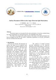
<strong>Phaeoacremonium</strong> aleophilum W. Gams,<br />
Crous, M. J. Wingf. et L. Mugnai.<br />
Mycologia 88:791 (1996). Fig. (1) A – I.<br />
Telemorph: Togninia minima (Tul.and C.<br />
Tul.) Berl., Icon. Fung. (Abellini) 3:9<br />
(1900).<br />
Cultural characters: colonies on MEA,<br />
reached a diameter <strong>of</strong> 22 mm after 20 days<br />
<strong>of</strong> incubation at 25°C. Flat, mostly felty<br />
texture with entire edge, Pale yellow color<br />
in above and in reverse. Colonies on PDA,<br />
reached a radial <strong>of</strong> 25.5 mm after 20 days.<br />
Flat, wooly texture with entire edge, Dark<br />
blond to brownish grey towards the edge<br />
above, in reverse pale brown to dark brown<br />
towards the edge.<br />
Aerial structure: Hyphae are verruculose,<br />
medium to pale brown, and 1.5 - 2.5 µm<br />
wide. Conidiophores are mostly short and<br />
usually unbranched, 0-3 septate. 17 - 29 µm<br />
long and 2 - 2.5 µm wide. The apical cell <strong>of</strong><br />
conidiophores usually produces one<br />
phialide. Phialides terminal or lateral,<br />
mostly monophialidic, smooth to<br />
verreculose, subhyline; type II and type III<br />
phialides are most common. Type I<br />
phialides are cylindrical occasionally wider<br />
at the base, 4 - 9 × 1 – 1.5 µm (av. 5 × 1.5)<br />
µm. Type II phialides are either elongateampulliform<br />
and attenuated at the base or<br />
are navicular, tapering towards the apex, 10-<br />
4<br />
Watertown, MA, USA) according to the<br />
following programs: An initial denaturation<br />
at 95°C for 3 min, after which 30<br />
cycles <strong>of</strong> de-naturation (2 min at 95°C),<br />
primer annealing (25 sec at 64°C) and<br />
primer extension (2 min at 72°C) were<br />
performed. A final extension was performed<br />
at 72°C for 10 min. Amplification reactions<br />
were conducted at least twice, in two<br />
separate experiments. For each isolate, 5 µl<br />
<strong>of</strong> PCR products were mixed with 7µl<br />
loading buffer and then analyzed by<br />
electrophoresis in 2% (w:v) agarose gels<br />
with 1xTBE buffer visualized by UV<br />
fluorescence.<br />
14 × 1.5-2.5 µm (av. 11 × 2) µm. Type III<br />
phialides are subcylindrical or elongate -<br />
ampulliform and attenuated at the base, 15 -<br />
20 × 1.5 - 2 µm (av. 18 × 2) µm. Conidia<br />
are mostly oblong-ellipsoidal or cylindrical,<br />
2.5 - 6 ×1 - 2 µm (av. 3.5 × 1.5) µm. This<br />
description was in agreement with the other<br />
investigations (Crous et al., 1996; Mostert et<br />
al., 2005b; and Mostert, 2006).<br />
<strong>Molecular</strong> detection <strong>of</strong> P.<br />
aleophilum<br />
- Genomic DNA isolation and<br />
purification.<br />
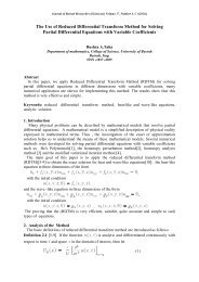
Suitable yields <strong>of</strong> genomic DNA were<br />
obtained from repeated experiments with an<br />
average yield <strong>of</strong> 1.5-6.70 µg/ml and a purity<br />
<strong>of</strong> about (1.6-1.8) determined by<br />
spectrophotometer ratio A 260/A 280. The<br />
molecular weight <strong>of</strong> DNA samples was<br />
estimated using 1% agarose gel<br />
electrophoresis containing DNA sample as<br />
control (Fig. 2,). Ratios above 2.0<br />
correspond to RNA contamination, while<br />
ratios below 1.6 suggest protein<br />
contamination (Sinha et al., 2001).<br />
- Species specific primers <strong>of</strong> P.<br />
aleophilum isolates.<br />
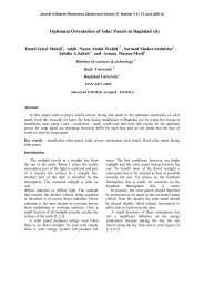
On the basis <strong>of</strong> the sequence data <strong>of</strong> the<br />
ITS regions, the primer pairs Pal1-Pal2 were<br />
designed by Tegli et al. (2000) to amplify<br />
specific DNA fragments using genomic
Journal <strong>of</strong> Basrah Researches ((Sciences)) Volume 37. Number 4. E ((2011))<br />
DNA from P. aleophilum (Pal) isolates.<br />
Two primer pair Pal1F-Pal2R specifically<br />
amplified a fragment <strong>of</strong> about 400 bp in<br />
three Pal tested isolates (DP1, DP2, and<br />
DP3), as shown in Fig. (3), but no<br />
amplification was detected when other<br />
isolates were tested.<br />
Based on morphological<br />
characteristics, these ten isolates were very<br />
close to each other, thus they were identified<br />
as P. aleophilum. However, the molecular<br />
detection confirmed that only three <strong>of</strong> them<br />
were related to P. aleophilum (DP1, DP2,<br />
and DP3). Thus the sequencer needed to<br />
obtain data <strong>of</strong> the ITS region to identify<br />
remaining species. To date, 22 species <strong>of</strong><br />
<strong>Phaeoacremonium</strong> have been isolated from<br />
5<br />
grapevines (Crous et al., 1996; Mostert et<br />
al., 2005b, 2006; Essakhi et al., 2008; and<br />
Gramaje et al., 2009). It was observed from<br />
previous studies that there were only six<br />
species <strong>of</strong> <strong>Phaeoacremonium</strong> spp. described<br />
depending on the morphological characters,<br />
whereas the other species were identified<br />
depending on the molecular analysis<br />
(Mostert et al., 2006; Essakhi et al., 2008;<br />
and Gramaje et al., 2009). In any case, it<br />
seems that the identification <strong>of</strong><br />
<strong>Phaeoacremonium</strong> species by their<br />
morphological and biological characteristics<br />
should be appropriately supported by<br />
sequence data <strong>of</strong> the ITS region. This work<br />
represents the first molecular detection <strong>of</strong> P.<br />
aleophilum by PCR assays in Iraq.
G<br />
Haleem, Abdullah & Jubraeel: <strong>Morphological</strong> <strong>And</strong> <strong>Molecular</strong> <strong>Identification</strong> <strong>of</strong> <strong>Phaeoacremonium</strong> aleophilum…<br />
A B<br />
D<br />
E<br />
H<br />
Fig. (1): <strong>Phaeoacremonium</strong> aleaophilum, A) Twenty- day old colony on MEA-left, and PDA-right B) Structures<br />
on the surface <strong>of</strong> and in MEA. Adelophialide with conidia. C) Mycelium with Phialides. D) Conidiophores. E)<br />
Type I phialide. F-G) Type II Phialides. H) Type III Phialides, I) Conidia. Scale bars: B,C, I=5µm; D – H =<br />
10µm<br />
6<br />
I<br />
F<br />
C
Journal <strong>of</strong> Basrah Researches ((Sciences)) Volume 37. Number 4. E ((2011))<br />
Fig. (2): Agarose gel electrophoresis 1% at 70 volt for 45 minutes. M represents unrestricted DNA as a<br />
standard molecular weight marker. Lane1- 10 Whole Genomic DNA <strong>of</strong> P. aleophilum isolates isolated<br />
from different locations <strong>of</strong> Duhok Governorate.<br />
bp<br />
1500<br />
1000<br />
900<br />
800<br />
700<br />
600<br />
500<br />
400<br />
300<br />
M 1 2 3 4 5 6 7 8<br />
Fig. (3). Agarose gel <strong>of</strong> the PCR products using primer pairs Pal1-Pal2. Lanes 1-3, P. aleophilum isolates<br />
(DP1, DP2, and DP3). Lane 8, negative control <strong>of</strong> sterile distilled water; lane M, 1Kb Plus DNA Ladder.<br />
References<br />
Aroca, A; R. Raposo; and P. Lunello.<br />
(2008). A biomarker for the<br />
identification <strong>of</strong> four<br />
<strong>Phaeoacremonium</strong> species using the<br />
-tubulin gene as a target sequence.<br />
Appl. Microbiol. Biotechnol.<br />
80:1131–1140.<br />
Auger, J.; I. Pérez; M. Esterio; V. Navia; W.<br />
D. Gubler and A. Eskalen. (2005).<br />
Fungi associated with grapevine<br />
wood decay and young vine decline<br />
in Chile. Phytopathol. Mediterr. 44:<br />
89–90 (abstract).<br />
Borges M. I.; M. O. Azevedo; R. Bonatelli;<br />
M. S. S. Felipe and S. Astolfi-Filho.<br />
(1990). A practical method for the<br />
preparation <strong>of</strong> total DNA from<br />
filamentous fungi. Fungal Genet.<br />
Newsl. 37.<br />
Crous P. W.; W. Gams; M. J. Wingfield and<br />
P. S. Van Wyk (1996).<br />
7<br />
<strong>Phaeoacremonium</strong> gen. nov.<br />
associated with wilt and decline<br />
diseases <strong>of</strong> woody hosts and human<br />
infections. Mycologia 88: 786–796.<br />
Crous, P. W. and W. Gams. (2000).<br />
Phaeomoniella chlamydospora<br />
gen. et. comb. nov., a causal<br />
organism <strong>of</strong> Petri grapevine decline<br />
and esca. Phytopathol. Mediterr. 39:<br />
112-118.<br />
Dupont, J.; W. Laloui; S. Magnin; P.<br />
Larignon and M. F. Roquebert.<br />
(2000). <strong>Phaeoacremonium</strong> viticola,<br />
a new species associated with Esca<br />
disease <strong>of</strong> grapevine in France.<br />
Mycologia 92:499–504.<br />
Dupont, J.; S. Magnin; C. De´sari and M.<br />
Gatica. (2002). ITS and -tubulin<br />
markers help delineate<br />
<strong>Phaeoacremonium</strong> species, and the<br />
occurrence <strong>of</strong> Pm. parasiticum in
Haleem, Abdullah & Jubraeel: <strong>Morphological</strong> <strong>And</strong> <strong>Molecular</strong> <strong>Identification</strong> <strong>of</strong> <strong>Phaeoacremonium</strong> aleophilum…<br />
grapevine disease in Argentina.<br />
Mycol. Res. 106:1143–1150.<br />
Eskalen, A.; S. N. Rooney and W. D.<br />
Gubler. (2005). Occurrence <strong>of</strong><br />
Togninia fraxinoiennsyl-vanica on<br />
esca-diseased grapevines (Vitis<br />
vinifera) and declining ash trees<br />
(Fraxinus latifolia) in California.<br />
Plant Dis. 89:528.<br />
Essakhi, S; L. Mugnai; P. W. Crous; J. Z.<br />
Groenewald; and G. Surico. (2008).<br />
<strong>Molecular</strong> and phenotypic<br />
characterization <strong>of</strong> novel<br />
<strong>Phaeoacremonium</strong> species<br />
associated with Petri disease and<br />
esca <strong>of</strong> grapevine. Persoonia<br />
21:119–134.<br />
Gramaje, D.; Armengol J.; Mohammadi H.;<br />
Banihashemi Z.; and Mostert L.<br />
(2009). Novel <strong>Phaeoacremonium</strong><br />
species associated with Petri disease<br />
and esca <strong>of</strong> grapevine in Iran and<br />
Spain. Mycologia 101: 920–929.<br />
Groenewald, M.; J. C. Kang; P. W. Crous<br />
and W. Gams. (2001). ITS and tubulin<br />
phylogeny <strong>of</strong><br />
<strong>Phaeoacremonium</strong> and<br />
Phaeomoniella species. Mycol. Res.<br />
105: 651–657.<br />
Larignon, P. and B. Dubos. (1997). Fungi<br />
associated with esca disease in<br />
grapevine. Eur. J. Plant Pathol. 103:<br />
147–157.<br />
Luque, J.; S. Martos and A. J. L. Phillips.<br />
(2005). Botryosphaeria viticola sp.<br />
nov. on grapevines: a new species<br />
with a Dothiorella anamorph.<br />
Mycologia 97:1111-1121.<br />
Mostert, L.; W. Gams and P. W. Crous.<br />
(2005a). New teleomorph findings<br />
for species in the genus<br />
<strong>Phaeoacremonium</strong>. Phytopathol.<br />
Mediterr. 44: 91 (abstract).<br />
Mostert, L.; J. Z. Groenewald; R. C.<br />
Summerbell; R. V.; D.A. Sutton; A.<br />
A. Padhye and P. W. Crous. (2005b).<br />
Species <strong>of</strong> <strong>Phaeoacremonium</strong><br />
8<br />
associated with human infections<br />
and environmental reservoirs in<br />
infected woody plants. J. Clin.<br />
Microbiol. 43: 1752–1767.<br />
Mostert, L. (2006). Phylogeny and<br />
taxonomy <strong>of</strong> <strong>Phaeoacremonium</strong> and<br />
its relatives, Ph.D. Thesis,<br />
Wagenningen University, The<br />
Netherlands.<br />
Mugnai, L.; A. Graniti and G. Surico.<br />
(1999). Esca (Black measles)<br />
and brown wood streaking: two<br />
old and elusive disease <strong>of</strong><br />
grapevines. Plant Dis. 83: 404-<br />
418.<br />
Rooney-Latham, S.; A. Eskalen and W.<br />
D. Gublcr. (2005a). Telemorph<br />
formation <strong>of</strong> <strong>Phaeoacremonium</strong><br />
aleophilum, cause <strong>of</strong> esca and<br />
grapevine decline in California<br />
Plant Dis. 89:177- 184.<br />
Rooney-Latham, S.; A. Eskalen and W. D.<br />
Gubler. (2005b). Occurrence <strong>of</strong><br />
Togninia minima perithecia in escaaffected<br />
vineyards in California.<br />
Plant Dis. 89: 807- 871.<br />
Sinha, R. P.; M. Dautz and D. P. Häder.<br />
(2001). A Simple and Efficient<br />
Method for the Quantitative Analysis<br />
<strong>of</strong> Thymine Dimers in<br />
Cyanobacteria, Phytoplankton and<br />
Macroalgae. Acta Protozoologica<br />
40: 187 – 195.<br />
Tegli, S.; E. Bertelli and G. Surico. (2000).<br />
Sequence analysis <strong>of</strong> ITS ribosomal<br />
DNA in five Pheoacremonium<br />
species and development <strong>of</strong> a PCRbased<br />
assay for the detection <strong>of</strong> P.<br />
chlamydosporum and P. aleophilum<br />
in grapevine tissue. Phytopathol.<br />
Mediterr. 39:134–149.<br />
Van Niekerk, J. M.; P. W. Crous; J. Z.<br />
Groenewald; P. H. Fourie and F.<br />
Halleen. (2004). DNA phylogeny,<br />
morphology and pathogenicity <strong>of</strong><br />
Botryosphaeria species on<br />
grapevines. Mycologia 96:781-798.