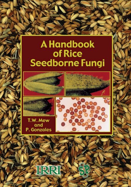
A Handbook of Rice Seedborne Fungi TW Mew and P ... - IRRI books
A Handbook of Rice Seedborne Fungi TW Mew and P ... - IRRI books
A Handbook of Rice Seedborne Fungi TW Mew and P ... - IRRI books
- No tags were found...

Create successful ePaper yourself
Turn your PDF publications into a flip-book with our unique Google optimized e-Paper software.
ContentsPrefaceVINTRODUCTION 1FUNCTIONS OF SEED HEALTH TESTING 3Cataloguing pathogens <strong>of</strong> crops 3Detection methods 3Post introduction measures 5THE MISSING LINK 6Epidemiology 6Disease <strong>and</strong> infection cycles 7Seed transmission 8Relationship between seedborne inoculum <strong>and</strong> 9disease development in the fieldInoculum level <strong>and</strong> inoculum thresholds 9Risk analysis 10Microorganisms associated with seed 11SEED HEALTH MANAGEMENT FOR CROP PRODUCTION 12IDENTIFICATION OF FUNGI DETECTED ON RICE SEED 13<strong>Seedborne</strong> fungi causing foliage diseases in rice 14Alternaria padwickii 14Bipolaris oryzae 17Cercospora janseana 21Microdochium oryzae 24Pyricularia oryzae 27<strong>Seedborne</strong> fungi causing stem, leaf sheath, <strong>and</strong> root diseases in rice 31Fusarium moniliforme 31Sarocladium oryzae 35<strong>Seedborne</strong> fungi causing grain <strong>and</strong> infloresence diseases in rice 38Curvularia sp. 38Fusarium solani 42Nigrospora sp. 44Phoma sorghina 47Pinatubo oryzae 51Tilletia barclayana 53Other fungi detected on rice seeds 57Acremoniella atra 58Acremoniella verrucosa 58Alternaria longissima 59iii
Alternaria longissima 59Alternaria tenuissima 59Aspergillus clavatus 60Aspergillus flavus-oryzae 60Aspergillus niger 61Chaetomium globosum 61Cladosporium sp. 62Curvularia eragrostidis 62Dreclslera hawaiiensis 63Epicoccum purpurascens 63Fusarium avenaceum 64Fusarium equiseti 65Fusarium larvarum 66Fusarium nivale 66Fusarium semitectum 67Gilmaniella humicola 67Memnoniella sp. 68Microascus cirrosus 68Monodictys putredinis 69Myrothecium sp. 70Nakataea sigmoidea 70Nectria haematococca 71Papularia sphaerosperma 71Penicillium sp. 72Pestalotia sp. 73Phaeoseptoria sp. 74Phaeotrichoconis crotolariae 74Pithomyces sp. 75Pyrenochaeta sp. 75Rhizopus sp. 76Septogloeum sp. 76Sordaria fimicola 77Spinulospora pucciniiphila 77Sterigmatobotrys macrocarpa 78Taeniolina sp. 78Tetraploa aristata 79Trichoderma sp. 79Trichothecium sp. 81Tritirachium sp. 81Ulocladium botrytis 80REFERENCES 82iv
PrefaceSeed health testing has become an important component<strong>of</strong> germplasm exchange between internationalcenters <strong>and</strong> national agricultural research <strong>and</strong> extensionsystem (NARES) partners. It assures the safemovement <strong>of</strong> seed, which is a carrier <strong>of</strong> pathogens,insect pests, <strong>and</strong> contaminants such as weed seeds.This h<strong>and</strong>book focuses on the importantseedborne fungi that cause diseases <strong>of</strong> the foliage,stem, leafsheath, root, grain, <strong>and</strong> inflorescence inrice. It provides information on more than 50 speciesthat have been detected in rice seed during routinetesting <strong>and</strong> analysis.Seed health testing is also a means <strong>of</strong> qualitycontrol that ensures the exchange <strong>of</strong> high-qualityseed among scientists or research centers. For thisreason, the Seed Health Unit at IFW functions as thegatekeeper <strong>of</strong> safe germplasm movement from <strong>and</strong>within <strong>IRRI</strong>, the Philippines, <strong>and</strong> outside.<strong>IRRI</strong> has always given high priority to the safety<strong>of</strong> germplasm exchange. It has worked closely withthe Philippine Plant Quarantine Service to achievethis objective. As the volume <strong>of</strong> germplasm exchangeincreased annually, a Seed Health Unit wasestablished in 1982 <strong>and</strong> a new laboratory was developed<strong>and</strong> deputized by the Philippine Plant Quarantineto undertake major activities on rice seed healthtesting for plant quarantine certification. Besides itsregular activities on seed health testing <strong>of</strong> ricegermplasm, the unit also <strong>of</strong>fers rice seed health trainingto NARES partners. To date, more than 100 scientistsfrom rice-growing countries worldwide havereceived such training.<strong>Rice</strong> seed, like seeds <strong>of</strong> other crops, carries alarge number <strong>of</strong> organisms, such as fungi describedin this book, bacteria, <strong>and</strong> nematodes. Theseseedborne organisms can be pathogens <strong>and</strong>saprophytes; many <strong>of</strong> the bacteria or fungi canied byrice seed are also potential biological control agentsagainst other rice pathogens. Some also function topromote seed germination <strong>and</strong> seedling vigor.In quarantine regulations, seedborne pathogens<strong>of</strong>ten serve as baniers to seed movement for research<strong>and</strong> for trade. Misunderst<strong>and</strong>ing arises because<strong>of</strong> insufficient biological <strong>and</strong> epidemiologicaldata to guide the development <strong>of</strong> plant quarantineregulations. Many seed-importing countries need thisinformation to determine whether seed carries thetargeted pathogens <strong>of</strong> "quarantine importance." Yetlittle is known about the pathogens carried by seed interms <strong>of</strong> crop damage <strong>and</strong> yield losses. Equally importantis the transmission <strong>of</strong> seedborne pathogens inrelation to disease establishment in the field <strong>and</strong> theconsequent effects on crop production. Not everypathogen carried by rice seed, for instance, is transmittedto the field when the seed is grown. The rate<strong>of</strong> transmission varies from one pathogen to another<strong>and</strong> in the same pathogen when the seed is sownunder different conditions for rice growth. These importantsubjects need further research.Since the early 1960s, IFW has conducted riceseed health testing to accomplish seed certification.As a result, the levels <strong>of</strong> rice seed infection causedby various microorganisms <strong>and</strong> the detection frequency<strong>of</strong> a given microorganism from different ricegrowingcountries are well documented. The informationprovided on seedborne fungi in this h<strong>and</strong>bookcan be used for teaching <strong>and</strong> as a reference whenconducting seed health testing in different laboratories.It should be used with reference to local conditions.
which take into account various production situations<strong>and</strong> ecology. In reality, it is not possible or desirableto obtain seed lots free from any organism (<strong>Mew</strong>1997).From a plant pathologist’s point <strong>of</strong> view, thereare missing links in documented information onseedborne pathogens. McGee (1995) pointed out theneed for accurate information on seed transmission<strong>of</strong> some key seedborne pathogens. We need to studythe epidemiology <strong>of</strong> seedborne pathogens in relationto disease development in the field. We need yieldloss data to estimate the risk <strong>of</strong> seedborne pathogens.Furthermore, we need to study the role <strong>of</strong> seed healthtesting to improve farmers’ pest management <strong>and</strong>crop production. To know whether common pathogenscarried by the seed pose a threat to crop production,we need to underst<strong>and</strong> disease epidemiology.Conventional seed health testing provides adequateinformation about the frequency <strong>of</strong> detection from theseed <strong>and</strong> levels <strong>of</strong> seed infection. We need to assesswhether these pathogens, upon detection, could betransmitted to the field when the seed is sown <strong>and</strong> ifthe disease that develops causes damage or injury toeffect yield loss. In scientific literature, this informationis not readily available or it needs to be confirmed.Very little research has been done in thisarea. Because <strong>of</strong> the increasing concern aboutseedborne pathogens, we need to underst<strong>and</strong> theirepidemiology. The initial inoculum is the key to underst<strong>and</strong>ingwhat causes an epidemic in a plant quarantinecontext. The threshold inoculum carried by aseed lot has to be defined in terms <strong>of</strong> its effect ontransmission <strong>and</strong> disease establishment. Detectionmethods <strong>and</strong> the potential role <strong>of</strong> nonpathogenic microorganisms,especially those possessing biologicalcontrol properties, must be studied <strong>and</strong> taken intoaccount.2
Functions <strong>of</strong> seed health testingSeed health testing is done to determine microbialinfection or contamination for quarantine purposes(e.g., international seed exchange or movement). Itidentifies the cause <strong>of</strong> seed infection that affects theplanting value <strong>of</strong> seed lots for seed certification byseed growers to supply seed to farmers. Seed testingaffects policies on seed improvement, seed trade,<strong>and</strong> plant protection. Neergard (1979) brought out theimportance <strong>of</strong> pathogens carried by seeds <strong>and</strong> thedisease potential assigned to pathogens.Several routine activities are undertaken duringseed health testing. These include dry seed inspection,the st<strong>and</strong>ard blotter test for seed infection <strong>and</strong>contamination, postentry planting for field inspection<strong>of</strong> undetected plant diseases <strong>of</strong> seedborne <strong>and</strong> seedcontaminatedpathogens, <strong>and</strong> certification. In seedmultiplication for export, crop inspection prior to seedharvest <strong>of</strong>fers an additional means to link seedbornepathogens <strong>and</strong> diseases <strong>of</strong> mother plants. All theseactivities provide preventive measures to eliminatethe introduction <strong>of</strong> undesirable pathogens into a regionor country. Seed health testing <strong>of</strong>fers a powerfultool for documenting microorganisms associated withseeds. Information on microorganisms, however,needs to be associated with a database on yield loss<strong>and</strong> information on pathogens that cause diseases.Catalouging pathogens <strong>of</strong> cropsFor rice, seed health testing has been done on morethan 500,000 seed lots following International SeedTesting Association (ISTA) rules (1985). A total <strong>of</strong>more than 80 fungi were detected on rice seeds(Table 1). The detection frequency varied. About 20species <strong>of</strong> fungal pathogens were detected from riceseed at any one time. Not all <strong>of</strong> them cause notablediseases in the field <strong>and</strong> it was not ascertainedwhether diseases were all seed-transmitted <strong>and</strong>, if so,what their transmission efficiency was. The role <strong>of</strong> arice seed in a fungus life cycle is not clear.Pyricularia oryzae, the rice blast pathogen, althoughconsidered a very important rice pathogen,has the lowest detection frequency. The level variedaccording to seed source. Except Fusariummoniliforme, the seedborne inoculum <strong>of</strong> the otherpathogens may not serve as an important source <strong>of</strong>secondary inoculum in the field. The infection level<strong>of</strong> P. oryzae is likely to be higher in temperate orsubtropical environments than in tropical environments.The data set provides insights into the occurrence<strong>of</strong> rice fungal pathogens. The detection frequency<strong>and</strong> infection level are very high for Alternariapadwickii (80–90%) (Fig. 1). In tropical Asia,stackburn, the disease it causes, is hardly observed inthe field.Detection methodsMany detection methods have been developed overthe years for various seedborne pathogens. We foundthe blotter test to be a common but efficient method<strong>of</strong> detecting seedborne fungal pathogens in rice seed.Following ISTA rules, the method involves plating400 seeds on some layers <strong>of</strong> moistened filter paper.Below is a list <strong>of</strong> the different detection methods usedin routine seed health testing. Descriptions <strong>of</strong> thesemethods can be found in the references listed (seepage 82).Seed health testing procedures involve techniquessuch as• Direct examination <strong>of</strong> dry seeds• Examination <strong>of</strong> germinated seeds• Examination <strong>of</strong> organisms removed by washing• Examination after incubation (both blotter <strong>and</strong>agar plates)• Examination <strong>of</strong> growing plants (for example,the seedling symptom test)• Embryo count methods• Molecular <strong>and</strong> serological techniquesOther methods include a selective medium forspecific pathogens. With advances in moleculartechniques, emphasis in fungal identification <strong>and</strong>taxonomy has changed from a morphological approach(for example, spore size <strong>and</strong> spore shape) toa more functional approach based on aspects <strong>of</strong> thelife cycle, mechanisms <strong>of</strong> spore production <strong>and</strong> release,DNA relationships, <strong>and</strong> physiological attributes.DNA analysis techniques such as the polymerasechain reaction (PCR), <strong>and</strong> r<strong>and</strong>om amplifiedpolymorphic DNA (RAPD) analysis are the mostcommonly used tools.These are powerful techniques for detecting <strong>and</strong>for establishing the relationship between the inocu-3
The missing linkEpidemiologyThere is little doubt that many pathogens areseedborne. Questions arise, however, on whether theintroduction <strong>of</strong> seedborne inoculum <strong>of</strong> these pathogenswould lead to the establishment <strong>of</strong> a disease inthe field or whether the field population <strong>of</strong> a fungalpathogen is derived from the seedborne inoculum.Pathogens <strong>of</strong> significance to quarantine suggestthe potential <strong>of</strong> seed transmission. They also relate tothe potential damage or yield loss caused by diseasesderived from the seedborne inoculum <strong>of</strong> the pathogen.However, there is very little accurate informationabout yield loss caused by rice diseases, <strong>and</strong>diseases derived from seedborne inoculum. Yieldloss caused by a pest outbreak or a disease epidemicis important in determining pathogens with quarantinesignificance. There are very few comprehensivestudies or databases on yield losses caused by pestsor pathogens in scientific literature. Studies conducted<strong>and</strong> documented by Savary et al (1996, 1997,1998, 2000a,b), Savary <strong>and</strong> Willocquet (1999), <strong>and</strong>Willocquet et al (1999a,b) are some <strong>of</strong> the most comprehensiveones on rice diseases. Using both survey<strong>and</strong> experimental data, they developed pest <strong>and</strong>pathogen pr<strong>of</strong>iles for different rice production situations(PS). Production situations refer to the set <strong>of</strong>environmental conditions—climatic, technical, social,economic, <strong>and</strong> biological—under which agriculturalproduction takes place. These were then relatedto yield losses with individual pests <strong>and</strong> pathogens,<strong>and</strong> also pest <strong>and</strong> pathogen pr<strong>of</strong>iles.Savary et al (1996, 1997, 1998, 1999) believethat by using such a systems approach combinedwith different statistical analyses, all these factorscould be captured by a limited number <strong>of</strong> variables,such as those that describe patterns <strong>of</strong> cropping practices,for example, method <strong>of</strong> crop establishment,amount <strong>of</strong> chemical fertilizer used, type <strong>of</strong> weedcontrol, <strong>and</strong> rice cultivar type (with or without diseaseresistance). In reality, farmers’ practices are, to alarge extent, reflections <strong>of</strong>, or adaptations to, social,physical, <strong>and</strong> biological environments. Injury pr<strong>of</strong>ilesrefer to the sequence <strong>of</strong> harmful organisms that mayoccur during the crop cycle. Many such organismsaffect rice. The number <strong>of</strong> processes by which a pestor pathogen may affect rice, however, is limited toless than 10, <strong>and</strong> injuries are <strong>of</strong>ten associated withone another. On this basis, yield losses caused byindividual injuries as well as by injury pr<strong>of</strong>iles establishthe importance <strong>of</strong> rice pests <strong>and</strong> diseases in specificPS at the regional level (Savary et al 2000a,b).The database identified sheath blight caused byRhizoctonia solani AG1 <strong>and</strong> brown spot caused byBipolaris oryzae as the two most important diseasesin rice in Asia, each responsible for 6% yield loss,whereas blast caused by Pyricularia grisea <strong>and</strong> bacterialblight caused by Xanthomonas oryzae pv.oryzae account for 1–3% <strong>and</strong> 0.1% yield losses, respectively.However, most rice cultivars planted byAsian farmers are resistant to these two diseases. Ifcultivars possess no resistance to these two diseases,yield losses are likely to be higher than current estimates.Other diseases, such as sheath rot, stem rot,<strong>and</strong> those known as sheath rot complex <strong>and</strong> graindiscoloration (Cottyn et al 1996a,b), are responsiblefor rice yield losses ranging from 0.1% to 0.5%. Allother diseases alone or in combination would notcause more than 0.5–1% yield losses based on estimates.Projected yield losses cause by various ricediseases under different production situations aregiven in Table 3.In seed health testing, detection frequencymeans the number <strong>of</strong> pathogens detected in a seedlot. Infection frequency refers to the number <strong>of</strong> seeds(based on 400 seeds tested) within a seed lot whichare infected (<strong>Mew</strong> <strong>and</strong> Merca 1992) <strong>and</strong> is equivalentto the inoculum level. In the epidemiologicalsense, no information is available to correlate detectionfrequency <strong>and</strong> infection frequency to seed transmission<strong>and</strong> disease establishment in the field. Still,there are other questions related to seedborne pathogensthat must be answered. In rice, in which mostfungal pathogens can be seedborne, <strong>and</strong> for whichcurrent farmer cultural practices have done little toimprove quality (a result <strong>of</strong> farm labor shortage <strong>and</strong>short turnaround time), what is introduced to the fieldwith seeds when the rice crop is planted? In seedproduction fields, it is necessary to practice diseasemanagement to produce disease-free seed?6
Table 3. Pathogen pr<strong>of</strong>iles closely associated with rice production situations (PS) <strong>and</strong> potential yield losses causedby rice diseases (adapted <strong>and</strong> modified from Savary et al 1998, Savary <strong>and</strong> Willocquet 1999).PS1 PS2 PS3 PS4 PS5 PS6 Yield loss(%)Actual yield (t ha –1 ) 4.8 4.6 3.5 6.7 3.8 3.9DiseaseBlast a L L M M 1–3Bacterial blight L L L L L 0.2Bakanae VL 0.0Brown spot L L VH H H 6.6Sheath blight VH VH M VH H H 6.4Sheath rot complex M M H M 0.5Grain discoloration M M H M 0.1Characteristics <strong>of</strong> environmentsMineral fertilizer m l l h m hFallow period l l m s m sDrought stress l l h l h mWater stress l l l h h hCrop establishment tr tr tr ds ds dsHerbicide use m l l m l lInsecticide use m m m m m m<strong>Fungi</strong>cide use l l l h h hPrevious crop rice rice w/b w/b rice riceaIn the surveys, rice varieties possessing resistance to blast <strong>and</strong> bacterial blight diseases. For characteristics <strong>of</strong> environments, m = moderate, h =high, l = low, tr = transplanted rice, ds = direct-seeded rice, s = short, w/b = wheat or barley. For diseases <strong>and</strong> grain discoloration, L = low, M = medium,H = high, VH = very high.Disease <strong>and</strong> infection cyclesFigure 2 shows how seedborne inoculum reinfectsthe seed during the development <strong>of</strong> a disease epidemic:seedborne inoculum → disease establishment→ disease development in the field (infection cycle)→ crop damage or yield loss (effect <strong>of</strong> seedborneinoculum) → reinfection <strong>of</strong> infestation <strong>of</strong> seed (potentialdissemination to other fields, regions, or countries).There is voluminous information on seedbornepathogens <strong>of</strong> various crops derived from routine seedhealth testing for either certification or issuance <strong>of</strong>phytosanitary certificates. Information on transmission<strong>of</strong> the pathogen from the infected or infestedseed to disease development in the field is scarce.Various factors that affect the infection cycle areweather conditions, cropping practices, resistance orsusceptibility <strong>of</strong> the variety, virulence <strong>of</strong> the pathogen,<strong>and</strong> amount <strong>of</strong> incoculum produced for secondaryspread <strong>and</strong> efficiency <strong>of</strong> the inoculum.It is <strong>of</strong>ten assumed that, for a pathogen to beseedborne, it must be seed-transmitted. McGee(1995) indicated that in only very few seedbornepathogens is the transmission clearly established.When conditions in the nursery bed <strong>and</strong> theecosystem where rice is grown re taken into account,there is inadequate documentation on plantquarantine to guide decision making. It is notknown under what specific conditions seedbornepathogens are transmitted to the crop at the seedlingstage. Blast caused by P. oryzae <strong>and</strong> bakanaecaused by F. moniliforme, are two <strong>of</strong> the betterknown diseases (Ou 1985). Once a disease is establishedin a crop, its intensity will depend on factorsthat influence the infection cycle. Climaticconditions <strong>and</strong> crop management practices arecrucial to disease development.In rice, the infection frequency <strong>of</strong> P. oryzae isvery low, yet the disease potential under a conduciveenvironment (e.g., upl<strong>and</strong>, subtropical, <strong>and</strong>temperate) is very high. Once seedlings are infectedfrom seedborne inoculum, even at a lowinfection rate, millions <strong>of</strong> conidia are produced forsecondary infection. On the other h<strong>and</strong>, seedborneF. moniliforme <strong>of</strong>ten induces bakanae with onlyone cycle <strong>of</strong> infection. Therefore, the initial inocu-7
<strong>Seedborne</strong>inoculumReinfection/infection <strong>of</strong> seedTransmission(Establishment)InfectedseedCrop damage/Injury (Impact)InoculumproductionDiseasedevelopmentInfectionefficiencyClimaticconditionsCroppingenvironmentsFig. 2. Diseases <strong>and</strong> infection cycles <strong>of</strong> a seedborne fungal disease <strong>and</strong> its effect.lum for F. moniliforme is important. Once theseedborne inoculum is minimized, the disease islikely to be controlled.Changes in crop cultivation methods <strong>and</strong> culturalpractices affect seedborne diseases. In traditionalmethods <strong>of</strong> cultivation, rice seedlings are raised in aseedbed with a saturated water supply. Because <strong>of</strong>the reduction in arable l<strong>and</strong> <strong>and</strong> the decreasing productivity<strong>of</strong> available agricultural l<strong>and</strong>, new methods<strong>of</strong> cultivation are being developed. These new methodsare conducive to the transmission <strong>and</strong> development<strong>of</strong> seedborne diseases previously consideredminor.In epidemiological research, seed transmission<strong>and</strong> establishment <strong>of</strong> disease derived from seedborneinoculum should be considered. These data are essentialfor assessing the importance <strong>of</strong> seedbornepathogens.Seed transmissionMcGee (1995) indicated that one <strong>of</strong> the missing linksin seed health testing is the lack <strong>of</strong> information onseed transmission. Based on postquarantine planting,one <strong>of</strong> the difficulties encountered is distinguishingbetween a disease that developed from inoculumderived from the seed <strong>and</strong> that from other sources.Polymerase chain reaction (PCR) DNA technologyis useful in this regard. Based on DNA fingerprinting,patterns <strong>of</strong> a pathogen population can be distinguishedfrom those <strong>of</strong> the pathogen manifesting adisease on the crop grown from the seed. Thiswould establish the transmission <strong>of</strong> the seedborneinoculum <strong>and</strong> its relation to the disease on the cropin the field. In routine disease monitoring <strong>of</strong> fieldcrops such as rice or other nursery crops, identifyingdisease foci in nursery beds may be an alternative.For rice, this appears feasible at the seedlingstage in the seedbed. A disease focus is a patch <strong>of</strong>crop with disease limited in space <strong>and</strong> time(Zadoks <strong>and</strong> van den Bosch 1994) <strong>and</strong> is likely tohave been caused by the initial source <strong>of</strong> inoculum.In Japan, the seedbox nursery for rice provides anideal means to identify the disease foci <strong>of</strong> single ordifferent seedborne pathogens. The paper towelmethod, a very common method for testing seedgermination, resulted in more seedling mortality<strong>and</strong> thus less germination than the seedbed method(seedbed with field soil) used in crop production(Table 4). The method used for assessing the effect<strong>of</strong> seedborne fungal pathogens on seed germinationvaries.8
Table 4. Germination (%) <strong>of</strong> untreated <strong>and</strong> treated seeds using paper towel <strong>and</strong> in-soil germination methods (400seeds each; r<strong>and</strong>omized complete block design).Normal a Abnormal Dead seedsVarieties Paper In-soil Paper In-soil Paper In-soiltowel test towel test towel testUntreatedIR62 79.7 ab 91.7 a 16.0 a 5.3 a 4.3 a 3.0 bSARBON 65.3 b 75.7 ab 20.3 a 12.0 a 14.3 a 12.3 abC22 94.0 a 84.0 ab 4.3 b 10.3 a 1.7 a 5.7 abBS1-10 68.0 b 72.7 b 18.3 a 11.0 a 13.7 a 16.3 aHot-water treatmentIR62 86.7 a 85.3 a 5.0 b 7.7 b 8.3 b 7.0 bSARBON 46.3 b 50.3 c 19.7 a 10.3 b 34.0 a 39.3 aC22 92.3 a 94.3 a 5.0 b 4.0 b 2.7 b 1.7 bBS1-10 76.3 a 67.7 b 13.7 ab 25.0 a 10.0 a 7.3 baIn a column under each treatment, means followed by a common letter are not significantly different at the 5% level by Duncan’s multiple rangetest.Relationship between seedborne inoculum<strong>and</strong> disease development in the fieldIn determining the importance <strong>of</strong> a seedborne pathogen,it is essential to relate inoculum production <strong>and</strong>the efficiency <strong>of</strong> the secondary spread to the inoculumthreshold <strong>and</strong> disease severity after establishment.For a monocyclic disease, initial infectionshould be closely related to the initial inoculum providedby the seed. For a polycyclic disease, a lowlevel <strong>of</strong> seedborne inoculum is adequate to begininfection from the seedbed to the main field, <strong>and</strong> increasedisease intensity if climatic or crop-growingconditions are favorable. For instance, in rice blastcaused by P. oryzae with low detection <strong>and</strong> infectionfrequencies, seed-carried inoculum is more importantin temperate or subtropical environments than ina tropical lowl<strong>and</strong> environment. In the former environments,the likelihood <strong>of</strong> seed-carried inoculumbeginning an infection <strong>and</strong> producing a sufficientamount <strong>of</strong> inoculum for secondary infection is higher(Ou 1985).Inoculum level <strong>and</strong> inoculum thresholdsIn seed health testing for certification, the inoculumthreshold <strong>of</strong> seedborne pathogens is defined as theamount <strong>of</strong> seed infection or infestation that can causea disease in the field under conducive conditions <strong>and</strong>lead to economic losses (Kuan 1988). We believethat this should mean a minimal amount <strong>of</strong> seed infectionor infestation. In principle <strong>and</strong> as Gabrielson(1988) indicated, one infected seed may give rise toone infected plant, but, under field conditions, this ishardly the case. The values <strong>of</strong> the inoculum thresholdfor different crop-pathogen combinations in differentcountries vary widely (Gabrielson 1988).Our experience with rice has shown that thepotential <strong>of</strong> a seedborne pathogen to cause a diseaseis determined by the type <strong>of</strong> pathogen in relation tothe crop growth environment. Under conditions in awet-bed nursery for rice seedlings, the likelihood <strong>of</strong> afungal pathogen beginning an infection appears lessthan under tropical conditions. Perhaps this is because<strong>of</strong> the microbial competition or antagonism.On the other h<strong>and</strong>, if the level <strong>of</strong> seedborne inoculumis high (we have not had it quantified), then the probability<strong>of</strong> it causing infection is also high. As one infectedseed begins one disease focus <strong>and</strong> this focalpoint exp<strong>and</strong>s, the probability <strong>of</strong> infection increases.In reality, disease establishment is affected by inoculumdensity <strong>and</strong> the crop cultivation environment.The more infected seeds there are (inoculum level),the higher the probability <strong>of</strong> having an infection.We have monitored detection levels <strong>of</strong> seedbornefungal pathogens from imported seed lots byplanting them in the field after seed treatment forpostentry plant quarantine observation. Diseases observedwere not related to seedborne pathogens(Table 2). Pathogens from harvested seeds fromthese plants were detected, but we are not surewhether these fungal pathogen populations were thesame as those carried by the original seed or if theycame from other sources in the field.9
For other fungal pathogens, there is a close relationbetween seed infection <strong>and</strong> infected plantsgrown from these seeds. An example is blackleg <strong>of</strong>crucifer caused by Phoma lingam (Leptosphaeriamaculans) (Gabrielson1983). The classical examplefrom Heald (1921) indicated that the sporeload <strong>of</strong>seeds was highly correlated to the percentage <strong>of</strong>smut appearing in the field.Inoculum thresholds vary according to culturalenvironments. In Japan, for instance, after rice cultivationbecame mechanized <strong>and</strong> seedlings wereraised indoors in seedboxes, the occurrence <strong>of</strong> manyseedborne fungal <strong>and</strong> bacterial pathogens increased.This is because the indoor conditions—high temperature<strong>and</strong> high humidity with artificial light—are veryfavorable for seedling disease development. As aresult, the inoculum threshold is lower than that <strong>of</strong>seedlings raised outdoors under a field nursery. Theinoculum becomes more efficient under certain conditions.Inoculum efficiency is determined by variousfactors. The type <strong>of</strong> disease <strong>and</strong> crop-growing environmentsare important. Gabrielson (1988) cautionedthat thresholds must be developed for average environmentalconditions <strong>of</strong> crop growth because theyare influenced by all factors affecting the epidemiology<strong>of</strong> each host-parasite combination. It is difficultto use a single threshold <strong>of</strong> a single disease for allcropping environments. There is no clear definitionon levels <strong>of</strong> threshold for the different pathogens detectedfrom the seed. In rice, different fungal pathogensare detected from the seed (Table 1) <strong>and</strong> all <strong>of</strong>them are distributed throughout the rice-growingcountries worldwide. Disease potential, however,depends on the rice ecosystem (upl<strong>and</strong>, rainfed, irrigated,tropical, subtropical, <strong>and</strong> temperate environments,<strong>and</strong> deepwater <strong>and</strong> tidal coastal areas), culturalconditions, <strong>and</strong> types <strong>of</strong> crop management <strong>and</strong>production. Whether there is a need to treat all diseasesthe same way or differently for different ecosystems<strong>and</strong> production levels needs careful study.There is a general agreement that the threshold levelfor a disease is zero in an area if it has not been reportedthere.Risk analysisRisk analysis should serve an important basis fordeveloping plant quarantine regulations. Risk analysisbased on seed health testing needs to consider thefollowing factors:1. type <strong>of</strong> pathogens2. role <strong>of</strong> seed in the life cycle <strong>of</strong> thepathogen3. disease or epidemic potential4. genetic variability <strong>of</strong> the pathogen5. type or site <strong>of</strong> initial infection6. kind <strong>of</strong> crop production environment (<strong>Mew</strong>1997)The risk <strong>of</strong> infection from seedborne pathogensis a function <strong>of</strong> risk probability <strong>and</strong> risk magnitude.Furthermore, risk probability is determined by introductionrisk, that is, the probability that a pathogenenters a region or a field through the seed, the epidemiologicalrisk, the probability that the pathogen establishesinfection through seedborne inoculum. Riskmagnitude is the potential consequence <strong>of</strong> an epidemiccaused by the pathogen. Consequences areconsidered from the viewpoint <strong>of</strong> yield loss. Seedhealth testing results provide actual data on a pathogenthat could potentially be introduced into a regionor a field. The risk magnitude can be computed froma yield loss database or from modeling. In rice, thiskind <strong>of</strong> database is available at <strong>IRRI</strong>. The yield lossdatabase provides an estimate <strong>of</strong> losses <strong>and</strong> “hazards”caused by a pathogen once the infection is establishedthrough seedborne inoculum.However, data are lacking on the transmissionefficiency <strong>of</strong> seedborne inoculum <strong>of</strong> many riceseedborne pathogens. A concerted effort is needed tocompile this information through international collaboration.Research on seed pathology provides thebasis for setting seed health testing policy, while informationon pest or pathogen risk provides a startingpoint for seed health testing on target organisms forplant quarantine regulations. Very limited or no financialsupport is available for this important area <strong>of</strong>activities.A yield loss database can estimate the “hazards”<strong>of</strong> a pathogen once an infection is establishedthrough the introduction <strong>of</strong> a seedborne inoculum.However, data on inoculum levels <strong>and</strong> thresholds arealso needed to develop realistic assessment or measurementprocedures for some important seedbornepathogens. Data on seed transmission <strong>of</strong> manypathogens <strong>and</strong> transmission efficiency <strong>of</strong> seedborneinoculum are currently not available.Although conventional seed health testing providesadequate information on detection frequency<strong>and</strong> infection levels <strong>of</strong> some pathogens, we need toassess whether these pathogens cause any real injuryto effect yield loss. In scientific literature, this informationis not readily available.10
Microorganisms associated with seedNot all microorganisms associated with seed arepathogens. Some microorganisms possess biologicalcontrol properties. The occurrence <strong>of</strong> nonpathogenicXanthomonas has further complicated the issue <strong>of</strong>seedborne bacterial pathogens. Cottyn et al (2001)<strong>and</strong> Xie et al (2001) proved that seedborne antagonisticbacteria are present in rice <strong>and</strong> promote seed germination<strong>and</strong> seedling vigor, <strong>and</strong> also suppress diseasewith an inoculum from the seed. Micr<strong>of</strong>loraassociated with the seed may be roughly categorizedinto pathogens <strong>and</strong> nonpathogens. The study byCottyn et al (2001), supported by the BelgiumAdminstration for Development Cooperation, <strong>and</strong>Xie et al (2001) showed that rice seed carries manybacteria belonging to 17 genera <strong>and</strong> over hundreds <strong>of</strong>species. Predominant were Enterobacteriacae(25%), Bacillus spp. (22%) <strong>and</strong> Pseudomonas spp.(14%). Other bacteria regularly present wereXanthomonas spp., Cellulomonas flavigena, <strong>and</strong>Clavibacter michiganense. We found that about 4%<strong>of</strong> the total bacterial population possesses biologicalcontrol properties against most seedborne pathogens.Also, seedling vigor was enhanced after soakingseeds in bacterial suspension. These studies showthat rice seed not only carries pathogens but alsoabundant microorganisms that act as biological controlagents. Whether they play a bigger role in cropproduction <strong>and</strong> disease management needs furtherresearch. More support should be given to this researcharea, which is a vital part <strong>of</strong> a farmers’ internalresource management for sustainable crop production<strong>and</strong> disease management.11
Identification <strong>of</strong> fungi detected on rice seedThe st<strong>and</strong>ard detection method used in identifyingfungi on rice seed at <strong>IRRI</strong> is given below. Figure 3shows the parts <strong>of</strong> a rice seed attacked by fungi.With this method, numerous fungi have been detectedon rice seed. The pr<strong>of</strong>ile <strong>of</strong> each fungus detectedis presented in the following pages.Methods <strong>and</strong> conditions <strong>of</strong> rice seed incubationfor microorganism detection are listed below.The International Rules for Seed Testing recommendthe blotter test for detecting seedborne fungi.The procedure involves these steps:1. Prepare materials (9.5-cm plastic petri dish,marking pencil, round blotter paper, distilledwater, sampling pan, forceps, seed sample).2. Label plates accordingly using a markingpencil.3. Place 2–3 pieces <strong>of</strong> moistened round blotterpaper in labeled plastic petri dishes.4. Sow 25 seeds per plate making sure thatseeds are sown equidistantly with 15 seeds onthe outer ring, 9 seeds at the inner ring, <strong>and</strong> 1seed in the middle.5. Incubate seeded plates at 21 °C under a 12-hlight <strong>and</strong> 12-h dark cycle. Light sources canbe near ultraviolet (NUV) light or daylightfluorescent tubes. The NUV light source canbe a 320–400 nm lamp, preferably PhilipsTLD 36W/08 or GE F 40 BL. Daylight fluorescenttubes can be Philips TL 40W/54 daylight or its equivalent.6. Examine each <strong>of</strong> the seeds after 5–7 d <strong>of</strong>incubation for fungal growth.Partition betweenlemma <strong>and</strong> paleaLemmaAwnSterile lemmasPaleaFig. 3. Parts <strong>of</strong> a rice seed.13
<strong>Seedborne</strong> fungi causing foliage diseases in riceAlternaria padwickii (Ganguly) Ellissyn. Trichoconis padwickii GangulyTrichoconiella padwickii (Ganguly) JainDisease caused: stackburna. SymptomsOn leaves—large oval or circular spots with apale brown center <strong>and</strong> distinct dark brown margin.Color <strong>of</strong> center eventually becomes white <strong>and</strong>bears minute black dots.On grains—pale brown to whitish spots with blackdots at the center <strong>and</strong> dark brown border.Roots <strong>and</strong> coleoptile <strong>of</strong> germinating seedlings—dark brown to black spots that eventually coalesce.Small, discrete, <strong>and</strong> black bodies areformed on the surface <strong>of</strong> the darkened area asdecay proceeds.b. Occurrence/distributionStackburn disease is widespread in most <strong>of</strong> therice-growing countries worldwide (Fig. 4).c. Disease historyThe disease was first reported in the U.S. It resemblesblack rust <strong>of</strong> wheat on rice leaves, butonly sclerotia <strong>and</strong> mycelium were observed.Later the fungus was observed in <strong>and</strong> on riceseeds.d. Importance in crop productionStackburn leaf spot disease is not considered tobe <strong>of</strong> economic importance. However, seed infectionresults in grain discoloration, which mayreduce germination <strong>and</strong> lower grain quality. Thedisease potential <strong>of</strong> stackburn is very low <strong>and</strong> theyield loss caused by A. padwickii in literaturemay be overestimated. The effect <strong>of</strong> infectedseed on seed germination is not yet properly assessed.Detection on seeda. Incubation period on blotterA. padwickii is easily observed on seeds using theblotter method 5 d after seeding on moistenedblotter <strong>and</strong> incubated under NUV at 21 °C. Thedetection frequency is about 67.1% on seedscoming from different regions (Fig. 5a,b).b. Habit characterSeed infected with A. padwickii after incubationshows abundant aerial mycelia, hairy to cottony,pr<strong>of</strong>usely branched, grayish or hyaline whenFig. 4. Occurence <strong>of</strong> stackburn (Ou 1985, Agarwal <strong>and</strong> Mathur 1988, EPPO 1997).14
Detection frequency (%)100806040200Detection level (%)10080East AsiaEuropeLatin AmericaSouth AsiaSoutheast AsiaSub-Saharan Africa60402001990 1991 1992 1993 1994 1995 1996 1997YearFig. 5. Detection level (a) <strong>and</strong> frequency (b) <strong>of</strong> Alternaria padwickii from imported untreated seeds, 1990-97.young, becoming creamy yellow when mature;pinkish to light violet pigmentation is produced onthe blotter; conidia are borne singly per conidiophore;darker than mycelia; sterile appendageprominent (Fig. 6a-c).c. Location on the seedA. padwickii is most <strong>of</strong>ten observed growing overthe entire seed surface (36%) (Fig. 7).Microscopic charactera. Mycelia—septate, pr<strong>of</strong>usely branched; hyalinewhen young, becoming creamy yellow when mature;branches arising at right angles from themain axis (Fig. 6d).b. Conidiophore—simple, not sharply distinguishablefrom mature hyphae, <strong>of</strong>ten swollen at the apex,hyaline when young, becoming creamy yellowwhen mature (Fig. 6e).c. Conidia—straight, shape varies from fusiform toobclavate <strong>and</strong> rostrate or in some caseselongately fusoid; with long sterile appendage; atfirst hyaline, becoming straw-colored to goldenbrown; thick-walled; 3–5 septate; constricted atthe septum; 4- to 5-celled, second cell from thebase larger than the rest <strong>of</strong> the cells (Fig. 6f).Measurements: 81.42–225.40 µ long includingappendage; 11.96–23.46 µ wide at the broadestpart <strong>and</strong> 2.99–5.52 µ wide at the center <strong>of</strong> the appendage(PSA); 83.95–203.78 µ long includingappendage; 9.66–17.48 µ wide in the broadest part<strong>and</strong> 3.45–5.75 µ wide at the middle <strong>of</strong> the appendage.Colony characters on culture media (Fig. 8)Colonies on potato dextrose agar (PDA) incubated atambient room temperature (ART) (28–30 °C) grow15
→ab→ef→f→cd →→Fig. 6. Habit character <strong>of</strong> Alternaria padwickii (Ganguly) Ellis on (a) whole seed (8X) <strong>and</strong> on sterile lemmas at (b)12.5X <strong>and</strong> (c) 25X. Photomicrograph <strong>of</strong> A. padwickii showing (d) mycelia, (e) conidiophore, <strong>and</strong> (f) conidia at 40X<strong>and</strong> stained with lactophenol blue.Observed frequency (%)403020100SterilelemmasAwnPartitionbet. lemma<strong>and</strong> paleaSeed partEntireseedLemma/paleaonly orbothFig. 7. Observed frequency <strong>of</strong> Alternaria padwickiioccurrence on seed part.Fig. 8. Plate culture <strong>of</strong> Alternaria padwickii Ellis showingcolony growths on potato dextrose agar (PDA), potatosucrose agar (PSA), <strong>and</strong> malt extract agar (MEA)incubated at ambient room temperature (ART), 21 °C,<strong>and</strong> 28 °C at 15 d after inoculation.16
moderately fast <strong>and</strong> attain a 4.32-cm diam in 5 d.They are slightly zonated, thickly felted, <strong>and</strong> grayish,becoming light outward. On the reverse side <strong>of</strong> theagar plate, the colony is azonated, black, <strong>and</strong> lighteroutward. At 21 °C under alternating 12-h NUV light<strong>and</strong> 12-h darkness, colonies grow moderately fast<strong>and</strong> attain a 4.14-cm diam in 5 d. They are azonated,becoming markedly zonated outward, felted, yellowishto greenish gray, with a 0.5-cm sterile white margin.On the reverse side <strong>of</strong> the agar plate, the colonyappears zonated <strong>and</strong> black <strong>and</strong> light outward. At 28°C under alternating 12-h light <strong>and</strong> 12-h darkness,colonies grow moderately fast <strong>and</strong> attain a 4.33-cmdiam in 5 d. They are zonated, felted, <strong>and</strong> greenishgray. On the reverse side <strong>of</strong> the agar plate, the colonyis zonated <strong>and</strong> black <strong>and</strong> yellowish brown outward.Colonies on potato sucrose agar (PSA) incubatedat ART (28–30 °C) grow moderately fast <strong>and</strong>attain a 4.18-cm diam in 5 d. They are deeply felted,zonated with an even margin, <strong>and</strong> gray. The colonyappears zonated <strong>and</strong> black on the reverse side <strong>of</strong> theagar plate. At 21 °C under alternating 12-h NUV <strong>and</strong>12-h darkness, colonies grow moderately fast <strong>and</strong>attain a 4.36-cm diam in 5 d. They are slightly zonatedwith a light gray submerged advancing margin,felted, <strong>and</strong> dark greenish gray. The colony appearsslightly zonated, black, <strong>and</strong> lighter outward on thereverse side <strong>of</strong> the agar plate. At 28 °C under alternating12-h light <strong>and</strong> 12-h darkness, colonies growmoderately fast <strong>and</strong> attain a 4.06-cm diam in 5 d.They are zonated, felted with a sinuate margin, yellowishto greenish gray, <strong>and</strong> lighter at the margins.The colony appears zonated <strong>and</strong> black, <strong>and</strong> yellowishbrown outward on the reverse side <strong>of</strong> the agarplate.Colonies on malt extract agar (MEA) incubatedat ART (28–30 °C) grow moderately fast <strong>and</strong> attain a4.53-cm diam in 5 d. They are zonated, felted, <strong>and</strong>light gray to gray. The colony appears zonated <strong>and</strong>black on the reverse side <strong>of</strong> the agar plate. At 21 °Cunder alternating 12-h NUV <strong>and</strong> 12-h darkness, coloniesgrow moderately fast <strong>and</strong> attain a 4.47-cm diamin 5 d. They are azonated, becoming markedly zonatedoutward, <strong>and</strong> white to yellowish gray <strong>and</strong> becominggray outward. The colony appears zonated<strong>and</strong> black with a light gray margin on the reverse side<strong>of</strong> the agar plate. At 28 °C under alternating 12-hfluorescent light <strong>and</strong> 12-h darkness, colonies growmoderately fast <strong>and</strong> attain a 4.90-cm diam in 5 d.They are zonated, felted, <strong>and</strong> greenish gray, becominggray at the margins. The colony appears slightlyzonated <strong>and</strong> black on the reverse side <strong>of</strong> the agarplate.Bipolaris oryzae (Breda de Haan) Shoem.syn. Drechslera oryzae (Breda de Haan) Subram. & JainHelminsthosporium oryzaeteleomorph: Cochliobolus miyabeanus (Ito & Kurib)Disease caused: brown spot (brown leaf spot orsesame leaf spot)Helminsthosporium blighta. SymptomsOn leaves—small <strong>and</strong> circular dark brown orpurple brown spots eventually becoming oval(similar to size <strong>and</strong> shape <strong>of</strong> sesame seeds) <strong>and</strong>brown spots with gray to whitish centers, evenlydistributed over the leaf surface; spots muchlarger on susceptible cultivars. A halo relating totoxin produced by the pathogen <strong>of</strong>ten surroundsthe lesions.On glumes—black or brown spots covering theentire surface <strong>of</strong> the seed in severe cases. Underfavorable environments, conidiophore <strong>and</strong> conidiamay develop on the spots, giving a velvety appearance.Coleoptile—small, circular, or oval brown spots.b. Occurrence/distributionBrown spot is distributed worldwide <strong>and</strong> reportedin all rice-growing countries in Asia, America,<strong>and</strong> Africa (Fig. 9). It is more prevalent in rainfedlowl<strong>and</strong>s <strong>and</strong> upl<strong>and</strong>s or under situations with abnormalor poor soil conditions.c. Disease historyThis fungus was first described in 1900 <strong>and</strong>named as Helminthosporium oryzae. In Japan, theteleomorph was found in culture <strong>and</strong> was namedOphiobolus miyabeanus. However, Drechslerdecided it belonged to Cochliobolus <strong>and</strong> renamed17
Fig. 9. Occurrence <strong>of</strong> brown spot (Ou 1985, Agarwal <strong>and</strong> Mathur 1988, EPPO 1997).it Cochliobolus miyabeanus. Because <strong>of</strong> the bipolargermination <strong>of</strong> the conidia, the anamorph <strong>of</strong> C.miyabeanus was changed to Bipolaris oryzae.d. Importance in crop productionBipolaris oryzae causes seedling blight, necroticspots on leaves <strong>and</strong> seeds, <strong>and</strong> also grain discoloration.Severely infected seeds may fail to germinate.Seedling blight is common on rice in bothrainfed lowl<strong>and</strong>s <strong>and</strong> upl<strong>and</strong>s. Under these riceproduction situations, brown spot can be a seriousdisease causing considerable yield loss. In history,the Bengal famine <strong>of</strong> 1942 is attributed to brownspot.Detection on seeda. Incubation period on blotterB. oryzae is easily observed on seeds using theblotter method 5 d after seeding on moistenedblotter incubated under NUV light at 22 °C. Thedetection frequency is about 56.7% on seeds comingfrom different regions (Fig. 10a,b).b. Habit characterThere are two types <strong>of</strong> fungal detection on riceseed: type I shows less conidia <strong>and</strong> abundantaerial mycelia, fluffy to cottony; gray, greenishgray to black; conidiophores are usually slender<strong>and</strong> hard to distinguish from main mycelia;conidia are darker than mycelia, borne singly onthe terminal portion <strong>of</strong> the hyphae.Type II shows abundant conidia <strong>and</strong> aerialmycelia are either absent or scanty. Conidiophoresare straight or flexuous, relatively long;simple, brown to dark brown, arising directly fromseed surface either solitary or in small groupsbearing conidia at the end <strong>and</strong>/or on the sides,usually with 3–5 conidia per conidiophore (Fig.11a-c).c. Location on seedB. oryzae is <strong>of</strong>ten observed on the entire seedsurface (about 32%) or on sterile lemmas (about29%) (Fig. 12).Microscopic charactera. Mycelium—gray to dark greenish gray, septate.b. Conidiophores—septate, solitary, or in smallgroups; straight or flexuous, sometimes geniculate(bent like a knee); simple; pale to mid-brown;bearing conidia at the end <strong>and</strong> on sides (Fig. 11d).c. Conidia—dark brown to olivaceous brown,obclavate, cymbiform, naviculart, fusiform,straight, or curved (slightly bent on one side). Thelargest conidia may have 13 pseudosepta with aprominent hilum or basal scar (Fig. 11e). Measurements:5–9 septate, 39.56–101.89 µ × 11.96–18
Detection frequency (%)120100East AsiaEuropeLatin AmericaSouth AsiaSoutheast AsiaSub-Saharan Africa806040200Detection level (%)1008060402001990 1991 1992 1993 1994 1995 1996 1997YearFig. 10. Detection frequency (a) <strong>and</strong> level (b) <strong>of</strong> Bipolaris oryzae from imported untreated seeds, 1990-97.16.10 µ (PDA); 4–11 septate, 43.47–101.43 µ ×12.19–16.10 µ (PSA); <strong>and</strong> 5–11 septate, 59.80–106.03 µ × 10.12–16.33 µ (MEA).Colony characters on culture media (Fig. 13)Colonies on PDA at ART (28–30 °C) grow slowly<strong>and</strong> attain a 3.38-cm diam in 5 d. They are azonatedwith sinuate margins, hairy at the center, becomingcottony toward the margin, yellowish gray at the center<strong>and</strong> gray toward the margin, <strong>and</strong> becoming grayisholive with age. The colony appears azonated <strong>and</strong>black on the reverse side <strong>of</strong> the agar plate. At 21 °Cunder alternating 12-h NUV light <strong>and</strong> 12-h darkness,colonies grow very slowly <strong>and</strong> attain a 2.38-cm diamin 5 d. They are fluffy, azonated with uneven margins,with olive gray aerial mycelia, becoming darkolive gray outward. The colony on the reverse side <strong>of</strong>the agar plate appears azonated <strong>and</strong> black. At 28 °Cunder alternating 12-h fluorescent light <strong>and</strong> 12-hdarkness, colonies grow very slowly <strong>and</strong> attain a2.53-cm diam in 5 d. They are fluffy with nil toscanty aerial mycelia, azonated with uneven margins,<strong>and</strong> olive black with 3.0-mm light gray advancingmycelia. The colony appears azonated <strong>and</strong> blackwith light gray margins on the reverse side <strong>of</strong> theagar plate.Colonies on PSA incubated at ART (28–30 °C)grow moderately fast <strong>and</strong> attain a 4.48-cm diam in 5d. They are fluffy, azonated with sinuate margins,<strong>and</strong> grayish yellow at the center, becoming dark olivegray outward. The colony appears azonated <strong>and</strong> oliveblack to black on the reverse side <strong>of</strong> the agar19
→deaebdedde→cFig. 11. Habit character <strong>of</strong> Bipolaris oryzae (Breda de Haan) Shoem. on (a) whole seed (10X), (b) sterile lemmas(40X), <strong>and</strong> (c) awn portion (40X). Photomicrograph <strong>of</strong> B. oryzae showing (d) conidiophore <strong>and</strong> (e) conidia at 10X <strong>and</strong>40X.Observed frequency (%)403020100SterilelemmasAwnPartitionbet. lemma<strong>and</strong> paleaEntireseedLemma/paleaonly orbothSeed partFig. 12. Observed frequency <strong>of</strong> Bipolaris oryzaeoccurrence on the seed.Fig. 13. Plate cultures <strong>of</strong> Bipolaris oryzae (Breda deHaan) Shoem. showing colony growths on PDA, PSA,<strong>and</strong> MEA incubated at ART, 21 °C, <strong>and</strong> 28 °C at 15 dafter inoculation.plate. At 21 °C under alternating 12-h NUV light <strong>and</strong>12-h darkness, colonies spread moderately fast <strong>and</strong>attain a 4.62-cm diam in 5 d. They are fluffy, zonatedwith sinuate margins, <strong>and</strong> dark olive gray with olivegray mycelial tufts <strong>and</strong> 4-mm grayish advancingmycelia. The colony appears slightly zonated to zonated,black, <strong>and</strong> becomes dark olive gray outward onthe reverse side <strong>of</strong> the agar plate. At 28 °C underalternating 12-h fluorescent light <strong>and</strong> 12-h darkness,colonies grow fast <strong>and</strong> attain a 5.10 cm diam in 5 d.20
They are feathery to slightly fluffy, zonated witheven to slightly uneven margins, <strong>and</strong> alternating oliveyellow <strong>and</strong> dark olive with 5-mm light yellow margins.On the reverse side <strong>of</strong> the agar plate, the colonyappears azonated to slightly zonated, black, <strong>and</strong> becomesdark greenish gray to olive black toward themargin.Colonies on MEA at ART (28–30 °C) grow veryslowly <strong>and</strong> attain a 2.29-cm diam in 5 d. Colonies arescanty with fluffy aerial mycelia, azonated with unevenmargins, <strong>and</strong> olive gray with grayish yellowaerial mycelia. The colony appears azonated <strong>and</strong>olive black on the reverse side <strong>of</strong> the agar plate. At21 °C under alternating 12-h NUV light <strong>and</strong> 12-hdarkness, colonies are restricted in growth <strong>and</strong> attaina 1.71-cm diam in 5 d. They are azonated withcrenate margins, velvety, <strong>and</strong> olive black with whiteto dark olive mycelial tufts. The colony appears zonated<strong>and</strong> olive black to black on the reverse side <strong>of</strong>the agar plate. At 28 °C under alternating 12-h fluorescentlight <strong>and</strong> 12-h darkness, colonies are restrictedin growth <strong>and</strong> attain a 1.71-cm diam in 5 d.They are azonated with crenate margins, velvetywith slightly fluffy centers, <strong>and</strong> dark greenish gray toolive black. The colony on the reverse side <strong>of</strong> theagar plate appears azonated <strong>and</strong> black.Cercospora janseana (Racib.) Const.syn. Cercospora oryzae Miyaketeleomorph: Sphaerulina oryzina HaraDisease caused: narrow brown leaf spota. SymptomsShort, linear, brown lesions most common onleaves but also occur on leaf sheaths, pedicels,<strong>and</strong> glumes.b. Occurrence/distributionThe disease has worldwide distribution (Fig. 14).c. Disease historyThe disease was first observed in North Americabefore 1910 but its detailed description was re-Fig. 14. Occurrence <strong>of</strong> narrow brown leaf spot (Ou 1985, Agarwal <strong>and</strong> Mathur 1988, EPPO 1997).21
ported in 1910 in Japan. The causal fungus wasnamed Cercospora oryzae. In 1982, the funguswas renamed as C. janseana.d. Importance in crop productionThe disease reduces effective leaf area <strong>of</strong> theplant <strong>and</strong> causes premature senescence <strong>of</strong> infectedleaves <strong>and</strong> sheaths. Together with leafscald, it may cause 0.1% yield loss across all riceproduction situations in Asia.Detection on seeda. Incubation on blotterUsing the blotter test, C. janseana can be observedon rice seed 7 d after incubation in NUVlight at 21 °C. The frequency <strong>of</strong> detection is
Observed frequency (%)120100806040200SterilelemmasAwnPartitionbet. lemma<strong>and</strong> paleaEntirese edLemma/paleaonly orbothSeed partFig. 16. Observed frequency <strong>of</strong> Cercospora janseanaoccurrence on the seed.Fig. 17. Plate cultures <strong>of</strong> Cercospora janseana (Racib)Const. showing colony growths <strong>of</strong> PDA, PJA, <strong>and</strong> VJAincubated at ART, 21 °C, <strong>and</strong> 28 °C at 15 d afterinoculation.Colony characters on culture media (Fig. 17)Colonies on PDA at ART (28–30 °C) grow veryslowly <strong>and</strong> attain a 2.40-cm diam in 17 d. They areazonated, plane to slightly felted, with sinuate margins,slightly radial furrows, <strong>and</strong> dark gray. Thecolony appears azonated with radial wrinkles <strong>and</strong>black on the reverse side <strong>of</strong> the agar plate. At 21 °Cunder alternating 12-h NUV light <strong>and</strong> 12-h darkness,colonies grow very slowly <strong>and</strong> attain a 2.60-cm diamin 17 d. They are zonated, plane to felted, with sinuatemargins <strong>and</strong> radial furrows, <strong>and</strong> gray <strong>and</strong> lightgray at the margins. The colony appears azonatedwith radial wrinkles <strong>and</strong> black on the reverse side <strong>of</strong>the agar plate. At 28 °C under alternating 12-h fluorescentlight <strong>and</strong> 12-h darkness, colonies are restrictedin growth <strong>and</strong> attain a 1.4-cm diam in 17 d.They are zonated, felted, with even to sinuate margins<strong>and</strong> deep radial furrows, <strong>and</strong> light gray. Thecolony appears azonated with wrinkles <strong>and</strong> black onthe reverse side <strong>of</strong> the agar plate.Colonies on prune juice agar (PJA) at ART (28–30 °C) grow very slowly <strong>and</strong> attain a 2.40-cm diam in17 d. They are azonated, plane, powdery to granularwith slightly radial furrows <strong>and</strong> even margins, <strong>and</strong>dark gray to gray <strong>and</strong> becoming light at the margins.The colony on the reverse side <strong>of</strong> the agar plate appearsazonated <strong>and</strong> black. At 21 °C under alternating12-h NUV light <strong>and</strong> 12-h darkness, colonies growslowly <strong>and</strong> attain a 3.10-cm diam in 17 d. They areslightly zonated, plane, granular, <strong>and</strong> gray with 0.5-cm white margins. The colony on the reverse side <strong>of</strong>the agar plate appears azonated <strong>and</strong> black with orangecoloration. At 28 °C under alternating 12-h fluorescentlight <strong>and</strong> 12-h darkness, colonies grow slowly<strong>and</strong> attain a 3.20-cm diam in 17 d. They are plane,zonated with radial furrows <strong>and</strong> sinuate margins, <strong>and</strong>light gray to gray. The colony on the reverse side <strong>of</strong>the agar plate appears azonated with radial wrinkles<strong>and</strong> black.Colonies on V-8 juice agar (VJA) at ART (28–30°C) are restricted in growth <strong>and</strong> attain a 2.20-cmdiam in 17 d. They are zonated, felted, with sinuatemargins <strong>and</strong> deep radial furrows, <strong>and</strong> light gray togray with dark gray margins. The colony on the reverseside <strong>of</strong> the agar plate appears azonated withwrinkles <strong>and</strong> black. At 21 °C under alternating 12-hNUV light <strong>and</strong> 12-h darkness, colonies grow veryslowly <strong>and</strong> attain a 2.30-cm diam in 17 d. They arezonated, felted, with even to sinuate margins <strong>and</strong>deep radial furrows, <strong>and</strong> light gray to dark gray. Thecolony on the reverse side <strong>of</strong> the agar plate appearsazonated with wrinkles <strong>and</strong> black. At 28 °C underalternating 12-h fluorescent light <strong>and</strong> 12-h darkness,colonies grow very slowly <strong>and</strong> attain a 2.30-cm diamin 17 d. They are zonated with deep radial furrows<strong>and</strong> sinuate margins, felted, <strong>and</strong> brownish gray. Thecolony on the reverse side <strong>of</strong> the agar plate isazonated with radial wrinkles <strong>and</strong> black.23
Microdochium oryzae (Hashioka & Yokogi) Samuels & Hallettsyn. Gerlachia oryzae (Hashioka & Yokogi) W. Gams.Rhychosporium oryzae Hashioka & Yokogiteleomorph: Monographella albescens (Thumen) Parkinson, Sivanesan & C. Boothsyn. Metasphaeria albescens Thum.Metasphaeria oryzae-sativae HaraMicronectriella pavgii R.A. SinghGriposphaerella albescens (Thumen) Von ArxDisease caused: leaf scalda. SymptomsLesions are usually observed on mature leaves.Characteristic symptoms include zonated lesionsthat start at leaf edges or tips. The lesion shape ismore or less oblong with light brown halos measuring1–5 cm long <strong>and</strong> 0.5–1 cm wide. Individuallesions enlarge <strong>and</strong> eventually coalesce. As lesionsbecome old, zonations fade.b. Occurrence/distributionLeaf scald has been reported in all rice-growingcountries worldwide (Fig. 18).c. Disease historyLeaf scald was first reported in 1955 in Japan,<strong>and</strong> the causal organism was named asRhynchosporium oryzae. However, the diseasewas known under different names. The causalfungus was confused with Fusarium nivale as theanamorph <strong>and</strong> with Micronectriella nivalis as theteleomorph. Later it was proved that the leaf scaldfungus is not F. nivale. The anamorph <strong>and</strong>teleomorph <strong>of</strong> the leaf scald fungus have undergonemany changes <strong>and</strong> are now known asGerlachia oryzae <strong>and</strong> Monographella albescens,respectively.d. Importance in crop productionLeaf scald is very common on rice in tropicalAsia. It is considered a relatively minor problemcausing little yield loss alone in rice production.Detection on seeda. Incubation period on blotterUsing the blotter test, M. oryzae can be observedon rice seeds 7 d after seeding <strong>and</strong> incubationunder NUV light at 21 °C. The detection frequencyis about 28.2% on seeds coming fromdifferent regions (Fig. 19a,b).b. Habit characterAerial mycelia are absent; light pinkish or lightorange to bright orange irregular massesFig. 18. Occurrence <strong>of</strong> leaf scald (Ou 1985, Agarwal <strong>and</strong> Mathur 1988).24
Frequency level (%)120100East AsiaEuropeLatin AmericaSouth AsiaSoutheast AsiaSub-Saharan Africa806040200Detection level (%)504030201001990 1991 1992 1993 1994 1995 1996 1997YearFig. 19. Detection frequency (a) <strong>and</strong> level (b) <strong>of</strong> Microdochium oryzae from imported untreated seeds, 1990-97.(pionnotes) varying in size <strong>and</strong> thickness are scatteredon seed surface (Fig. 20a-c).c. Location on seedM. oryzae is most likely observed growing onsterile lemmas <strong>of</strong> the rice seed (about 55%) (Fig.21).Microscopic characterConidia (epispore)—borne on superficial stromataarising on lesions, bow-shaped; single-celled whenyoung, 2-celled when mature; one septum; occasionally2–3 septate; not considered at septum; thinwalled,hyaline, pink in mass, hyaline under the microscope(Fig. 22d). Measurements: 8.97–17.48 µ ×2.53–5.98 µ (PDA); 8.51–18.17 µ × 6.21–8.51 µ(PSA); <strong>and</strong> 10.36–15.64 µ × 2.30–5.52 µ (MEA).Colony characters on culture media (Fig. 22)Colonies on PDA at ART (28–30 °C) grow moderatelyfast, thinly spreading, <strong>and</strong> attain a 4.20-cm diamin 5 d. They are evenly zonated with uneven margins,orange with scanty, white aerial mycelia, <strong>and</strong>somewhat pressed to the media. Colonies appear wetat the center, spreading outward with age. Thecolony on the reverse side <strong>of</strong> the agar plate appearsevenly zonated <strong>and</strong> light orange. At 21 °C under alternating12-h NUV light <strong>and</strong> 12-h darkness, coloniesare thinly spreading, grow moderately fast, <strong>and</strong> attain25
abcdFig. 20. Habit character <strong>of</strong> Microdochium oryzae (Hashioka <strong>and</strong> Yokogi) Sam. <strong>and</strong> Hal. showing orange pionnoteson the seed surface at (a) 8X, (b) 12.5X, <strong>and</strong> (c) 20X. cPhotomicrograph <strong>of</strong> M. oryzae showing lunar-shaped conidia d(40X).Observed frequency (%)6050403020100SterilelemmasAwnPartitionbet. lemma<strong>and</strong> paleaEntirese edLemma/paleaonly orbothSeed partFig. 21. Observed frequency <strong>of</strong> Microdochium oryzaeoccurrence on the seed.Fig. 22. Plate cultures <strong>of</strong> Microdochium oryzae(Hashioka <strong>and</strong> Yokogi) Samuels <strong>and</strong> Hallet showingcolony growths on PDA, PSA, <strong>and</strong> MEA incubated atART, 21 °C, <strong>and</strong> 28 °C at 5 d after inoculation.26
a 4.90-cm diam in 5 d. They are evenly zonated,orange with white aerial mycelia that appear to bepressed to the media, <strong>and</strong> wet at the center. Thecolony on the reverse side <strong>of</strong> the agar plate isslightly zonated <strong>and</strong> orange to light orange outward.At 28 °C under alternating 12-h fluorescent light <strong>and</strong>12-h darkness, colonies grow moderately fast <strong>and</strong>attain a 5.0-cm diam in 5 d. They are orange withoutaerial mycelia. Colonies appear wet <strong>and</strong> slightlyzonated with radial furrows <strong>and</strong> even margins. Thecolony on the reverse side <strong>of</strong> the agar plate isslightly zonated with radial wrinkles <strong>and</strong> orange,becoming light outward.Colonies on PSA at ART (28–30 °C) are thinlyspreading, grow moderately fast, <strong>and</strong> attain a 4.87-cm diam in 5 d. They are azonated with even margins<strong>and</strong> light orange with scarce white aerial mycelia.White mycelial tufts are produced as coloniesage. The colony on the reverse side <strong>of</strong> the agarplate appears azonated <strong>and</strong> light orange. At 21 °Cunder alternating 12-h NUV light <strong>and</strong> 12-h darkness,colonies are thinly spreading, grow moderately fast,<strong>and</strong> attain a 4.48-cm diam in 5 d. They are slightlyzonated at the center, with even margins, <strong>and</strong> lightorange with scarce white aerial mycelia that arepressed to the media. The colony on the reverseside <strong>of</strong> the agar plate is slightly zonated at the center<strong>and</strong> light orange. At 28 °C under alternating 12-hfluorescent light <strong>and</strong> 12-h darkness, colonies growmoderately fast <strong>and</strong> attain a 5.89-cm diam in 5 d.They are zonated at the center with about 1.5-cmsubmerged advancing mycelia <strong>and</strong> sinuate margins.Colonies are orange with white, densely floccoseaerial mycelia. The colony on the reverse side <strong>of</strong> theagar plate appears zonated at the center <strong>and</strong> orange.Colonies on MEA at ART (28–30 °C) are thinlyspreading, grow moderately fast, <strong>and</strong> attain a 4.93-cm diam in 5 d. They are zonated, with even margins,<strong>and</strong> orange with scarce white aerial myceliathat are somewhat pressed to the media. The coloniesappear wet. The colony on the reverse side <strong>of</strong>the agar plate is zonated <strong>and</strong> orange, At 21 °C underalternating 12-h NUV light <strong>and</strong> 12-h darkness, coloniesare thinly spreading, grow moderately fast, <strong>and</strong>attain a 5.67-cm diam in 5 d. They are zonated, withfew radial furrows <strong>and</strong> even margins, <strong>and</strong> orangewith scarce white aerial mycelia that are somewhatpressed to the media. The colonies appear wet at thecenter <strong>and</strong> spread outward with age. The colony onthe reverse side <strong>of</strong> the agar plate appears zonatedwith few radial wrinkles <strong>and</strong> orange. At 28 °C underalternating 12-h fluorescent light <strong>and</strong> 12-h darkness,colonies are thinly spreading, grow fast, <strong>and</strong> attain a6.03-cm diam in 5 d. They are zonated with serratedmargins, orange with scarce white aerial myceliathat are somewhat pressed to the media, especially atthe center, <strong>and</strong> become wet with age. The colony onthe reverse side <strong>of</strong> the agar plate appears zonatedwith few radial wrinkles <strong>and</strong> orange.Pyricularia oryzae Cav.syn. Pyricularia grisea (Cooke) Sacc.Pyricularia griseaPyricularia oryzae CavaraDactylaria oryzae (Cav.) SawadTrichothecium griseum Cooketeleomorph: Magnaporthe grisea (Hebert) BarrCeratospaeria grisea HebertPhragmoporthe grisea (Hebert) MonodDisease caused: blasta. SymptomsThe fungus can infect rice plants at any growthstage although it is more frequent at the seedling<strong>and</strong> flowering stage.On the leaves—Initially, lesions appear as smallwhitish or grayish specks that eventually enlarge<strong>and</strong> become spindle-shaped necrotic spots withbrown to reddish brown margins. The size, shape,<strong>and</strong> color <strong>of</strong> the spots vary depending upon thesusceptibility <strong>of</strong> the variety <strong>and</strong> environmentalconditions.On the panicle base—Infected tissue shrivels <strong>and</strong>turns black. It breaks easily at the neck <strong>and</strong> hangsdown.On the nodes—Infected nodes rot <strong>and</strong> turn black.27
. Occurrence/distribution<strong>Rice</strong> blast is widely distributed in all rice-growingcountries (Fig. 23). It is most prevalent in temperatesubtropical environments <strong>and</strong> also in ricegrown in upl<strong>and</strong> conditions.c. Disease historyRecords <strong>of</strong> this disease can be traced back to asearly as 1637 in China. It was reported in 1704 inJapan, in 1828 in Italy, <strong>and</strong> in 1907 in South Carolina,USA. In India, it was first recorded in 1913.Its causal fungus, Pyricularia oryzae, was namedin 1891 in Italy. It was recently renamed P. griseabut P. oryzae has widespread usage.d. Importance in crop productionBlast is generally considered as the principal disease<strong>of</strong> rice because <strong>of</strong> its wide distribution <strong>and</strong>destruction in causing crop failure <strong>and</strong> epidemics.The epidemic potential is very high under favorableconditions where susceptible cultivars areplanted. Blast may cause total crop failure but,because resistant cultivars are grown widely inmajor rice production environments, it accountsfor 1–3% yield loss across all rice production situationsin Asia.Detection on seeda. Incubation period on blotterUsing the blotter test, P. oryzae can be observedon rice seeds 3–4 d after incubation in NUV lightat 21 °C. The detection frequency is about 9.9%on seeds coming from different regions (Fig.24a,b).b. Habit characterAerial mycelia are rarely present or in most casesabsent. If present, mycelia are branched, hyalineto olivaceous. If aerial mycelium is absent, conidiophoresarise directly from the seed surface singlyor in small groups or bundles. They are moderatelylong, simple, <strong>and</strong> light brown. Conidia arehyaline, pale olive or grayish, <strong>and</strong> bornesympodially (Fig. 25a-c).c. Location on seedP. oryzae is observed mostly on sterile lemmas <strong>of</strong>the seed (91%) (Fig. 26).Microscopic charactersa. Mycelium—septate, branched, <strong>and</strong> hyaline.b. Conidiophores—simple to rarely branched, moderatelylong, septated, light brown, slightly thickenedat the base with denticles at the apex (Fig.25d).c. Conidia (sympodulosphores)—pyriform toobclavate, hyaline to pale olive; usually 2 septate,rarely 1 or 3 septate (observed in PJA); apex narrow,base rounded with a prominent appendage orhilum (Fig. 25e). Measurements: 15.64–22.54 µ ×7.82–10.81 µ (PDA); 16.56–31.97 µ × 8.51–12.42µ (PJA); <strong>and</strong> 15.18–25.76 µ × 7.13–10.81 µ(VJA).Fig. 23. Occurrence <strong>of</strong> blast (Ou 1985, CMI 1981).28
Detection frequency (%)9080East AsiaEuropeLatin AmericaSouth AsiaSoutheast AsiaSub-Saharan Africa706050403020100Detection level (%)353025201510501990 1991 1992 1993 1994 1995 1996 1997YearFig. 24. Detection frequency (a) <strong>and</strong> level (b) <strong>of</strong> Pyricularia oryzae from imported untreated seeds, 1990-97.Colony characters on culture media (Fig. 27)Colonies on PDA at ART (28–30 °C) grow veryslowly <strong>and</strong> attain a 3.04-cm diam in 5 d. They areazonated, slightly felted, with aerial mycelia that arewhite with brownish gray portions, even margins, <strong>and</strong>with about 1-mm advancing mycelia submerged.Saltation <strong>of</strong> colonies is observed in some plates. Thecolony on the reverse side <strong>of</strong> the agar plate is zonated<strong>and</strong> black, turning light outward. At 21 °C underalternating 12-h NUV light <strong>and</strong> 12-h darkness, coloniesgrow very slowly <strong>and</strong> attain a 2.17-cm diam in 5d. They are zonated, slightly felted with even margins,<strong>and</strong> brownish gray. The colony on the reverseside <strong>of</strong> the agar plate appears zonated <strong>and</strong> black witha lighter color outward. At 28 °C under alternating12-h fluorescent light <strong>and</strong> 12-h darkness, coloniesgrow very slowly <strong>and</strong> attain a 2.78-cm diam in 5 d.They are azonated <strong>and</strong> become zonated toward themargin, floccose to slightly felted. Aerial mycelia arewhite with brownish gray portions. The colony on thereverse side <strong>of</strong> the agar plate is azonated <strong>and</strong> becomeszonated near the margins, <strong>and</strong> appears whitewith greenish gray centers.Colonies on PJA at ART (28–30 °C) grow veryslowly <strong>and</strong> attain a 2.87-cm diam in 5 d. They areazonated, becoming zonated toward the margins,velvety, becoming granular with age, with 0.8-cmsubmerged advancing mycelia, gray to light gray29
→abcdee→→e→→Fig. 25. Habit character <strong>of</strong> Pyricularia oryzae Cav. On sterile lemmas at (a) 10X, (b) 40X, <strong>and</strong> (c) 50X. Photomicrograph<strong>of</strong> P. oryzae showing (d) conidiophore <strong>and</strong> (e) conidia at 40X.Observed frequency (%)100806040200SterilelemmasAwnPartitionbet. lemma<strong>and</strong> paleaSeed partEntireseedLemma/paleaonly orbothFig. 26. Observed frequency <strong>of</strong> Pyricularia oryzaeoccurrence on the seed.Fig. 27. Plate cultures <strong>of</strong> Pyricularia oryzae showingcolony growths on PDA, PJA, <strong>and</strong> VJA, incubated at ART,21 °C, <strong>and</strong> 28 °C at 15 d after inoculation.near the margins. The colony on the reverse side <strong>of</strong>the agar plate appears azonated <strong>and</strong> dark purplishgray. At 21 °C under alternating 12-h NUV light <strong>and</strong>12-h darkness, colonies grow very slowly <strong>and</strong> attain a2.50-cm diam in 5 d. They are zonated, velvety with0.5-cm submerged advancing mycelia, <strong>and</strong> gray,becoming dark gray outward. The colony on the reverseside <strong>of</strong> the agar plate is zonated <strong>and</strong> dark purplishgray, becoming lighter outward. At 28 °C underalternating 12-h fluorescent light <strong>and</strong> 12-h darkness,colonies grow very slowly <strong>and</strong> attain a 3.17-cm diam in 5 d. They appear slightly zonated <strong>and</strong>30
gray, becoming dark near the margins with 1-cm submergedadvancing mycelia. The colony on the reverseside <strong>of</strong> the agar plate is slightly zonated <strong>and</strong>dark purplish gray, becoming lighter outward.Colonies on VJA incubated at ART (28–30 °C)grow very slowly <strong>and</strong> attain a 2.96-cm diam in 5 d.They appear azonated, felted with even margins, <strong>and</strong>white with light gray portions. The colony on the reverseside <strong>of</strong> the agar plate appears azonated <strong>and</strong>brown-purple. At 21 °C under alternating 12-h NUVlight <strong>and</strong> 12-h darkness, colonies grow very slowly<strong>and</strong> attain a 2.04-cm diam in 5 d. They appear zonated,slightly floccose to felted with radial furrows,<strong>and</strong> gray, becoming light gray toward the margins.The colony on the reverse side <strong>of</strong> the agar plate iszonated with radial wrinkles <strong>and</strong> brown-purple,becoming lighter toward the margin. At 28 °C underalternating 12-h fluorescent light <strong>and</strong> 12-h darkness,colonies grow very slowly <strong>and</strong> attain a 2.96-cmdiam in 5 d. They appear azonated, becomingslightly zonated toward the margin, slightly feltedwith radial furrows <strong>and</strong> even margins, <strong>and</strong> gray,becoming lighter outward. The colony on the reverseside <strong>of</strong> the agar plate is azonated, becomingzonated toward the margins, with radial wrinkles<strong>and</strong> brown-purple, becoming lighter outward.<strong>Seedborne</strong> fungi causing stem, leaf sheath, <strong>and</strong> root diseases in riceFusarium moniliforme Sheld.syn. Fusarium heterosporum NeesFusarium verticillioides (Sacc.) NirenbergLisea fujikuroi Sawadateleomorph: Gibberella fujikuroi (Sawada) S. ItoGibberella moniliformis Winel<strong>and</strong>Gibberella moniliformeDisease caused: bakanae, foot rota. SymptomsThe most conspicuous <strong>and</strong> common symptomsare the bakanae tillers or seedlings—an abnormalelongation <strong>of</strong> seedlings that are thin <strong>and</strong> yellowishgreen. These can be observed in the seedbed <strong>and</strong>in the field. In mature crops, infected plants mayhave a few tall, lanky tillers with pale green flagleaves; leaves dry up one after the other frombelow <strong>and</strong> eventually die. If the crop survives,panicles are empty.b. Occurrence/distributionThe disease is widely distributed in all rice-growingcountries (Fig. 28). The pathogen detected inAfrica is closely associated with that from maize<strong>and</strong> sorghum.c. Disease historyThis disease has been known since 1828 in Japan.In India, the disease was described as causingfoot rot in 1931. Fujikuroi found the teleomorph<strong>and</strong> the fungus was placed in the genusGibberella as G. fujikuroi with Fusariummoniliforme as its anamorph.d. Importance in crop productionThe disease can be observed in seedbeds <strong>and</strong> inthe field. Infected seedlings are either taller thannormal seedlings or stunted. Infected matureplants eventually wither <strong>and</strong> die. When suchplants reach the reproductive stage, they bearempty panicles. Across different rice productionsituations, bakanae can cause 0.01% yield loss inAsia.Detection on seeda. Incubation on blotterUsing the blotter test, F. moniliforme can be observedon rice seeds 5 d after incubating seedsunder NUV light at 21 °C. The detection frequencyis about 28.1% on seeds coming fromdifferent regions (Fig. 29a,b).b. Habit characterThere are abundant aerial mycelia, floccose t<strong>of</strong>elted, with loose <strong>and</strong> abundant branching, dirtywhite to peach. The conidiophores terminate infalse heads <strong>and</strong> dirty white to peach pionnotesmay be present (Fig. 30a-c).31
Fig. 28. Occurrence <strong>of</strong> bakanae (Ou 1985, Agarwal <strong>and</strong> Mathur 1988, EPPO 1997).c. Location on seedF. moniliforme is most likely observed on the entirerice seed (about 57%) (Fig. 31).Microscopic charactera. Mycelia—hyaline, septated (Fig. 30d).b. Microconidiophore—single, lateral, subulatephialides formed from aerial hyphae, taperingtoward the apex (Fig. 30e).c. Macroconidiophore—consisting <strong>of</strong> a basal cellbearing 2–3 phialides that produce macroconidia.d. Microconidia—hyaline, fusiform, ovate or clavate;slightly flattened at both ends; one- or twocelled;more or less agglutinated in chains, <strong>and</strong>remain joined or cut <strong>of</strong>f in false heads (Fig. 30f).Measurements: 2.53–16.33 µ × 2.30–5.75 µ(PDA); 5.06–14.26 µ × 1.61–4.83 µ (PSA); <strong>and</strong>4.60–10.35 µ × 1.61–4.83 µ (OA, oatmeal agar).e. Macroconidia—hyaline, inequilaterally fusoid;slightly sickle-shaped or almost straight; thinwalled;narrowed at both ends, occasionally bentinto a hook at the apex <strong>and</strong> with a distinct foot cellat the base; 3–5 septate, usually 3 septate, rarely6–7 septate; formed in salmon orangesporodochia or pionnotes (Fig. 30g). Measurements:18.86–40.71 µ × 2.76–4.60 µ (PDA);16.10–35.42 µ × 2.07–4.60 µ (PSA); <strong>and</strong> 21.39–39.56 µ × 2.53–4.60 µ (OA).Colony characters on culture media (Fig. 32)Colonies on PDA at ART (28–30 °C) grow moderatelyfast <strong>and</strong> attain a 5.20-cm diam in 5 d. They areslightly zonated; floccose to slightly felted, <strong>and</strong> becomepowdery with age; white tinged with pink at thecenter. The colony appearance on the reverse side <strong>of</strong>the agar plate is slightly zonated <strong>and</strong> white with a purplishcenter. At 21 °C under alternating 12-h NUVlight <strong>and</strong> 12-h darkness, colonies grow slowly <strong>and</strong> attaina 3.72-cm diam in 5 d. They are slightly zonated,cottony to slightly felted with submerged advancingmargins <strong>and</strong> pink. The colony on the reverse <strong>of</strong> theagar plate appears slightly zonated <strong>and</strong> dark pink <strong>and</strong>light toward the margin. At 28 °C under alternating 12-h fluorescent light <strong>and</strong> 12-h darkness, colonies growmoderately fast <strong>and</strong> attain a 5.10-cm diam in 5 d.They are zonated <strong>and</strong> appear cottony to slightly feltedwith sinuate margins, purplish at the center <strong>and</strong> lightoutward. Saltation <strong>of</strong> colonies is occasionally observedin some plates. The colony on the reverse side<strong>of</strong> the agar plate is zonated <strong>and</strong> purple <strong>and</strong> light purpleoutward.32
Detection frequency (%)10080East AsiaEuropeLatin AmericaSouth AsiaSoutheast AsiaSub-Saharan Africa6040200Detection level (%)60504030201001990 1991 1992 1993 1994 1995 1996 1997YearFig. 29. Detection frequency (a) <strong>and</strong> level (b) <strong>of</strong> Fusarium moniliforme from imported untreated seeds, 1990-97.Colonies on PSA at ART (28–30 °C) are spreading<strong>and</strong> grow moderately fast, attaining a 5.30-cmdiam in 5 d. They are azonated, floccose with serratemargins, <strong>and</strong> white tinged with pink at the center.The colony on the reverse side <strong>of</strong> the agar plate isslightly zonated <strong>and</strong> white with a purplish tinge at thecenter. At 21 °C under alternating 12-h NUV light<strong>and</strong> 12-h darkness, colonies grow moderately fast<strong>and</strong> attain a 5.34-cm diam in 5 d. They are slightlyzonated, floccose to slightly felted with serrate margins,<strong>and</strong> purplish at the center <strong>and</strong> light purple outward.The colony on the reverse side <strong>of</strong> the agarplate appears slightly zonated <strong>and</strong> white to creamy.At 28 °C under alternating 12-h fluorescent light <strong>and</strong>12-h darkness, colonies grow moderately fast <strong>and</strong>attain a 5.90-cm diam in 5 d. They are slightly zonated,densely floccose with serrate margins, <strong>and</strong>white; they later become creamy at the center. Thecolony on the reverse side <strong>of</strong> the agar plate is slightlyzonated <strong>and</strong> white to creamy.Colonies on OA at ART (28–30 °C) grow moderatelyfast <strong>and</strong> attain a 5.78-cm diam in 5 d. Theyare azonated <strong>and</strong> floccose to deeply felted with evento slightly sinuate margins. Colonies are white <strong>and</strong>become purple at the center. The colony on the reverseside <strong>of</strong> the agar plate appears azonated <strong>and</strong>white with dark purple centers. At 21 °C under alternatingNUV light <strong>and</strong> 12-h darkness, colonies growslowly <strong>and</strong> attain a 4.93-cm diam in 5 d. They arezonated, slightly felted with even margins, <strong>and</strong> whiteto purple. Saltation <strong>of</strong> colonies was observed in someplates. The colony on the reverse side <strong>of</strong> the agar33
ab→d→→→→f→→eg→→→→cFig. 30. Habit character <strong>of</strong> Fusarium moniliforme Sheld. on (a) whole seed (10X) <strong>and</strong> (b) awn portion (17X). (c)Mycelial growth <strong>of</strong> F. moniliforme showing false heads (49X). Photomicrograph <strong>of</strong> F. moniliforme showing (d)mycelia, (e) microconidiophores, (f) microconidia, <strong>and</strong> (g) macroconidia at 40X <strong>and</strong> stained with lactophenol blue.→Observed frequency (%)120100806040200SterilelemmasAwnPartitionbet. lemma<strong>and</strong> paleaSeed partEntireseedLemma/paleaonly orbothFig. 31. Observed frequency <strong>of</strong> Fusarium moniliformeoccurrence on the seed.Fig. 32. Plate cultures <strong>of</strong> Fusarium moniliforme Sheld.showing colony growths on PDA, PSA, <strong>and</strong> oatmeal agar(OA) incubated at ART, 21 °C, <strong>and</strong> 28 °C at 15 d afterinoculation.plate is zonated <strong>and</strong> white to dark purple. At 28 °Cunder alternating 12-h fluorescent light <strong>and</strong> 12-hdarkness, colonies grow moderately fast <strong>and</strong> attain a5.73-cm diam in 5 d. They are slightly zonated, floccosewith even margins, <strong>and</strong> white with light purplishcoloration at the center. The colony on the reverseside <strong>of</strong> the agar plate appears slightly zonated <strong>and</strong>dark purple <strong>and</strong> lighter outward.34
Sarocladium oryzae (Sawada) W. Gams & D. Hawks.syn. Acrocylindrium oryzae Saw.Sarocladium attenuatum W. Gams & D. Hawks.Disease caused: sheath rota. SymptomsLesions start at the uppermost leaf sheath enclosingyoung panicles as oblong or irregular spots,with brown margins <strong>and</strong> gray center or brownishgray throughout. Spots enlarge <strong>and</strong> coalesce coveringmost <strong>of</strong> the leaf sheath. Panicles remainwithin the sheath or may partially emerge. Affectedleaf sheaths have abundant whitish powderymycelium. The pathogen infects rice plantsat all growth stages, but it is most destructive afterthe booting stage.b. Occurrence/distributionSarocladium oryzae is present in all rice-growingcountries worldwide (Fig. 33). Sheath rot has becomemore prevalent in recent decades <strong>and</strong> isvery common in rainfed rice or rice during therainy season.c. Disease historySawada first described sheath rot <strong>of</strong> rice in 1922from Taiwan. He named the causal organism asAcrocylindrium oryzae. In 1975, Gams <strong>and</strong>Hawksworth reclassified the causal organism asSarocladium oryzae after comparing their isolateswith those <strong>of</strong> Sawada.d. Importance in crop productionDensely planted fields <strong>and</strong> those infested by stemborers are susceptible to S. oryzae infection. Thefungus tends to attack leaf sheaths enclosingyoung panicles, which retards or aborts the emergence<strong>of</strong> panicles. Seeds from infected paniclesbecome discolored <strong>and</strong> sterile, thereby reducinggrain yield <strong>and</strong> quality.Detection on seeda. Incubation period on blotterUsing the blotter test, S. oryzae can be observedon rice seeds 7 d after seeding <strong>and</strong> incubated underNUV light conditions at 21 °C. The detectionfrequency is about 21.3% on seeds coming fromdifferent regions (Fig. 34a,b).b. Habit characterThe mycelia are white, sparsely branched, septated,scanty to moderate, creeping close to theFig. 33. Occurrence <strong>of</strong> sheath rot (Ou 1985, EPPO 1997).35
Detection frequency (%)120100East AsiaEuropeLatin AmericaSouth AsiaSoutheast AsiaSub-Saharan Africa806040200Detection level (%)353025201510501990 1991 1992 1993 1994 1995 1996 1997YearFig. 34. Detection frequency (a) <strong>and</strong> level (b) <strong>of</strong> Sarocladium oryzae from imported untreated seeds, 1990-97.seed surface, rarely becoming aerial. Conidiophoresare very short with conidia collected in aslime drop that are globose <strong>and</strong> shiny (Fig. 35a-c).c. Location on seedS. oryzae is mostly observed on the entire seed(about 46%) <strong>and</strong> on the lemma <strong>and</strong>/or palea(about 31%) (Fig. 36).Microscopic charactera. Mycelia—white, sparsely branched, septate (Fig.35d).b. Conidiophores—slightly thicker than the vegetativehyphae, simple, or branched either once ortwice; terminal branches tapering at the tip (Fig.35e).c. Conidia—hyaline, smooth, single-celled, cylindricalwith rounded ends; straight, sometimes slightlycurved, formed singly (Fig. 35f). Measurements:2.07–8.74 µ × 1.15–3.68 µ (PDA); 4.14–8.28 µ ×1.38–3.68 µ (PSA); <strong>and</strong> 4.14–7.13 µ ×1.38–3.91 µ(MEA).Colony characters on culture media (Fig. 37)Colonies on PDA at ART (28–30 °C) are restricted ingrowth <strong>and</strong> attain a 4.33-cm diam in 15 d. They areazonated, plane, velvety with even margins, <strong>and</strong> pale36
abcefdfFig. 35. Habit character <strong>of</strong> Sarocladium oryzae (Sawada) W. Gams. <strong>and</strong> D. Hawks. on (a) whole seed (10X), (b) awnportion (32X), <strong>and</strong> (c) sterile lemmas (50X) showing minute, shiny, <strong>and</strong> globose false heads. Photomicrograph <strong>of</strong> S.oryzae showing (d) mycelia, (e) conidiophores, <strong>and</strong> (f) conidia at 40X <strong>and</strong> stained with lactophenol blue.Observed frequency (%)50403020100SterilelemmasAwnPartitionbet. lemma<strong>and</strong> paleaEntirese edLemma/paleaonly orbothSeed partFig. 36. Observed frequency <strong>of</strong> Sarocladium oryzaeoccurrence on the seed.Fig. 37. Plate cultures <strong>of</strong> Sarocladium oryzae (Sawada)W. Gams <strong>and</strong> D. Hawks. showing colony growths onPDA, PSA, <strong>and</strong> MEA incubated at ART, 21 °C, <strong>and</strong> 28 °Cat 15 d after inoculation.37
orange. On the reverse side <strong>of</strong> the agar plate, thecolony looks azonated <strong>and</strong> yellowish brown. At 21 °Cunder alternating 12-h NUV light <strong>and</strong> 12-h darkness,colonies are restricted in growth <strong>and</strong> attain a 3.96-cmdiam in 15 d. They are azonated, plane, velvety witheven to slight sinuate margins, pale orange, <strong>and</strong>moisture is produced with age. The colony on thereverse side <strong>of</strong> the agar plate appears azonated <strong>and</strong>yellowish brown. At 28 °C under alternating 12-hfluorescent light <strong>and</strong> 12-h darkness, colonies are restrictedin growth <strong>and</strong> attain a 4.23-cm diam in 15 d.They are zonated, plane, velvety with sinuate margins,<strong>and</strong> pale orange. On the reverse side <strong>of</strong> the agarplate, the colony appears slightly zonated <strong>and</strong> yellowishbrown.Colonies on PSA at ART (28–30 °C) are restrictedin growth <strong>and</strong> attain a 4.80-cm diam in 15 d.They are slightly zonated, slightly felted with sinuatemargins, <strong>and</strong> pale orange. The colony on the reverseside <strong>of</strong> the agar plate appears slightly zonated <strong>and</strong>pale yellow-orange. At 21 °C under alternating 12-hNUV light <strong>and</strong> 12-h darkness, colonies are restrictedin growth <strong>and</strong> attain a 4.21-cm diam in 15 d. Theyare azonated, plane, slightly velvety, with a fewslight radial furrows, sinuate margins, <strong>and</strong> pale orange.The colony appears azonated, with a few radialwrinkles <strong>and</strong> pale yellow-orange on the reverseside <strong>of</strong> the agar plate. At 28 °C under alternating 12-hfluorescent light <strong>and</strong> 12-h darkness, colonies are restrictedin growth <strong>and</strong> attain a 4.05-cm diam in 15 d.They are slightly zonated, slightly felted with a fewslight radial furrows in some plates, with slightly sinuateto even margins, <strong>and</strong> pale orange; moisture isproduced with age. The colony appears slightly zonatedwith a few slight radial wrinkles <strong>and</strong> pale yellow-orangeon the reverse side <strong>of</strong> the agar plate.Colonies on MEA at ART (28–30 °C) are restrictedin growth <strong>and</strong> attain a 4.84-cm diam in 15 d.They are slightly zonated, plane, velvety, <strong>and</strong> paleorange. The colony appears slightly zonated <strong>and</strong> dullorange with pale yellow-orange margins on the reverseside <strong>of</strong> the agar plate. At 21 °C under alternating12-h NUV light <strong>and</strong> 12-h darkness, colonies arerestricted in growth <strong>and</strong> attain a 4.37-cm diam in 15d. They are zonated, felted with slight sinuate margins,<strong>and</strong> pale orange. On the reverse side <strong>of</strong> the agarplate, the colony appears azonated <strong>and</strong> dull orangewith pale yellow-orange margins. At 28 °C underalternating 12-h fluorescent light <strong>and</strong> 12-h darkness,colonies are restricted in growth <strong>and</strong> attain a 4.47-cmdiam in 15 d. They are plain to felted, zonated at thecenter, <strong>and</strong> become azonated toward the margins,with slight radial furrows <strong>and</strong> slight sinuate margins.Colonies are pale orange <strong>and</strong> whitish toward the margins.On the reverse side <strong>of</strong> the agar plate, the colonyappears zonated with a few radial wrinkles <strong>and</strong> dullorange with pale yellow-orange margins.<strong>Seedborne</strong> fungi causing grain <strong>and</strong> inflorescence diseases in riceCurvularia sp.Disease caused: black kernela. SymptomsBlack discoloration on grains.b. Occurrence/distributionCurvularia sp. is frequently isolated from discoloredrice grains. Several species have been reportedon rice from different countries (Fig. 38),but C. lunata <strong>and</strong> C. geniculata are the most commonones.c. Importance in crop productionCurvularia sp. causes little or no yield loss undernormal rice production situations. Infected grains,after being polished, may produce black kernels,thus reducing their market value.Detection on seeda. Incubation period on blotterOn blotters incubated under NUV light at 21 °C,the fungus can be observed growing on rice seed7 d after seeding. The detection frequency isabout 70.6% on seeds coming from different regions(Fig. 39a,b).b. Habit characterAerial mycelia are scanty or absent; if present,they are light brown to brown with abundantbranching. Conidiophores are solitary or in groups;dark brown; straight, sometimes bent; simple;arising directly from the seed surface. Conidia areborne more or less at the tip in a whorl or in thickpanicles (Fig. 40a-c).38
Fig. 38. Occurrence <strong>of</strong> black kernel (Ou 1985, CABI/EPPO 1997).c. Location on seedCurvularia sp. is observed most <strong>of</strong>ten on thelemma <strong>and</strong>/or palea (about 66%) <strong>of</strong> the seed (Fig.41).Microscopic charactera. Mycelia—septated, branched, subhyaline to lightbrown, in some cases dark brown (Fig. 40d).b. Conidiophores—dark brown, unbranched, septate,sometimes bent <strong>and</strong> knotted at the tip (Fig. 40e).c. Conidia—dark brown, boat-shaped, rounded at thetip, mostly a little constricted at the base; withhilum scarcely or not at all protuberant, smoothwalled,light to dark brown, with three septa; the2nd cell is larger than the 1st, 3rd, <strong>and</strong> 4th cells;bent on the 2nd cell; borne at the tip, arranged in awhorl one over another or more or less spirallyarranged or in thick panicles (Fig. 40f). Measurements:16.33–24.84 µ × 7.36–13.34 µ (PDA);17.48–27.37 µ × 8.28–13.11 µ (PSA); <strong>and</strong> 15.64–26.91 µ × 7.36–12.65 µ (MEA).Colony characters on culture media (Fig. 42)Colonies on PDA at ART (28–30 °C) grow fast <strong>and</strong>attain an 8.40-cm diam in 5 d. Colonies are zonated<strong>and</strong> felted; the conidial area is greenish gray with a 3-mm sterile advancing margin <strong>and</strong> abundant grayishmycelial tufts. The colony appearance on the reverseside <strong>of</strong> the agar plate is zonated <strong>and</strong> gray. At 21 °Cunder alternating 12-h NUV light <strong>and</strong> 12-h darkness,colonies grow moderately fast <strong>and</strong> attain a 5.94-cmdiam in 5 d. They are cottony to slightly felted withslight radial furrows <strong>and</strong> 5-mm even, sterile margins,zonated, <strong>and</strong> black, becoming gray toward the margins.The colony appearance on the reverse side <strong>of</strong>the agar plate appears zonated <strong>and</strong> black at the center<strong>and</strong> lighter outward. At 28 °C under alternating12-h fluorescent light <strong>and</strong> 12-h darkness, coloniesgrow very fast <strong>and</strong> attain an 8.30-cm diam in 5 d.Colonies are zonated, hairy to slightly felted, <strong>and</strong> theconidial area is olive gray to greenish gray with a 2-mm sterile white margin. The colony appearance onthe reverse side <strong>of</strong> the agar plate is slightly zonated<strong>and</strong> black.Colonies on PSA at ART (28–30 °C) grow veryfast <strong>and</strong> attain an 8.50-cm diam in 5 d. They areslightly zonated, slightly cottony, <strong>and</strong> somewhat depressedto the media. The conidial area is black with3-mm sterile white margins becoming black asconidia are produced. The colony on the reverse side<strong>of</strong> the agar plate appears zonated <strong>and</strong> slightly black.At 21 °C under alternating 12-h NUV light <strong>and</strong> 12-hdarkness, colonies grow fast <strong>and</strong> attain a 6.91-cmdiam in 5 d. They are zonated, slightly cottony, with a39
Detection frequency (%)120100East AsiaEuropeLatin AmericaSouth AsiaSoutheast AsiaSub-Saharan Africa806040200Detection level (%)1008060402001990 1991 1992 1993 1994 1995 1996 1997YearFig. 39. Detection frequency (a) <strong>and</strong> level (b) <strong>of</strong> Curvularia sp. from imported untreated seeds, 1990-97.black conidial area with 3-mm sterile white margins.The colony on the reverse side <strong>of</strong> the agar plate appearszonated, black, <strong>and</strong> light outward. At 28 °Cunder alternating 12-h fluorescent light <strong>and</strong> 12-hdarkness, colonies grow very fast <strong>and</strong> attain a 9.0-cmdiam in 5 d. They are zonated, slightly cottony, <strong>and</strong>the conidial area is black with 5-mm sterile whitemargins turning black as conidia are produced. Thecolony on the reverse side <strong>of</strong> the agar plate appearsslightly zonated, black, <strong>and</strong> light outward.Colonies on MEA at ART (28–30 °C) grow veryfast <strong>and</strong> attain an 8.17-cm diam in 5 d. They aremarkedly zonated, felted, <strong>and</strong> greenish gray. Thecolony on the reverse side <strong>of</strong> the agar plate appearszonated <strong>and</strong> black. At 21 °C under alternating 12-hNUV light <strong>and</strong> 12-h darkness, colonies grow moderatelyfast <strong>and</strong> attain a 6.53-cm diam in 5 d. They arezonated, brown, <strong>and</strong> light outward. The colony on thereverse side <strong>of</strong> the agar plate appears zonated <strong>and</strong>brown, becoming lighter outward. At 28 °C underalternating 12-h fluorescent light <strong>and</strong> 12-h darkness,colonies grow very fast <strong>and</strong> attain an 8.23-cm diamin 5 d. They are zonated, slightly felted, olivaceousbrown, <strong>and</strong> become light outward with 2-mm sterilewhite margins. The colony on the reverse side <strong>of</strong> theagar plate appears slightly zonated <strong>and</strong> black.40
abd→→→e→f→f→d→cFig. 40. Habit character <strong>of</strong> Curvularia sp. on (a) whole seed (10X), (b) sterile lemmas (20X), <strong>and</strong> (c) lemma (32X).Photomicrograph <strong>of</strong> Curvularia sp. showing (d) mycelia, (e) conidiophores, <strong>and</strong> (f) conidia at 40X.Observed frequency (%)706050403020100SterileglumesAwnPartitionbet. lemma<strong>and</strong> paleaSeed partEntireseedLemma/paleaonly orbothFig. 41. Observed frequency <strong>of</strong> Curvularia sp. occurrenceon the seed.Fig. 42. Plate cultures <strong>of</strong> Curvularia lunata (Wakker)Boedijin showing colony growths on PDA, PSA, <strong>and</strong> MEAincubated at ART, 21 °C, <strong>and</strong> 28 °C at 15 d afterinoculation.41
Fusarium solani (Mart.) Sacc.syn. Fusisporium solani MartiusFusarium javanicum KoordersFusarium solani var. martii (Apel & Wollenw.) Wollenw.Fusarium solani var. striatum (Sherbakov) Woellenw.teleomorph: Hyphomyces solaniNectria haematococca var. brevicona (Wollenw.) GerlachDisease caused: none reported in ricea. Occurrence/distributionF. solani is a rice seedborne pathogen that occursin low frequency. The fungus may be involved ingrain discoloration. However, it is detected onseed coming from different regions <strong>and</strong> rice ecosystems.Detection on seeda. Incubation on blotterUsing the blotter test, F. solani can be observedon rice seeds 5 d after incubation in NUV light at21 °C. The detection frequency is
dabe→→→→→fcghFig. 43. Habit character <strong>of</strong> Fusarium solani (Mart.) Sacc. on (a) whole seed (11X) <strong>and</strong> sterile lemmas at (b) 21X <strong>and</strong>(c) 40X showing (d) pionnotes <strong>and</strong> (e) false heads. Photomicrograph <strong>of</strong> F. solani showing (f) conidiophore, (g)macroconidia, <strong>and</strong> (h) microconidia at 40X stained with lactophenol blue.Observed frequency (%)706050403020100SterileglumesAwnPartitionbet. lemma<strong>and</strong> paleaSeed partEntireseedLemma/paleaonly orbothFig. 44. Observed frequency <strong>of</strong> Fusarium solanioccurrence on the seed.Fig. 45. Plate cultures <strong>of</strong> Fusarium solani (Mart.) Sacc.showing colony growths on PDA, PSA, <strong>and</strong> OA incubatedat ART, 21 °C, <strong>and</strong> 28 °C at 15 d after inoculation.43
verse side <strong>of</strong> the agar plate is zonated. The color isdark reddish brown. At 21 °C under alternating 12-hNUV light <strong>and</strong> 12-h darkness, colonies spread relativelyfast <strong>and</strong> attain a 5.16-cm diam in 5 d. They areazonated; aerial mycelia are nil to scanty, slightlypressed to the media, powdery, becoming creamywith age, <strong>and</strong> light yellow at the center, becomingdull reddish brown n with light gray margins. Thecolony on the reverse side <strong>of</strong> the agar plate appearsazonated <strong>and</strong> dull reddish brown. At 28 °C underalternating 12-h fluorescent light <strong>and</strong> 12-h darkness,colonies spread relatively fast <strong>and</strong> attain a 5.28-cmdiam in 5 d. They are zonated with even margins <strong>and</strong>aerial mycelia are scanty <strong>and</strong> hairy, with 6-mm submergedadvancing mycelia. The color is dull brownto brown, becoming lighter toward the margin. Thecolony on the reverse side <strong>of</strong> the agar plate appearszonated with a dull reddish brown color.Nigrospora sp.teleomorph: Khushia oryzae Huds.Disease caused: minute leaf <strong>and</strong> grain spota. SymptomsPresence <strong>of</strong> numerous minute black pustules(
Fig. 46. Occurrence <strong>of</strong> minute leaf <strong>and</strong> grain spot (EPPO 1997).Detection frequency (%)120100East AsiaEuropeLatin AmericaSouth AsiaSoutheast AsiaSub-Saharan Africa806040200Detection level (%)60504030201001990 1991 1992 1993 1994 1995 1996 1997YearFig. 47. Detection frequency (a) <strong>and</strong> level (b) <strong>of</strong> Nigrospora sp. from imported untreated seeds, 1990-97.45
ab→→f→→ce→d→Fig. 48. Habit character <strong>of</strong> Nigrospora sp. on (a) whole seed (10X), (b) sterile lemmas (32X), <strong>and</strong> (c) plea (32X)showing shiny, black, <strong>and</strong> globose conidia. Photomicrograph <strong>of</strong> Nigrosopra sp. showing (d) mycelia, (e) conidiophore,<strong>and</strong> (f) conidia at 40X.Observed frequency (%)706050403020100SterilelemmasAwnPartitionbet. lemma<strong>and</strong> paleaSeed partEntireseedLemma/paleaonly orbothFig. 49. Observed frequency <strong>of</strong> Nigrospora sp. occurrenceon the seed.Fig. 50. Plate cultures <strong>of</strong> Nigrospora sp. showing colonygrowths on PDA, PSA, <strong>and</strong> MEA incubated at ART, 21°C, <strong>and</strong> 28 °C at 15 d after inoculation.46
<strong>of</strong> the agar plate appears slightly zonated <strong>and</strong>black.Colonies on PSA at ART (28–30 °C) growmoderately fast <strong>and</strong> attain a 6.38-cm diam in 5 d.They are slightly zonated <strong>and</strong> densely floccosewith sinuate margins, black, <strong>and</strong> later become lighttoward the margins. The colony on the reverse side<strong>of</strong> the agar plate appears slightly zonated <strong>and</strong>black. At 21 °C under alternating 12-h NUV light<strong>and</strong> 12-h darkness, colonies grow fast <strong>and</strong> attain a7.78-cm diam in 5 d. They are slightly zonated,densely floccose, black, <strong>and</strong> lighter toward themargins. The colony on the reverse side <strong>of</strong> the agarplate appears zonated <strong>and</strong> black. At 28 °C underalternating 12-h fluorescent light <strong>and</strong> 12-h darkness,colonies grow fast <strong>and</strong> attain a 7.56-cm diamin 5 d. They are slightly zonated, densely floccose,black, <strong>and</strong> become lighter outward with sinuatemargins. The colony <strong>of</strong> the reverse side <strong>of</strong> the agarplate appears slightly zonated <strong>and</strong> black.Colonies on MEA at ART (28–30 °C) grow veryfast <strong>and</strong> attain an 8.00-cm diam in 5 d. They are zonated,slightly floccose to felted, grayish to black, <strong>and</strong>become lighter outward. The colony on the reverseside <strong>of</strong> the agar plate appears zonated <strong>and</strong> black. At21 °C under alternating 12-h NUV light <strong>and</strong> 12-hdarkness, colonies grow moderately fast <strong>and</strong> attain a6.53-cm diam in 5 d. They are zonated, slightlyfelted, <strong>and</strong> alternating white <strong>and</strong> light gray. Thecolony <strong>of</strong> the reverse side <strong>of</strong> the agar plate appearszonated, dark gray, <strong>and</strong> light toward the margins. At28 °C under alternating 12-h fluorescent light <strong>and</strong> 12-h darkness, colonies spread fast <strong>and</strong> attain a 9.00-cmdiam in 5 d. They are zonated, felted, <strong>and</strong> alternatinggray <strong>and</strong> light gray. The colony on the reverse side <strong>of</strong>the agar plate appears zonated <strong>and</strong> dark gray.Phoma sorghina (Sacc.) Boerema et alteleomorph: Mycosphaerella holci TehonDisease caused: glume blighta. SymptomsLesions are initially small, oblong, <strong>and</strong> brown,then gradually enlarge <strong>and</strong> coalesce, becomingwhitish with small black dots.The fungus infects glumes during the second orthird week following emergence <strong>of</strong> panicles.When infection occurs early, no grain is formed.On the other h<strong>and</strong>, when infection is late, grainsare partially filled or become discolored <strong>and</strong>brittle.b. Occurrence/distributionThis is a disease <strong>of</strong> the grain. It is widely distributed<strong>and</strong> the pathogen is <strong>of</strong>ten detected on riceseed from different regions <strong>and</strong> rice productionecosystems (Fig. 51).c. Disease historyThe disease was first reported in the U.S. <strong>and</strong>Japan <strong>and</strong> later reported in other rice-growingareas as well. The fungus P. sorghina has beenreported under different names, includingPhyllosticta glumarum (Ell. & Tracy) Miyake,Ph. oryzina Padw., <strong>and</strong> Ph. glumicola (Speg.)Hara, <strong>and</strong> Phoma oryzicola Hara withTrematosphaerella oryzae (Miyake) Padw. as itsteleomorph.d. Importance in crop productionThe disease is a relatively minor problem <strong>of</strong> rice.Together with other diseases <strong>of</strong> the grain, the lossis
Fig. 51. Occurrence <strong>of</strong> glume blight (EPPO 1997).b. Pycnidia—dark brown, globose or subglobosewith protruding ostioles (Fig. 53d).c. Conidia—oblong to ovoid, hyaline to slightly pigmented,single-celled (Fig. 53e). Measurements:2.99–6.21 µ × 1.84–3.68 µ (PDA); 3.68–8.74 µ ×1.84–7.59 µ (PSA); <strong>and</strong> 3.45–6.67 µ × 1.38–4.60 µ(MEA).Colony characters on culture media (Fig. 55)Colonies on PDA at ART (28–30 °C) spread veryfast <strong>and</strong> attain a 7.03-cm diam in 5 d. They areslightly zonated with even margins, thickly felted,<strong>and</strong> brownish gray. The colony on the reverse side <strong>of</strong>the agar plate appears zonated <strong>and</strong> is brownish graywith brownish black spots. At 21 °C under alternating12-h NUV light <strong>and</strong> 12-h darkness, colonies spreadvery fast <strong>and</strong> attain a 7.73-cm diam in 5 d. They arezonated with even margins, felted, dull yellowishbrown at the center, <strong>and</strong> greenish gray outward with4-mm light brownish gray advancing margins. Thecolony on the reverse side <strong>of</strong> the agar plate appearszonated <strong>and</strong> dark reddish brown at the center <strong>and</strong>reddish orange outward. At 28 °C under alternating12-h fluorescent light <strong>and</strong> 12-h darkness, coloniesgrow very fast <strong>and</strong> attain an 8.11-cm diam in 5 d.They are zonated, cottony to felted with even margins,<strong>and</strong> grayish olive <strong>and</strong> light gray outward. On thereverse side <strong>of</strong> the agar plate, the colony appearszonated <strong>and</strong> alternating brownish black <strong>and</strong> dull yellow-orange.Colonies on PSA at ART (28–30 °C) spreadvery fast <strong>and</strong> attain a 7.52-cm diam in 5 d. They areazonated <strong>and</strong> fluffy to slightly floccose with evenmargins. The color is grayish yellow-brown at thecenter, becoming dark grayish yellow to grayish yellow.On the reverse side <strong>of</strong> the agar plate, the colonyappears slightly zonated to zonated <strong>and</strong> is dull yellowishbrown to brownish black with dull yelloworangemargins. At 21 °C under alternating 12-hNUV light <strong>and</strong> 12-h darkness, colonies spread veryfast <strong>and</strong> attain a 7.58-cm diam in 5 d. They areazonated to slightly zonated, felted to cottony witheven margins, <strong>and</strong> grayish yellow <strong>and</strong> gray outward.On the reverse side <strong>of</strong> the agar plate, the colony appearszonated, black at the center, <strong>and</strong> alternatingdark olive <strong>and</strong> grayish olive outward. At 28 °C underalternating 12-h fluorescent light <strong>and</strong> 12-h darkness,colonies spread very fast <strong>and</strong> attain a 7.70-cm diamin 5 d. They are azonated with even margins, cottonyto fluffy, <strong>and</strong> olive black, becoming light gray outward.On the reverse side <strong>of</strong> the agar plate, colonyappears zonated. The color is black, becomingbrownish black to dull yellow brown toward the margins.Colonies on MEA at ART (28–30 °C) spreadvery fast <strong>and</strong> attain a 6.68-cm diam in 5 d. They are48
Detection frequency (%)120100East AsiaEuropeLatin AmericaSouth AsiaSoutheast AsiaSub-Saharan Africa806040200Detection level (%)7060504030201001990 1991 1992 1993 1994 1995 1996 1997YearFig. 52. Detection frequency (a) <strong>and</strong> level (b) <strong>of</strong> Phoma sp. from imported untreated seeds, 1990-97.azonated <strong>and</strong> loosely floccose to hairy with evenmargins. Aerial mycelia are pressed to the media,olive black at the center, <strong>and</strong> become grayish olive toolive yellow outward. On the reverse side <strong>of</strong> the agarplate, the colony appears zonated. It is dark olive atthe center <strong>and</strong> becomes grayish olive to olive yellowtoward the margins. At 21 °C under alternating 12-hNUV light <strong>and</strong> 12-h darkness, colonies are thin butspread very fast <strong>and</strong> attain a 6.89-cm diam in 5 d.They are azonated with even margins. Aerial myceliaare pressed to the media. The color is dark olivegreen at the center <strong>and</strong> grayish olive outward. On thereverse side <strong>of</strong> the agar plate, the colony appearsazonated. The color is olive black at the center <strong>and</strong>dark olive outward. At 28 °C under alternating 12-hfluorescent light <strong>and</strong> 12-h darkness, colonies spreadthinly but very fast <strong>and</strong> attain a 7.10-cm diam in 5 d.They are azonated to slightly zonated, hairy toloosely floccose with even submerged margins, <strong>and</strong>grayish olive. On the reverse side <strong>of</strong> the agar plate,the colony appears slightly zonated <strong>and</strong> is dark olive,becoming lighter outward.49
abece→dFig. 53. Habit character <strong>of</strong> Phoma sorghina (Sacc.) on (a) whole seed (10X), (b) palea <strong>and</strong> lemma (25X), <strong>and</strong> (c)sterile lemmas (40X) showing dark, globose to subglobose, ostiolate pycnidia. Photomicrograph <strong>of</strong> P. sorghinashowing (d) pycnidia <strong>and</strong> (e) conidia at 40X.Observed frequency (%)403020100SterilelemmasAwnPartitionbet. lemma<strong>and</strong> paleaSeed partEntireseedLemma/paleaonly orbothFig. 54. Observed frequency <strong>of</strong> Phoma sp. occurrenceon the seed.Fig. 55. Plate cultures <strong>of</strong> Phoma sp. showing colonygrowths on PDA, PSA, <strong>and</strong> MEA incubated at ART, 21°C, <strong>and</strong> 28 °C at 15 d after inoculation.50
Pinatubo oryzae Man<strong>and</strong>har & <strong>Mew</strong>Disease caused: seed rota. SymptomsGray lesions on plumules.b. Occurrence/distributionPinatubo oryzae is frequently encountered in riceseed grown in the Philippines <strong>and</strong> other countriesoccurring on both germinated <strong>and</strong> nongerminatingseeds (Fig. 56).c. Disease historyThis fungus was previously identified as Verticilliumcinnabarinum <strong>and</strong> re-identified as Pinatubooryzae in 1996. It has been detected from riceseeds since 1982. It is highly probable that theorganism was already present earlier but there areno literatures to document any efforts to identifythe fungus.d. Importance in crop productionThis fungus is a rice grain pathogen. It is minor inimportance to rice production.→Detection on seeda. Incubation period on blotterUsing the blotter test, P. oryzae can be observedon rice seeds 5 d after incubation in NUV light at21 °C. The detection frequency is about 25.2% onrice seeds coming from different regions.b. Habit characterAerial mycelia are sparse to abundant <strong>and</strong> whitewith loose <strong>and</strong> abundant branching. Conidia areborne terminally <strong>and</strong> arranged in a flower-likemanner. Pionnotes present on the seed surfaceare wet <strong>and</strong> creamy to pink or they hang under athin cover <strong>of</strong> aerial hyphae (Fig. 57 a-c).c. Location on seedP. oryzae is most <strong>of</strong>ten observed over the entireseed surface (about 46%) (Fig. 58).Microscopic charactera. Mycelia—hyaline, septate (Fig. 57d).b. Conidiophore—simple or branched, short, septatewith denticles at the terminal portion (Fig. 57e).c. Conidia—elongately oval, single- to 2-celled,very rarely 3-celled; pointed at the basal portion<strong>and</strong> rounded at the apical portion, hyaline (Fig.57f). Measurements: 5.06–10.81 µ × 2.70–6.21 µ(PDA); 5.75–12.88 µ × 2.76–5.98 µ (PSA); <strong>and</strong>5.75–11.73 µ × 2.53–5.06 µ (MEA).Colony characters on culture media (Fig. 59)Colonies on PDA at ART (28–30 °C) grow relativelyfast <strong>and</strong> attain a 5.02-cm diam in 5 d. They are zonatedwith even margins, floccose, <strong>and</strong> reddish or-Fig. 56. Occurrence <strong>of</strong> Pinatubo oryzae on rice seed from different countries received at <strong>IRRI</strong> (<strong>IRRI</strong>-SHU unpublisheddata 1990-97).51
ab→→→fefcdFig. 57. Habit character <strong>of</strong> Pinatubo oryzae Man<strong>and</strong>har <strong>and</strong> <strong>Mew</strong> on (a) whole seed (12X), (b) sterile lemmas (50X),<strong>and</strong> (c) awn portion (50X). Photomicrograph <strong>of</strong> P. oryzae showing (d) mycelia, (e) conidiophore, <strong>and</strong> (f) conidia at40X <strong>and</strong> stained with lactophenol blue.Observed frequency (%)6050403020100SterilelemmasAwnPartitionbet. lemma<strong>and</strong> paleaEntireseedLemma/paleaonly orbothSeed partFig. 58. Observed frequency <strong>of</strong> Pinatubo oryzaeoccurrence on the seed.Fig. 59. Plate cultures <strong>of</strong> Pinatubo oryzae Man<strong>and</strong>har<strong>and</strong> <strong>Mew</strong> showing colony growths on PDA, PSA, <strong>and</strong>MEA incubated at ART, 21 °C, <strong>and</strong> 28 °C at 15 d afterinoculation.52
ange with orange. The pionnotes are wet. On thereverse side <strong>of</strong> the agar plate, the colony appearsslightly zonated <strong>and</strong> becomes azonated toward themargins. The color is orange to light yellow-orangetoward the margins. At 21 °C under alternating 12-hNUV light <strong>and</strong> 12-h darkness, colonies grow fast <strong>and</strong>attain a 6.26-cm diam in 5 d. They are azonated witheven margins <strong>and</strong> pale orange to grayish red.Pionnotes are wet <strong>and</strong> appear as reddish orangesmall dots. The colony appears azonated to slightlyzonated on the reverse side <strong>of</strong> the agar plate. Thecolor is orange interspersed with dull reddish brown.At 28 °C under alternating 12-h fluorescent light <strong>and</strong>12-h darkness, colonies grow relatively fast <strong>and</strong> attaina 5.20-cm diam in 5 d. They are zonated witheven margins, floccose, <strong>and</strong> alternating pale orange<strong>and</strong> orange with pale orange mycelial tufts. Thecolony appears slightly zonated to zonated <strong>and</strong> isalternating orange <strong>and</strong> light yellow in color on thereverse side <strong>of</strong> the agar plate.Colonies on PSA at ART (28–30 °C) grow relativelyfast <strong>and</strong> attain a 5.40-cm diam in 5 d. They areslightly zonated with even margins, slightly felted,<strong>and</strong> orange to light yellow-orange, becoming reddishbrown with age. The colony appears slightly zonatedon the reverse side <strong>of</strong> the agar plate. The color isorange to bright reddish brown, becoming light yellow-orangetoward the margins. At 21 °C under alternating12-h NUV light <strong>and</strong> 12-h darkness, coloniesgrow relatively fast <strong>and</strong> attain a 5.70-cm diam in 5 d.They are azonated with even margins <strong>and</strong> slight radialfurrows, felted, <strong>and</strong> pale orange with shiny reddishorange pionnotes. The colony appearsazonated to slightly zonated <strong>and</strong> alternating yelloworange<strong>and</strong> orange on the reverse side <strong>of</strong> the agarplate. At 28 °C under alternating 12-h fluorescentlight <strong>and</strong> 12-h darkness, colonies grow relativelyfast <strong>and</strong> attain a 5.33-cm diam in 5 d. They areazonated to slightly zonated with even margins,slightly felted to fluffy, becoming wet with age, <strong>and</strong>pale orange to orange. The colony appears slightlyzonated <strong>and</strong> orange on the reverse side <strong>of</strong> the agarplate.Colonies on MEA at ART (28–30 °C) growmoderately fast <strong>and</strong> attain a 4.92-cm diam in 5 d.They are azonated with even margins, floccose,<strong>and</strong> light yellow-orange, becoming powdery withage. On the reverse side <strong>of</strong> the agar plate, thecolony appears slightly zonated <strong>and</strong> pale yellow toyellow. At 21 °C under alternating 12-h NUV light<strong>and</strong> 12-h darkness, colonies grow relatively fast <strong>and</strong>attain a 5.18-cm diam in 5 d. They are azonatedwith even margins <strong>and</strong> thin aerial mycelia that arepressed to the media. The color is pale orange. Thecolony appears azonated <strong>and</strong> dull yellow-orange onthe reverse side <strong>of</strong> the agar plate. At 28 °C underalternating 12-h fluorescent light <strong>and</strong> 12-h darkness,colonies grow moderately fast <strong>and</strong> attain a 4.77-cmdiam in 5 d. They are zonated with even margins.Aerial mycelia are thin <strong>and</strong> pressed to the media.The color is pale orange <strong>and</strong> becomes powderywith age. On the reverse side <strong>of</strong> the agar plate, thecolony appears slightly zonated to zonated <strong>and</strong> thecolor is pale orange.Tilletia barclayana (Bref.) Sacc. & Syd.syn. Neovossia barclayana Bref.Tilletia horrida Tak.Neovossia horrida (Tak.) Padw. & KahnDisease caused: kernel smut (bunt)a. SymptomsInfected grains show very small black pustules orstreaks bursting through the glumes. When infectionis severe, rupturing glumes produce a shortbeak-like outgrowth or the entire grain is replacedby powdery black mass <strong>of</strong> smut spores.b. Occurrence/distributionThe disease is known to occur in many countriesworldwide (Fig. 60).c. Disease historyIn 1986, the causal fungus <strong>of</strong> this disease wasoriginally called Tilletia horrida. Later it wasidentified as Neovossia barclayana. Further studiesmade placed it in the genus Tilletia; it is nowknown as Tilletia barclayana.d. Importance in crop productionThe disease can be observed in the field at themature stage <strong>of</strong> the rice plant. It is consideredeconomically unimportant, causing stunting <strong>of</strong>53
seedlings <strong>and</strong> reduction in tillers <strong>and</strong> yield whensmutted seeds are planted.Detection on seeda. Dry seed inspectionInfection on seeds can be detected by direct inspectionusing a stereo binocular microscope (Fig.61a,b).b. Habit characterAerial mycelia absent. Dull black <strong>and</strong> globoseteliospores scattered on seed surface <strong>and</strong> cotyledon(Fig. 62a-c).c. Microscopic characterTeliospores are globose to subglobose, light todark brown with spines, <strong>and</strong> measure 22.5–26.0 µ× 18.0–22.0 µ (Fig. 62d).Fig. 60. Occurrence <strong>of</strong> kernel smut (Ou 1985, CMI 1991).54
Detection frequency (%)7060East AsiaEuropeLatin AmericaSouth AsiaSoutheast AsiaSub-Saharan Africa50403020100Detection level (%)1008060402001990 1991 1992 1993 1994 1995 1996 1997YearFig. 61. Detection frequency (a) <strong>and</strong> level (b) <strong>of</strong> Tilletia barclayana from imported untreated seeds, 1990-97.55
abcdFig. 62. Habit character <strong>of</strong> Tilletia barclayana (Bref.) Sacc. <strong>and</strong> Syd. (Duran <strong>and</strong> Fischer) on (a) whole seed (16X),(b) palea (40X), <strong>and</strong> (c) cotyledon <strong>and</strong> portion <strong>of</strong> lemma (40X). Photomicrograph <strong>of</strong> T. barclayana showing (d)teliospores (40X).56
Other fungi detected on rice seedsOther fungi are detected on rice seeds from different countries. Some, such as Nakataea sigmoidea, theconidial state <strong>of</strong> Sclerotium rolfsii, cause stem rot, which is important in specific ecosystems. However, most<strong>of</strong> these “other fungi” are not known to cause diseases <strong>of</strong> economic importance. These fungi are listed inTable 5 <strong>and</strong> photomicrographs <strong>of</strong> their habit <strong>and</strong> microscopic characters are shown.Table 5. List <strong>of</strong> other fungi detected on rice seeds.<strong>Fungi</strong> Incidence aAcremoniella atra +Acremoniella verrucosa +Alternaria longissima ++A. tenuissima +Aspergillus clavatus +A. flavus-oryzae ++A. niger ++Chaetomium globosum +Cladosporium sp. ++Curvularia eragrostidis +Drechslera hawaiiensis +Epicoccum purpurascens +Fusarium avenaceum +F. equiseti +F. larvarum +F. nivale +F. semitectum +++Gilmaniella humicola +Memnoniella sp. +Microascus cirrosus +Monodictys putredinis +Myrothecium sp. +Nakataea sigmoidea ++Nectria heamatococca +Papularia sphaerosperma +Penicillium sp. ++Pestalotia sp. +Phaeoseptoria sp. +Phaeotrichoconis crotolariae +Pithomyces sp. ++Pyrenochaeta sp. +Rhizopus sp. ++Septogloeum sp. +Sordaria fimicola +Spinulospora pucciniiphila +Sterigmatobotrys macrocarpa +Taeniolina sp. +Tetraploa aristata +Trichoderma sp. +Trichothecium sp. +Tritirachium sp. +Ulocladium botrytis +a+ = low, ++ = moderate, +++ = frequent.57
abeedefcfFig. 63. Habit character <strong>of</strong> Acremoniella atra (Corda) Sacc. on (a) whole seed (10X) <strong>and</strong> palea at (b) 25X <strong>and</strong> (c) 40X.Photomicrograph <strong>of</strong> A. atra showing (d) mycelia, (e) conidiophores, <strong>and</strong> (f) conidia at 10X <strong>and</strong> stained withlactophenol blue.abd→→→ecdFig. 64. Habit character <strong>of</strong> Acremoniella verrucosa Tognini on (a) whole seed (12X) <strong>and</strong> awn area at (b) 25X <strong>and</strong> (c)50X. Photomicrograph <strong>of</strong> A. verrucosa showing (d) conidiophores <strong>and</strong> (e) conidia at 40X.58
abecdFig. 65. Habit character <strong>of</strong> Alternaria longissima Deighton & MacGarvie on (a) whole seed (10X), (b) lemma (40X),<strong>and</strong> (c) awn (40X). Photomicrograph <strong>of</strong> A. longissima showing (d) conidiophore <strong>and</strong> (e) conidia at 40X.abcdFig. 66. Habit character <strong>of</strong> Alternaria tenuissima (Kunze ex Pers.) Wiltshire on awn area at (a) 9X, (b) 25X, <strong>and</strong> (c)50X. Photomicrograph <strong>of</strong> A. tenuissima showing (d) conidia (40X).59
ab→→ecdFig. 67. Habit character <strong>of</strong> Aspergillus clavatus Desmazieres on palea <strong>and</strong> lemma at (a) 10X, (b) 25X, <strong>and</strong> (c) 40X.Photomicrograph <strong>of</strong> A. clavatus showing (d) conidiophore <strong>and</strong> (e) conidial head at 40X <strong>and</strong> stained with lactophenolblue.abfcdeFig. 68. Habit character <strong>of</strong> Aspergillus flavus-oryzae Thom & Raper on (a) whole seed (8X) <strong>and</strong> lemma at (b) 16X <strong>and</strong>(c) 30X. Photomicrograph <strong>of</strong> A. flavus-oryzae showing (d) conidiophore, (e) conidial head showing sterigmata, <strong>and</strong>(f) conidia at 40X.60
addbcFig. 69. Habit character <strong>of</strong> Aspergillus niger van Tiegh. showing black conidial heads on whole seed at (a) 9X <strong>and</strong>(b) 25X. Photomicrograph <strong>of</strong> A. niger showing (c) portion <strong>of</strong> conidiophore <strong>and</strong> (d) conidial head showing sterigmata<strong>and</strong> conidia at 40X.abdcFig. 70. Habit character <strong>of</strong> Chaetomium globosus Kunze. Fr. showing dark, ostiolate, sub-globose peritheciumwith brown flexuous hairs at (a) awn area (40X) <strong>and</strong> (b) sterile lemma (50X). Photomicrograph <strong>of</strong> C. globosumshowing (c) perithecium with hairs (40X) <strong>and</strong> (d) mature ascospores (10X).61
→→→→ab→d→→e→aFig. 71. Habit character <strong>of</strong> Cladosporium sp. on (a) embryonal area (10X) <strong>and</strong> sterile lemmas at (b) 40X <strong>and</strong> (c) 50X.Photomicrograph <strong>of</strong> Cladosporium sp. showing portion <strong>of</strong> (d) conidiophores <strong>and</strong> (e) conidia at 40X.abddd→→ee→cFig. 72. Habit character <strong>of</strong> Curvularia eragrostidis (P. Henn.) Meyer on (a) whole seed (9X) <strong>and</strong> lemma at (b) 25X <strong>and</strong>(c) 50X. Photomicrograph <strong>of</strong> C. eragrostidis showing (d) conidiophore <strong>and</strong> (e) conidia at 40X.62
→→→abffedeecfFig. 73. Habit character <strong>of</strong> Drechslera hawaiiensis Subram. & Jain on (a) whole seed (9X) <strong>and</strong> lemma at (b) 25X <strong>and</strong>(c) 50X. Photomicrograph <strong>of</strong> D. hawaiiensis showing (d) mycelia, (e) conidiophores, <strong>and</strong> (f) conidia at 40X.a→bd→→cFig. 74. Habit character <strong>of</strong> Epicoccum purpurascens Ehrenb. ex Schlect. on sterile lemmas showing sporodochiaat (a) 10X, (b) 25X, <strong>and</strong> (c) 50X. Photomicrograph <strong>of</strong> E. purpurascens showing (d) golden brown conidia (40X).63
abcFig. 75. Habit character <strong>of</strong> Fusarium avenaceum (Corda ex Fr.) Sacc. on whole seed at (a) 12X <strong>and</strong> (b) 40X showingwhite floccose aerial mycelia with pionnotal-like sporodochia. Photomicrograph <strong>of</strong> F. avenaceum showing (c)conidia (40X) stained with lactophenol blue.64
abaFig. 76. Habit character <strong>of</strong> Fusarium equiseti (Corda) Sacc. showing light orange pionnotes on (a) whole seed (10X)<strong>and</strong> (b) sterile lemmas <strong>and</strong> palea <strong>and</strong> lemma (25X). Photomicrograph <strong>of</strong> F. equiseti showing (c) falcate conidia(40X) stained with lactophenol blue.65
a→→→→→→af→db→fce→Fig. 77. Habit character <strong>of</strong> Fusarium larvarum Fuckel on embryonal area showing slimy yellowish pionnote at (a)10X, (b) 18X, <strong>and</strong> (c) 35X. Photomicrograph <strong>of</strong> F. larvarum showing (d) mycelia, (e) conidiophore, <strong>and</strong> (f) conidia(40X) stained with lactophenol blue.→→abcdFig. 78. Habit character <strong>of</strong> Fusarium nivale Ces. showing sporodochia on embryonal area at (a) 12X, (b) 25X, <strong>and</strong>(c) 40X. Photomicrograph <strong>of</strong> F. nivale showing (d) conidia (40X) stained with lactophenol blue.66
abdcdeeFig. 79. Habit character <strong>of</strong> Fusarium semitectum Berk. & Rav. on (a) whole seed (10X), (b) awn portion (25X), <strong>and</strong>(c) lemma (50X). Photomicrograph <strong>of</strong> F. semitectum showing (d) conidiophores <strong>and</strong> (e) macroconidia at 40X <strong>and</strong>stained with lactophenol blue.abdcdFig. 80. Habit character <strong>of</strong> Gilmaniella humicola Barron on sterile lemmas at (a) 9X, (b) 25X, <strong>and</strong> (c) 50X.Photomicrograph <strong>of</strong> G. humicola showing (d) conidia at 40X.67
→abf→→→f→e→→edc→dFig. 81. Habit character <strong>of</strong> Memmoniella sp. on sterile lemmas at (a) 10X, (b) 25X, <strong>and</strong> (c) 40X. Photomicrograph<strong>of</strong> Memmoniella sp. showing (d) conidiophores, (e) phialides, <strong>and</strong> (f) conidia at 40X.abecdFig. 82. Habit character <strong>of</strong> Microascus cirrosus showing dark, globose, ostiolate, with cylindrical neck ascoma on(a) whole seed (8X) <strong>and</strong> lemma <strong>and</strong> sterile lemmas at (b) 19X <strong>and</strong> (c) 40X. Photomicrograph <strong>of</strong> M. cirrosus showing(d) portion <strong>of</strong> ascoma <strong>and</strong> (e) mature ascopores at 40X.68
abc→→→d →Fig. 83. Habit character <strong>of</strong> Monodictys putredinis (Wallr.) Hughes showing blackish brown aerial mycelia <strong>and</strong>almost black conidia on whole seed at (a) 10X <strong>and</strong> (b) 50X. Photomicrograph <strong>of</strong> M. putredinis showing (c)conidiophore <strong>and</strong> (d) conidia at 40X.69
→→→→→→→→→→→→ab→cdFig. 84. Habit character <strong>of</strong> Myrothecium sp. on palea <strong>and</strong> lemma at (a) 10X, (b) 25X, <strong>and</strong> (c) 50X showing cushionlikesporodochia with marginal hyaline setae. Photomicrograph <strong>of</strong> Myrothecium sp. showing (d) elongately ovoidconidia (40X) stained with lactophenol blue.adbcFig. 85. Habit character <strong>of</strong> Nakataea sigmoidea Hara on sterile lemmas <strong>and</strong> pedicel at (a) 8X <strong>and</strong> (b) 50X.Photomicrograph <strong>of</strong> N. sigmoidea showing (c) conidiophore <strong>and</strong> (d) conidia at 40X.70
→→→abdcFig. 86. Habit character <strong>of</strong> Nectria haematococca Berk. & Broome showing superficial, red-orange, globoseascoma at (a) 20X <strong>and</strong> (b) 40X. Photomicrograph <strong>of</strong> N. haematococca showing (c) cross section <strong>of</strong> ascoma (10X)<strong>and</strong> (d) ascopores (40X) stained with lactophenol blue.abdcFig. 87. Habit character <strong>of</strong> Papularia sphaerosperma (Pers.) Hohnel. showing minute, black, round conidia at (a)8X, (b) 25X, <strong>and</strong> (c) 50X. Photomicrograph <strong>of</strong> P. sphaerosperma showing (d) conidia (63X).71
abfecdFig. 88. Habit character <strong>of</strong> Penicillium sp. on (a) whole seed (10X), (b) sterile lemmas <strong>and</strong> lemma (18X), <strong>and</strong> (c) awn(50X). Photomicrograph <strong>of</strong> Penicillium sp. showing (d) branched conidiophore, (e) phialides, <strong>and</strong> (f) conidia at OIO.72
abcFig. 89. Habit character <strong>of</strong> Pestalotia sp. showing subepidermal acervuli with conidial mass on sterile lemmas at(a) 12X <strong>and</strong> (b) 30X. Photomicrograph <strong>of</strong> Pestalotia sp. showing (c) septated conidia with 2–3 hyaline appendages(40X).73
→→→→→ab→decFig. 90. Habit character <strong>of</strong> Phaeoseptoria sp. on sterile lemmas at (a) 10X, (b) 25X, <strong>and</strong> (c) 50X. Photomicrograph<strong>of</strong> Phaeoseptoria sp. showing (d) portion <strong>of</strong> pycnidia <strong>and</strong> (e) conidia at 40X.abdceFig. 91. Habit character <strong>of</strong> Phaeotrichoconis crotalariae (Salam & Rao) Subram. on (a) palea <strong>and</strong> lemma (25X), (b)sterile lemmas (40X), <strong>and</strong> (c) awn area (40X). Photomicrograph <strong>of</strong> P. crotalriae showing (d) portion <strong>of</strong> conidiophore(40X) <strong>and</strong> (e) conidia with large dark brown scar at the base (40X).74
abdceFig. 92. Habit character <strong>of</strong> Pithomyces sp. on (a) whole seed (9X) <strong>and</strong> palea <strong>and</strong> lemma at (b) 18X <strong>and</strong> (c) 40X.Photomicrograph <strong>of</strong> Pithomyces sp. showing (d) mycelia <strong>and</strong> (e) conidia at 40X.bafecdFig. 93. Habit character <strong>of</strong> Pyrenochaeta sp. showing dark <strong>and</strong> globose pycnidia with bristles on (a) whole seed(9X) <strong>and</strong> palea at (b) 25X <strong>and</strong> 40X. Photomicrograph <strong>of</strong> Pyrenochaeta sp. showing portion <strong>of</strong> (d) pycnidia, (e)bristles, <strong>and</strong> (f) 1-celled conidia at 40X.75
→ag→→fg→→b→e→cdFig. 94. Habit character <strong>of</strong> Rhizopus sp. showing gray sporangia on (a) whole seed (9X), (b) lemma (25X), <strong>and</strong> (c)embryonal area (40X). Photomicrograph <strong>of</strong> Rhizopus sp. showing (d) rhizoids, (e) sporangiophores, (f) sporangium,<strong>and</strong> (g) sporangiospores (10X).→→→ →→a→b→→cdFig. 95. Habit character <strong>of</strong> Septogloeum sp. on sterile lemmas at (a) 10X, (b) 25X, <strong>and</strong> (c) 50X.Photomicrograph <strong>of</strong> Septogloeum sp. showing (d) conidia at 40X.76
→→→→abef→cdFig. 96. Habit character <strong>of</strong> Sordaria fimicola (Roberge ex Desmaz.) Ces. at (a) 9X, (b) 25X, <strong>and</strong> (c) 50X.Photomicrograph <strong>of</strong> S. fimicola showing (d) perithecium <strong>and</strong> (e) ascus with ascospores at 10X. (f) Ascus withascospores at 40X.abdcFig. 97. Habit character <strong>of</strong> Spinulospora pucciniiphila Deighton on (a) whole seed (10X), (b) sterile lemma area(25X), <strong>and</strong> (c) embryonal side (50X). Photomicrograph <strong>of</strong> S. pucciniiphila showing (d) bulbils (40X).77
→→→→abcdFig. 98. Habit character <strong>of</strong> Sterigmatobotrys macrocarpa (Corda) Hughes on (a) whole seed (10X), (b) portion <strong>of</strong>sterile lemmas (40X), <strong>and</strong> (c) awn portion (40X). Photomicrograph <strong>of</strong> S. macrocarpa showing (d) conidia at 40X.→abcdFig. 99. Habit character <strong>of</strong> Taeniolina sp. on sterile lemmas at (a) 12X, (b) 25X, <strong>and</strong> (c) 40X. Photomicrograph <strong>of</strong>Taeniolina sp. showing (d) multiseptate conidia (40X).78
abcdFig. 100. Habit character <strong>of</strong> Tetraploa aristata Berk. & Br. on (a) whole seed (12X), (b) palea <strong>and</strong> lemma (25X), <strong>and</strong>(c) palea (50X). Photomicrograph <strong>of</strong> T. aristata showing (d) conidia with septate appendages (40X).abcdFig. 101. Habit character <strong>of</strong> Trichoderma sp. on sterile lemma <strong>and</strong> palea <strong>and</strong> lemma at (a) 10X, (b) 25X, <strong>and</strong> (c) 40X.Photomicrograph <strong>of</strong> Trichoderma sp. showing (d) conidia borne in small terminal clusters (40X).79
abecedFig. 102. Habit character <strong>of</strong> Trichothecium sp. on (a) whole seed (9X), (b) embryonal area (25X), <strong>and</strong> (c) lemma(50X). Photomicrograph <strong>of</strong> Trichothecium sp. showing (d) conidiophore <strong>and</strong> (e) conidia at 40X <strong>and</strong> stained withlactophenol blue.ab→→ecdFig. 103. Habit character <strong>of</strong> Tritirachium sp. on (a) whole seed (10X), (b) palea <strong>and</strong> lemma (18X), <strong>and</strong> (c) awn (50X).Photomicrograph <strong>of</strong> Tritirachium sp. showing (d) branched conidiophore <strong>and</strong> (e) conidia at 40X.80
→→→→→→a→→→bddcFig. 104. Habit character <strong>of</strong> Ulocladium botrytis Preuss. at (a) 30X <strong>and</strong> (b) 50X. Photomicrograph <strong>of</strong> U. botrytisshowing (c) conidiophores <strong>and</strong> (d) conidia at 40X.81
ReferencesAgarwal PC, Mathur SB. 1988. <strong>Seedborne</strong> diseases<strong>of</strong> rice. Copenhagen. Danish Government Institute<strong>of</strong> Seed Pathology for Developing Countries.104 p.CMI. 1981. Distribution maps <strong>of</strong> plant diseases. No.51, edition 6. Wallingford (UK): CAB International.CMI. 1991. Distribution maps <strong>of</strong> plant diseases. No.75, edition 4. Wallingford (UK): CAB International.Cottyn B, Cerez MT, Van Outryve MF, Barroga J,Swings J, <strong>Mew</strong> <strong>TW</strong>. 1996a. Bacterial diseases<strong>of</strong> rice. I. Pathogenic bacteria associated withsheath rot complex <strong>and</strong> grain discoloration <strong>of</strong>rice in the Philippines. Plant Dis. 80(4):429-437.Cottyn B, Van Outryve MF, Cerez MT, De CleeneM, Swings J, <strong>Mew</strong> <strong>TW</strong>. 1996b. Bacterial diseases<strong>of</strong> rice. II. Characterization <strong>of</strong> pathogenicbacteria associated with sheath rot complex <strong>and</strong>grain discoloration <strong>of</strong> rice in the Philippines.Plant Dis. 80(4):438-445.Cottyn B, Regalado E, Lanoot B, De Cleene M, <strong>Mew</strong><strong>TW</strong>, Swings J. 2000. Bacterial populations associatedwith rice seed in the tropical environment.Phytopathology 91(3):282-292.EPPO. 1997. EPPO PQR database. Paris (France):EPPO.Gabrielson RL. 1983. Blackleg disease <strong>of</strong> cruciferscaused by Leptosphaeria maculans (Phomalingam) <strong>and</strong> its control. Seed Sci. Technol.11:749-780.Gabrielson RL. 1988. Inoculum thresholds <strong>of</strong>seedborne pathogens: fungi. Phytopathology78:868-872.Heald FD. 1921. The relation <strong>of</strong> spore load to thepercent <strong>of</strong> stinking smut appearing in the crop.Phytopathology 11:269-278.ISTA. 1985. International rules for seed testing rules,1985. Seed Sci. Technol. 13:299-355.Kahn RP, Mathur SB. 1999. Containment facilities<strong>and</strong> safeguards for exotic plant pathogens <strong>and</strong>pests. St. Paul, MN: APS Press.Kuan TL. 1988. Inoculum thresholds <strong>of</strong> seedbornepathogens: overview. Phytopathology 78:867-868.Man<strong>and</strong>har JB, <strong>Mew</strong> <strong>TW</strong>. 1996. Pinatubo oryzaegen. et sp. nov. <strong>and</strong> its identity during routinetests <strong>of</strong> rice seeds. Mycotaxon 60:201-212.McGee DC. 1995. Epidemiological approach to diseasemanagement through seed technology.Annu. Rev. Phytopathol. 33:445-466.<strong>Mew</strong> <strong>TW</strong>. 1997. Developments in rice seed healthtesting policy. In: Hutchins JD, Reeves JC, editors.Seed health testing: progress towards the21st century. London (UK): CABI. p 129-138.<strong>Mew</strong> <strong>TW</strong>, Merca SD. 1992. Detection frequency <strong>of</strong>fungi <strong>and</strong> nematode pathogens from rice seed.In: Manalo PL et al, editors. Plant quarantine inthe ‘90s <strong>and</strong> beyond. Kuala Lumpur (Malaysia):ASEAN Planti. p 193-206.<strong>Mew</strong> <strong>TW</strong>, Bridge J, Hibino H, Bonman JM, MercaSD. 1988. <strong>Rice</strong> pathogens <strong>of</strong> quarantine importance.In: <strong>Rice</strong> seed health. Los Baños (Philippines):International <strong>Rice</strong> Research Institute.p 101-115.<strong>Mew</strong> <strong>TW</strong>, Swings J. 2000. Xanthomonas. Encycl.Microbiol. 4:921-929.Neergard P. 1979. Seed pathology. London (UK):The Macmillan Press Ltd. 1025 p.Ou SH. 1985. <strong>Rice</strong> diseases. 2nd ed. Kew, Surrey(Engl<strong>and</strong>): Commonwealth Mycological Institute.380 p.Savary S, Elazegui F, Teng PS. 1996. A survey portfoli<strong>of</strong>or the characterization <strong>of</strong> rice pest constraints.<strong>IRRI</strong> Discussion Paper Series No. 18.Los Baños (Philippines): International <strong>Rice</strong> ResearchInstitute. 32 p.Savary S, Srivastava RK, Singh HM, Elazegui FA.1997. A characterization <strong>of</strong> rice pests <strong>and</strong> quantification<strong>of</strong> yield loses in the rice-wheat system<strong>of</strong> India. Crop Prot. 16:387-398.Savary S, Elazegui FA, Teng PS. 1998. Assessingthe representativeness <strong>of</strong> data on yield lossesdue to rice diseases in tropical Asia. Plant Dis.82(6):705-709.Savary S, Willocquet L. 1999. Characterization, impact,<strong>and</strong> dynamics <strong>of</strong> rice pests <strong>and</strong> pathogensin tropical Asia <strong>and</strong> prioritization <strong>of</strong> rice researchin plant protection: a report to <strong>IRRI</strong>’sBoard <strong>of</strong> Trustees.Savary S, Willocquet L, Elazegui FA, Teng PS, DuPV, Zhu D, Tang Q, Huang S, Lin X, SinghHM, Srivastava RK. 2000a. <strong>Rice</strong> pest constraintsin tropical Asia: characterization <strong>of</strong> injurypr<strong>of</strong>iles in relation to production situations.Plant Dis. 84(3):341-356.82
Savary S, Willocquet L, Elazegui FA, Castilla NP,Teng PS. 2000b. <strong>Rice</strong> pests constraints in tropicalAsia: quantification <strong>of</strong> yield losses due torice pests in a range <strong>of</strong> production situations.Plant Dis. 84(3):357-369.Willocquet L, Fern<strong>and</strong>ez L, Singh HM, SrivastavaRK, Rizvi SMA, Savary S. 1999. Further testing<strong>of</strong> a yield loss simulation model for rice in differentproduction situations. I. Focus on ricewheatsystem environments. Int. <strong>Rice</strong> Res.Notes 24(2):26-27.Willocquet L, Fern<strong>and</strong>ez L, Savary S. 1999. Furthertesting <strong>of</strong> a yield loss simulation model for ricein different production situations. II. Focus onwater-stressed environments. Int <strong>Rice</strong> Res.Notes 24(2):28-29.Xie GL, Pamplona RS, Cottyn B, Swings J, <strong>Mew</strong><strong>TW</strong>. 2001. <strong>Rice</strong> seed: source <strong>of</strong> bacteria antagonisticagainst rice pathogens. In: <strong>Mew</strong> <strong>TW</strong>,Cottyn B, editors. Seed health <strong>and</strong> seed associatedmicroorganisms for rice disease management.Limited Proceedings No. 6. Los Baños(Philippines): International <strong>Rice</strong> Research Institute.p 9-17.Zadoks JC, van den Bosch F. 1994. On spread <strong>of</strong>plant disease: a theory on foci. Ann. Rev.Phytopathol. 32:503-522.83