Aphanocladium macrosporum sp. nov. from Taiwan - Academia Sinica
Aphanocladium macrosporum sp. nov. from Taiwan - Academia Sinica
Aphanocladium macrosporum sp. nov. from Taiwan - Academia Sinica

You also want an ePaper? Increase the reach of your titles
YUMPU automatically turns print PDFs into web optimized ePapers that Google loves.
Chen Bot. Bull. et al. Acad. — A Sin. new (1999) <strong>sp</strong>ecies 40: of 333-336 <strong>Aphanocladium</strong> <strong>from</strong> <strong>Taiwan</strong> 333<br />
<strong>Aphanocladium</strong> <strong>macro<strong>sp</strong>orum</strong> <strong>sp</strong>. <strong>nov</strong>. <strong>from</strong> <strong>Taiwan</strong><br />
Jin-Liang Chen 1,3 , W.S. Lin 1 , and S.S. Tzean 2<br />
1Department of Ho<strong>sp</strong>ital and Health Care Administration, Chia-Nan College of Pharmacy and Science, Tainan<br />
717, <strong>Taiwan</strong><br />
2Department of Plant Pathology, National <strong>Taiwan</strong> University, Taipei 106, <strong>Taiwan</strong><br />
(Received October 7, 1998; Accepted March 12, 1999)<br />
Abstract. A <strong>sp</strong>ecies of <strong>Aphanocladium</strong> was isolated <strong>from</strong> rotten bark in Kuohsing, Nantou, <strong>Taiwan</strong>. Its distinguishing<br />
characteristics are slender, acicular phialide-like conidiogenous cells and solitary, large, smooth, hyaline conidia. As<br />
these morphological characteristics differ <strong>from</strong> all other known <strong>sp</strong>ecies in the genus, this isolate can be recognized as a<br />
new <strong>sp</strong>ecies, <strong>Aphanocladium</strong> <strong>macro<strong>sp</strong>orum</strong> J.L. Chen, W.S. Lin and S.S. Tzean.<br />
Keywords: <strong>Aphanocladium</strong> <strong>macro<strong>sp</strong>orum</strong> <strong>sp</strong>. <strong>nov</strong>.; Hyphomycetes; Taxonomy; <strong>Taiwan</strong>.<br />
Introduction<br />
Gams (1971) erected the genus <strong>Aphanocladium</strong> to include<br />
three <strong>sp</strong>ecies, A. album (Preuss) W. Gams<br />
(Basionym: Acremonium album Preuss), A. aranearum<br />
(Petch) W. Gams (Basionym: Acremonium aranearum<br />
Petch) and A. meliolae (Hansf.) W. Gams (Basionym:<br />
Oo<strong>sp</strong>ora meliolae Hansf.). In 1973, the generic conception<br />
of <strong>Aphanocladium</strong> was revised as having solitary<br />
conidia borne on phialide-like conidiogenous cells, and<br />
A. <strong>sp</strong>ectabile W. Gams was named as a new <strong>sp</strong>ecies<br />
(Gams, 1973). <strong>Aphanocladium</strong> album (Preuss) W. Gams<br />
was designated the type <strong>sp</strong>ecies. Later, a further three<br />
<strong>sp</strong>ecies, A. tomentosum Arambarri, A. aranearum<br />
(Petch) W. Gams var. sinense J.D. Chen and A.<br />
dimorphum J.D. Chen were added to the genus<br />
(Arambarri, 1981; Chen et al., 1984; 1985; Petch, 1932)<br />
bringing the total number of <strong>sp</strong>ecies in <strong>Aphanocladium</strong><br />
to seven. Three <strong>sp</strong>ecies—A. album, A. aranearum var.<br />
sinense and A. dimorphum—are parasitic on Agaricus<br />
bi<strong>sp</strong>orus (Lange) Sing and cause disease in the mushroom.<br />
<strong>Aphanocladium</strong> aranearum var. sinense is capable of<br />
infecting other mushroom <strong>sp</strong>ecies including Hericium<br />
erinaceus (Bull.) Pers., Lentinus edodes (Berk.) Sing.<br />
and Pleurotus ostreatus (Jacq. ex Fr.) Quél. (Chen et al.,<br />
1984). <strong>Aphanocladium</strong> dimorphum has two distinct<br />
types of conidial morphs, but conidial size was not described<br />
or measured (Chen et al., 1985).<br />
During a taxonomic study of hyphomycetes,<br />
Duteromycotina, <strong>from</strong> rotten leaf litter in <strong>Taiwan</strong>, an interesting<br />
fungus was isolated <strong>from</strong> rotten bark in<br />
Kuohsing, Nantou County. The general morphological<br />
3 Corre<strong>sp</strong>onding author. Tel: 06-266-4911 ext. 284; Fax: 06-266-<br />
7322; E-mail: ccl51911@ms29.hinet.net<br />
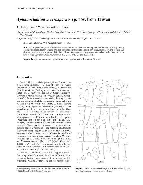
Figure 1. <strong>Aphanocladium</strong> <strong>macro<strong>sp</strong>orum</strong>. Characteristics of its<br />
conidiophores and conidia on oat meal agar.
334 Botanical Bulletin of <strong>Academia</strong> <strong>Sinica</strong>, Vol. 40, 1999<br />
characteristics of this isolate fit the generic concept of<br />
<strong>Aphanocladium</strong>. Phialide, conidial morphology and dimensions<br />
easily distinguish this isolate <strong>from</strong> other known<br />
<strong>sp</strong>ecies in the genus. Thus, a new <strong>sp</strong>ecies,<br />
<strong>Aphanocladium</strong> <strong>macro<strong>sp</strong>orum</strong> J.L. Chen, W.S. Lin and<br />
S.S. Tzean is proposed.<br />
Materials and Methods<br />
Samples were collected <strong>from</strong> rotten bark in Kuohsing,<br />
Nantou County during October, 1996 and incubated in<br />
moist chambers (plastic boxes, 30 × 20 × 12 cm, with<br />
three layers of moistened papers) to encourage fungal<br />
<strong>sp</strong>orulation. Pure culture was established by isolating a<br />
single <strong>sp</strong>ore or <strong>sp</strong>ores with a sterile glass microneedle<br />
on 3% agar. A piece of agar containing isolated <strong>sp</strong>ores<br />
was cut out and transferred to oat meal agar (OMA) slants<br />
or plates under a stereomicroscope. Details of fungal<br />
morphology and conidiogenesis were studied and<br />
recorded. The fungus was illustrated using a drawing tube<br />
and photographed using an Olympus light microscope<br />
(BX50). The taxonomic systems of Barron (1968),<br />
Hughes (1953), Tubaki (1963), Ellis (1971) and Saccardo<br />
(1882-1931) were used for identification. Both live cultures<br />
and dried <strong>sp</strong>ecimens were deposited in the Herbarium<br />
of the Chen-Fungi-Collection (Herb. CFC).<br />
Species Descriptions<br />
<strong>Aphanocladium</strong> <strong>macro<strong>sp</strong>orum</strong> J.L. Chen, W.S. Lin, and<br />
S.S. Tzean <strong>sp</strong>. <strong>nov</strong>. (Figures 1-2)<br />
Figure 2. <strong>Aphanocladium</strong> macro<strong>sp</strong>roum. A-B, phialide-like conidiogenous cells. A. Bar = 50 µm; B. Bar = 20 µm. C-D, globose,<br />
subglobose or ellipsoidial conidia. Bars = 20 µm.
Chen et al. — A new <strong>sp</strong>ecies of <strong>Aphanocladium</strong> <strong>from</strong> <strong>Taiwan</strong> 335<br />
Coloniae in OMA effusae, floccosae, albae; reversae<br />
albae; Mycelium fere superficiale, ex hyphis ramosis,<br />
septatis, lenibus, hyalinis, 1.6-8.0 µm latum compositum;<br />
Cellulae conidiogenae terminales vel laterales,<br />
monoblastae, directae, simplices vel fasciculatae,<br />
aciculares, laeves, hyalinae, 20.0-67.2 × 0.6-1.6 µm, cum<br />
collum ad apicem; Conidia solitaria, globosa ad<br />
subglobosa vel ellipsoidea, lenia, hyalina, 5.2-14.1 µm<br />
longus, 4.8-13.6 µm latus.<br />
In Cortice putrido, Kuohsing, Nantou, 13-X-96;<br />
Holotypus, Herb. CFC-2.<br />
Colonies on Oat meal Agar effuse, floccose, white; reverse<br />
white; Mycelium mostly superficial, composed of<br />
branched, septate, smooth, hyaline, 1.6-8.0 µm wide<br />
hyphae; Conidiogenous cells terminal or lateral,<br />
monoblastic, simple or fasciculate, acicular, with a collar<br />
at the apex, smooth, hyaline, 20.0-67.2 × 0.6-1.6 µm;<br />
Conidia solitary, globose to subglobose or ellipsoidal,<br />
smooth, hyaline, 5.2-14.1 µm long, 4.8-13.6 µm wide.<br />
Isolated <strong>from</strong>: rotten bark, Kuohsing, Nantou, Oct. 13<br />
1996. CTN-69.<br />
Chen et al. (1984, 1985) have made two comparative<br />
tables detailing the morphological characteristics of six<br />
<strong>sp</strong>ecies of <strong>Aphanocladium</strong>. The six <strong>sp</strong>ecies were<br />
compared and discussed including A. aranearum var.<br />
sinense, A. dimorphum, A. album, A. aranearum, A.<br />
meliolae and A. <strong>sp</strong>ectabile. The remaining <strong>sp</strong>ecies not<br />
included in the tables is A. tomentosum Arambarri, which<br />
is the only synnematous <strong>sp</strong>ecies of <strong>Aphanocladium</strong> and<br />
was collected <strong>from</strong> fallen leaves of Nothofagus pumilio<br />
in Argentina. The conidiogenous cells of A. tomentosum<br />
differ <strong>from</strong> those of A. <strong>macro<strong>sp</strong>orum</strong> as they are shorter<br />
and broader (11.0-13.5 × 2.4-3.0 µm) and produce<br />
smaller conidia (8.0-9.6 × 1.8-4.5 µm). Conidiogenous<br />
cells of A. <strong>macro<strong>sp</strong>orum</strong> are more slender and longer<br />
(20.0-67.2 × 0.6-1.6 µm) than those of all other known<br />
<strong>sp</strong>ecies in the genus, and the conidia, which are globose<br />
or subglobose, are the largest (5.2-14.1 × 4.8-13.6 µm)<br />
among the <strong>sp</strong>ecies. The conidial morphology of A. album<br />
is similar to that of A. <strong>macro<strong>sp</strong>orum</strong>, but the conidia of<br />
A. album are smaller (2.8-5.5 µm long, 1.7-3.7 µm wide).<br />
Conidial shape in other <strong>sp</strong>ecies of <strong>Aphanocladium</strong> is<br />
described as obovoid, ellipsoidal, short-ellipsoidal or<br />
ovoid. The unique characteristics of A. macro<strong>sp</strong>erum<br />
clearly distinguish it <strong>from</strong> all other members of<br />
<strong>Aphanocladium</strong>.<br />
Acknowledgments. We thank the National Science Council<br />
(NSC-86-2314-B-041-008) for funding this research.<br />
Literature Cited<br />
Arambarri, A.M. 1981. Micoflora de La hojarasca de Nothofagus<br />
obliqua de Y N. pumilio. I. Boln. Soc. Argent. Bot. 20(1-<br />
2): 19-30.<br />
Barron, G.L. 1968. The Genera of Hyphomycetes <strong>from</strong> Soil. The<br />
Williams and Wilkins Co., Baltimore.<br />
Chen, J.D., J.G. Liu, S.S. Chen, F.J. Cai, and Z.C. Zhang. 1984.<br />
Studies on <strong>Aphanocladium</strong> aranearum (Petch) Gams var.<br />
sinense var. <strong>nov</strong>. — A fungus parasitic on Agaricus<br />
bi<strong>sp</strong>orus (Lange) Sing. Acta Mycol. Sin. 3(2): 96-101<br />
Chen, J.D., J.G. Liu, S.S. Chen, F.J. Cai, and Z.C. Zhang. 1985.<br />
A new <strong>sp</strong>ecies of <strong>Aphanocladium</strong> on Agaricus bi<strong>sp</strong>orus.<br />
Acta Mycol. Sin. 4(4): 227-233.<br />
Ellis, M.B. 1971. Dematiaceous Hyphomycetes. Commonwealth<br />
Mycological Institute. Kew. Surrey, England.<br />
Gams, W. 1971. Cephalo<strong>sp</strong>roiumartige Schimmelpilze<br />
(Hyphomycetes). Gustar Fischer Verlag, Stuttgart, 262 pp.<br />
Gams, W. 1973. Phialides with solitary conidia? Remarks on<br />
conidium ontogeny in some hyphomycetes. Persoonia 7(2):<br />
161-169.<br />
Hughes, S.J. 1953. Conidiophores, conidia and classification. Can.<br />
J. Bot. 31: 577-659.<br />
Petch, T. 1932. Notes on Entomogenous Fungi. Trans. Br. Mycol.<br />
Soc. 16: 242-243.<br />
Saccardo, P.A. 1882-1931. Sylloge Fungorum Omnium<br />
Cognitorum. 25 volumes. Pavia, Italy.<br />
Tubaki, K. 1963. Taxonomic study of Hyphomycetes. Annu. Rep.<br />
Inst. Ferment. Osaka 1: 25-54.
336 Botanical Bulletin of <strong>Academia</strong> <strong>Sinica</strong>, Vol. 40, 1999<br />
<strong>Aphanocladium</strong> <strong>macro<strong>sp</strong>orum</strong> <strong>sp</strong>. <strong>nov</strong>.<br />
Aphanocldium <strong>macro<strong>sp</strong>orum</strong><br />
J.L. Chen, W.S. Lin and S.S. Tzean (conidiogenous<br />
cells; phialide-like) (solitary)<br />
<strong>macro<strong>sp</strong>orum</strong><br />
(<strong>Aphanocladium</strong> <strong>macro<strong>sp</strong>orum</strong> <strong>sp</strong>. <strong>nov</strong>.)