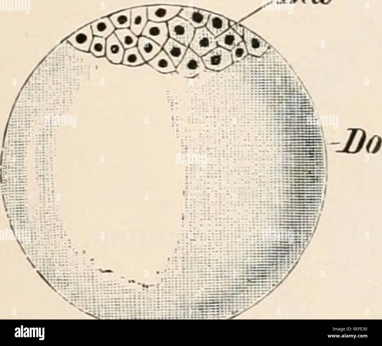
. Comparative anatomy of vertebrates. Anatomy, Comparative; Vertebrates. FIG. 3.—BLASTOSPHERE. BD, blastoderm ; FH, segmentation cavity. derm. Consisting primarily of a single layer of cells, the blasto- derm later on becomes two-layered, and then three-layered. From the relative position of these, they are spoken of respectively as the outer, middle, and inner germinal layers, or as ecto- derm (epiblast), mesoderm (mesoUast*), and endoderm (hypoblasf). The mode of distribution of the yolk-particles in the ovum, and an increase in their amount, result in certain modifications of the primitive
Image details
Contributor:
The Book Worm / Alamy Stock PhotoImage ID:
REFE30File size:
7.1 MB (224.8 KB Compressed download)Releases:
Model - no | Property - noDo I need a release?Dimensions:
1730 x 1444 px | 29.3 x 24.5 cm | 11.5 x 9.6 inches | 150dpiMore information:
This image is a public domain image, which means either that copyright has expired in the image or the copyright holder has waived their copyright. Alamy charges you a fee for access to the high resolution copy of the image.
This image could have imperfections as it’s either historical or reportage.
. Comparative anatomy of vertebrates. Anatomy, Comparative; Vertebrates. FIG. 3.—BLASTOSPHERE. BD, blastoderm ; FH, segmentation cavity. derm. Consisting primarily of a single layer of cells, the blasto- derm later on becomes two-layered, and then three-layered. From the relative position of these, they are spoken of respectively as the outer, middle, and inner germinal layers, or as ecto- derm (epiblast), mesoderm (mesoUast*), and endoderm (hypoblasf). The mode of distribution of the yolk-particles in the ovum, and an increase in their amount, result in certain modifications of the primitive form of segmentation as de- scribed above. Yolk is an inert substance, and its pre- sence tends to hinder or even entirely to prevent segmenta- tion in those parts of the ovum in which it is abundant. When the whole ovum undergoes division, the segmentation is known as entire or lioloblastic; when division is restricted to part of the ovum only, the segmentation is said to be partial or mcro- blasticl (Fig. 4). The question as to the origin of the germinal layers, on account of its im- portant signification, is one of the most burning problems in morphology, and as yet we cannot arrive at any full and satisfactory conclusion on the subject. It may, however, be stated that in all Vertebrates the blastosphere passes—or did so in earlier times—into a stage called the gastrula, which is retained in an unmodified form only in the lowest Vertebrate (Amphioxus, cf. p. 14). The gastrula is derived primitively from the blastula by the walls of the latter (Fig. 3) becoming pushed in or invaginated at one part, thus giving rise to a double-walled sac (Fig. 5). The outer wall then represents the ectoderm, which serves as an organ of protection and sensation, while the inner, or endoderm, encloses 1 In holoblastic segmentation the resulting cells are approximately equal in the Lancelet and in Mammals (with the exception of Monotremes and some Marsupials); and unequal in the Cyclostome