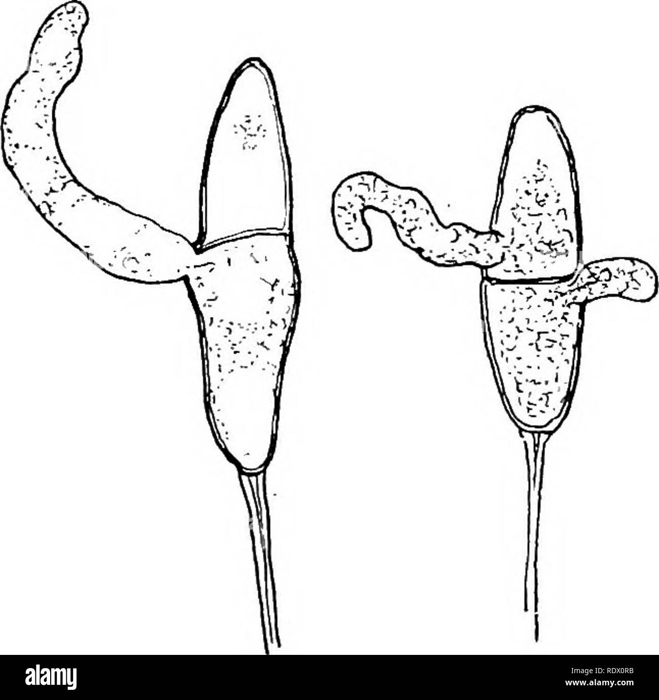
. Fungi, ascomycetes, ustilaginales, uredinales. Fungi. 198 PROTOBASIDIOMYCETES [CH. (sporidium) is formed and receives the nucleus and cytoplasm of the cell from which it arose. In Coleosporiuni, Ochropsora, and Chrysospora, nuclear division and septation take place within the teleutospore wall, and the basidiospores are budded out from it, so that the teleutospore cell becomes the basidium directly; in the majority of cases, however, the structure of the teleutospore is not such as readily to allow further growth, and development takes place after the extrusion of the contents as a tubular o
Image details
Contributor:
The Book Worm / Alamy Stock PhotoImage ID:
RDX0RBFile size:
7.2 MB (164.1 KB Compressed download)Releases:
Model - no | Property - noDo I need a release?Dimensions:
1586 x 1576 px | 26.9 x 26.7 cm | 10.6 x 10.5 inches | 150dpiMore information:
This image is a public domain image, which means either that copyright has expired in the image or the copyright holder has waived their copyright. Alamy charges you a fee for access to the high resolution copy of the image.
This image could have imperfections as it’s either historical or reportage.
. Fungi, ascomycetes, ustilaginales, uredinales. Fungi. 198 PROTOBASIDIOMYCETES [CH. (sporidium) is formed and receives the nucleus and cytoplasm of the cell from which it arose. In Coleosporiuni, Ochropsora, and Chrysospora, nuclear division and septation take place within the teleutospore wall, and the basidiospores are budded out from it, so that the teleutospore cell becomes the basidium directly; in the majority of cases, however, the structure of the teleutospore is not such as readily to allow further growth, and development takes place after the extrusion of the contents as a tubular outgrowth, the so-called promycelium, surrounded only by a delicate membrane (fig. 167). The nucleus migrates into this structure and here nuclear division takes place, transverse septa are formed and the basidiospores are produced. But it must be noted that the nucleus and cytoplasm of the young basidium are those of the teleutospore cell, whether development takes place within the original wall or by means of a promycelium. When the basidiospore germinates its germ-tube penetrates through the cuticle of the host and forms a mycelium of uninucleate cells bearing spermogonia and aecidia. The spermogonium is usually found on the adaxial side of the leaf; it consists of a group of more or less parallel, unbranched, upwardly directed hyphae, arising from a small-celled tangle below the epidermis or cuticle of the host. In the majority of cases the outer hyphae of each group elongate to form paraphyses, so that the spermogonium is restricted in extent, and acquires a flask-shaped or pyriform outline; the paraphyses push up through the ruptured epidermis of the host to project at a narrow ostiole (fig. 168 b). In. Fig. 167. Gymnosporangium clavariaeforme Rees; germinating teleutospores; x 666.. Please note that these images are extracted from scanned page images that may have been digitally enhanced for readability - coloration and appearance of these illustrations may not perfectly