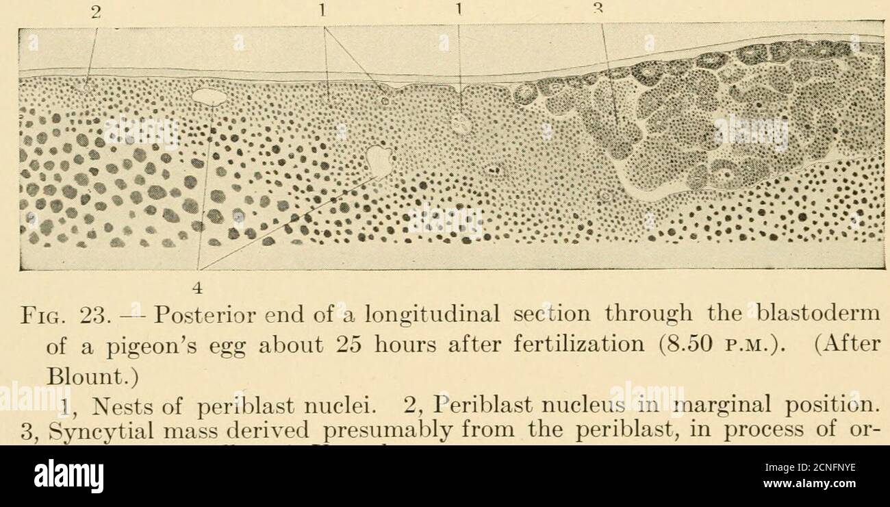
. The development of the chick; an introduction to embryology . r of the blastoderm of a pigeons egg, 14^ hours after fertilization (10.30 a.m.). (After Blount.) 1, Marginal cells. 2, Marginal periblast. 3, Nuclei of the subgerminalperiblast. 50 THE DEVELOPMENT OF THE CHICK a ring around the blastoderm. It persists during the expansionof the blastoderm over the surface of the yolk. The blastoderm now begins to expand, owing largely, at first,to additions of cells to its margin cut off from the periblast. Thecentral as well as the marginal periblast contributes to the blas-toderm, but the forme
Image details
Contributor:
Reading Room 2020 / Alamy Stock PhotoImage ID:
2CNFNYEFile size:
7.2 MB (388.2 KB Compressed download)Releases:
Model - no | Property - noDo I need a release?Dimensions:
2236 x 1118 px | 37.9 x 18.9 cm | 14.9 x 7.5 inches | 150dpiMore information:
This image could have imperfections as it’s either historical or reportage.
. The development of the chick; an introduction to embryology . r of the blastoderm of a pigeons egg, 14^ hours after fertilization (10.30 a.m.). (After Blount.) 1, Marginal cells. 2, Marginal periblast. 3, Nuclei of the subgerminalperiblast. 50 THE DEVELOPMENT OF THE CHICK a ring around the blastoderm. It persists during the expansionof the blastoderm over the surface of the yolk. The blastoderm now begins to expand, owing largely, at first, to additions of cells to its margin cut off from the periblast. Thecentral as well as the marginal periblast contributes to the blas-toderm, but the former appears to be rapidly used up. The mar-ginal periblast, which is commonly called the germ-wall fromthis stage, on the other hand grows at its periphery while it addscells to the blastoderm centrally, and thus it moves out in thewhite yolk, building up the margin of the blastoderm at the sametime. The original group of central cells appears to correspondapproximately to the pehucid area; the additions from the germ-wall would thus constitute the opaque area.. eanization into cells. 4, Vacuoles fe Some phases of these processes are illustrated in Figs. 23 and24. In the vertical section. Fig. 23, the surface of the germ-wall next the blastoderm is indented as though for the formationof superficial cells. Along the steep central margin of the germ-wall groups of cells are apparently being cut off and added tothe cellular blastoderm. In the horizontal section, Fig. 24, theprocess of cellularization at the central margin of the germ-wallis apparently proceeding rapidly. The superficial cells thus added to the margin of the cellularblastoderm become continuous with the ectoderm, and thedeeper layers later form the yolk-sac entoderm which becomescontinuous with the embryonic entoderm secondarily. We can DEVELOPMENT PRIOR TO LAYING 51 thus distinguish a syncytial, more peripheral, and a cellular, morecentral, portion of the germ-wall.